診斷方法 (Diagnostic Approaches)
臨床評估與外眼檢查
- 由於結膜 (conjunctiva) 容易直接觀察,腫瘤與其他病灶通常在相對早期即可被辨認與診斷。
- 經驗豐富的眼科醫師往往可藉由仔細的外眼檢查 (external ocular examination) 與裂隙燈生物顯微鏡檢查 (slit-lamp biomicroscopy) 做出準確診斷。
- 必須評估球結膜 (bulbar)、瞼結膜 (palpebral)、上下穹窿結膜 (forniceal conjunctiva) 以及角膜 (cornea) 是否有腫瘤侵犯,並應將病灶拍照存證,以記錄腫瘤及其邊界。
- 鱗狀細胞 (squamous) 或黑色素細胞 (melanocytic) 腫瘤對角膜的侵犯可能很細微,表現為灰色的表面混濁 (gray surface opacity)。
切片與組織學診斷
- 結膜或角膜腫瘤的確定診斷需要組織學檢查 (histologic examination)。
- 小型(≤ 4 個鐘點位 (clock hours) 的角膜緣 (limbal) 腫瘤,或基底徑 ≤ 15 mm)、無症狀、外觀為良性的腫瘤通常採觀察追蹤,並在有生長或惡性變化證據時施行切片。
- 對於有症狀或疑似惡性的小型腫瘤,建議完整切除(切除性切片,excisional biopsy)。
- 對於大型病灶(> 4 個鐘點位的角膜緣腫瘤,或基底徑 > 15 mm),建議施行切取性切片 (incisional biopsy),因為完整切除可能嚴重損害眼表 (ocular surface),或根本無法完整切除。
- 廣泛的結膜切除會減少杯狀細胞 (goblet cells) 的數量,進而干擾角膜的濕潤。由此產生的乾眼 (dry eye) 使病人易發生角膜潰瘍 (corneal ulceration) 與疼痛性的視力喪失。
- 切取性切片亦適用於以放射治療 (radiotherapy)、化學治療 (chemotherapy) 或局部方式(如冷凍治療 cryotherapy 或局部化學治療 topical chemotherapy)處理的腫瘤,例如淋巴樣腫瘤 (lymphoid tumors)、轉移性腫瘤 (metastatic tumors),以及部分鱗狀細胞癌 (squamous cell carcinoma) 與原發性後天性黑色素沉著 (primary acquired melanosis, PAM) 的病例。
- 脫落細胞學 (exfoliative cytology) 在選定的病例中可作為有用的輔助手段,但其僅能提供病灶淺層的資訊,無法反映侵犯的深度。
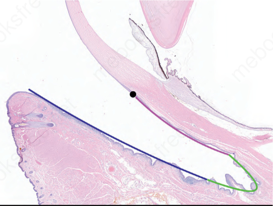
圖 27-1:正常眼部結構:一件眼眶內容物剜除標本 (exenteration specimen) 的矢狀切面 (sagittal section),呈現結膜的不同區域——角膜緣 (limbus) 為黑色、球結膜 (bulbar conjunctiva) 為粉紅色、穹窿 (fornix) 為綠色、瞼(瞼板)結膜 (palpebral (tarsal) conjunctiva) 為深藍色。
Fig. 27.1 Normal eye structure: a sagital section of an exanteration specimen illustrates the different regions of the conjunctiva – limbus in black, bulbar conjunctiva in pink, fornix in green and palpebral (tarsal) conjunctiva in dark blue.
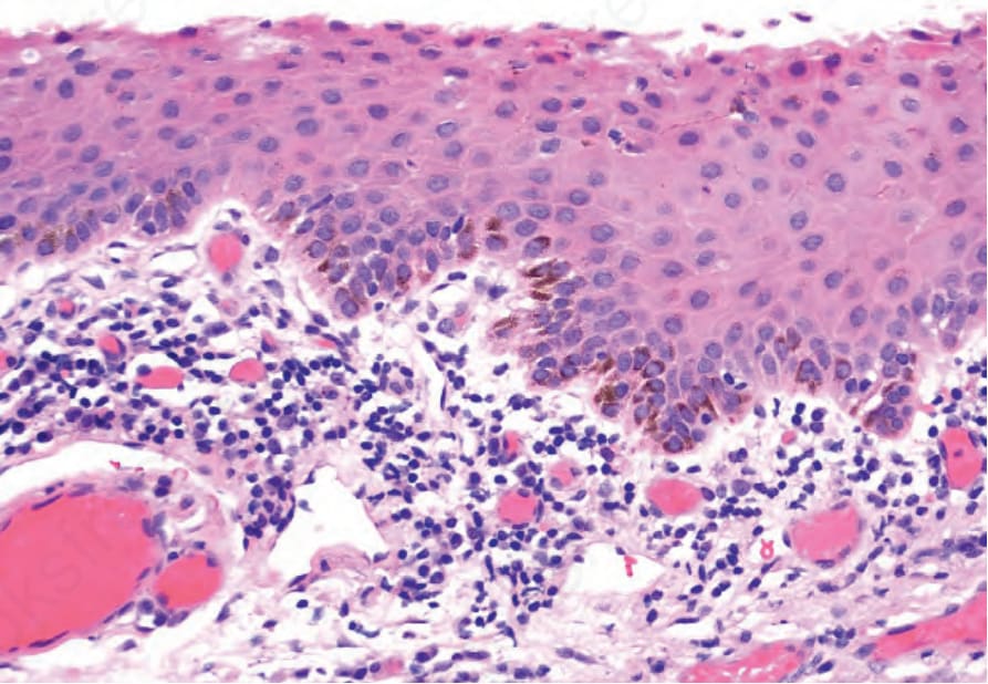
圖 27-2:正常角膜緣 (limbus):組織學顯示非角化性鱗狀上皮 (nonkeratinizing squamous epithelium) 與 Vogt 柵欄 (palisades of Vogt),基底上皮 (basal epithelium) 含黑色素沉著,並散在上皮內樹突狀黑色素細胞 (intraepithelial dendritic melanocytes)。
Fig. 27.2 Normal limbus: histology shows nonkeratinizing squamous epithelium and palisades of Vogt with melanin pigmentation of the basal epithelium and scattered intraepithelial dendritic melanocytes.
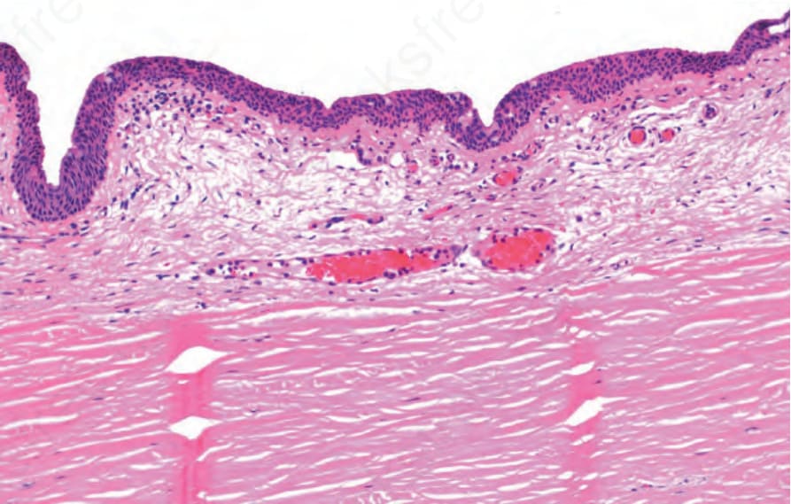
圖 27-3:正常球結膜 (bulbar conjunctiva):組織學顯示鱗狀黏膜 (squamous mucosa) 內散在杯狀細胞 (goblet cells),固有層 (substantia propria) 為疏鬆結締組織 (loose connective tissue)。
Fig. 27.3 Normal bulbar conjunctiva: histology shows squamous mucosa with scattered goblet cells and loose connective tissue in the substantia propria.
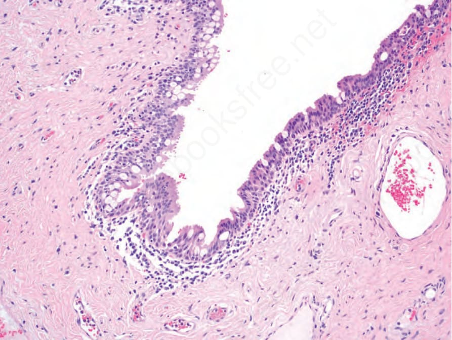
圖 27-4:正常穹窿 (fornix):組織學顯示偽複層柱狀上皮 (pseudostratified columnar epithelium) 含眾多杯狀細胞 (goblet cells),固有層 (substantia propria) 內可見淋巴球 (lymphocytes) 與漿細胞 (plasma cells)。
Fig. 27.4 Normal fornix: histology shows pseudostratified columnar epithelium with numerous goblet cells and lymphocytes and plasma cells in the substantia propria.