Diagnostic approaches
Diagnostic approaches
Because the conjunctiva is readily visible, tumors and other lesions are usually recognized and diagnosed at a relatively early stage. An experienced ophthalmologist can often make an accurate diagnosis with careful external ocular examination and slit-lamp biomicroscopy. The bulbar, palpebral, and upper and lower forniceal conjunctiva, as well as the cornea, must be evaluated for tumor involvement, and lesions should be photographed to document the tumors and their margins. Corneal involvement by squamous or melanocytic neoplasia may be subtle, and appear as gray surface opacity.
Definitive diagnosis of conjunctival or corneal tumors requires histologic examination. Small (≤ 4 clock hours limbal tumor or ≤ 15 mm basal
1364 Tumors of the conjunctiva
only provides information about superficial layers of the lesion and not the degree of invasiveness.4
dimension), asymptomatic, benign appearing tumors are usually observed, and biopsied when there is evidence of growth or malignant change. For small tumors that are symptomatic or suspected to be malignant, complete removal (excisional biopsy) is recommended. For large lesions (> 4 clock hour limbal tumor or > 15 mm basal dimension), incisional biopsy is recommended because complete removal can severely compromise the ocular surface or may not be possible. Extensive conjunctival resection decreases the number of goblet cells, which interferes with corneal wetting. The resultant dry eye predisposes the patient to corneal ulceration and painful loss of vision.3 Incisional biopsy is also appropriate for tumors that are treated with radiotherapy, chemotherapy, or local means such as cryotherapy or topical chemotherapy, such as lymphoid tumors, metastatic tumors, and some cases of squamous cell carcinoma and primary acquired melanosis (PAM). Exfoliative cytology can be a helpful adjunct in select cases, but
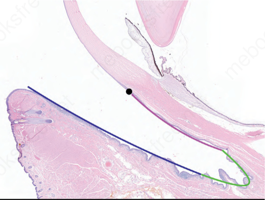
Fig. 27.1 Normal eye structure: a sagital section of an exanteration specimen illustrates the different regions of the conjunctiva – limbus in black, bulbar conjunctiva in pink, fornix in green and palpebral (tarsal) conjunctiva in dark blue.
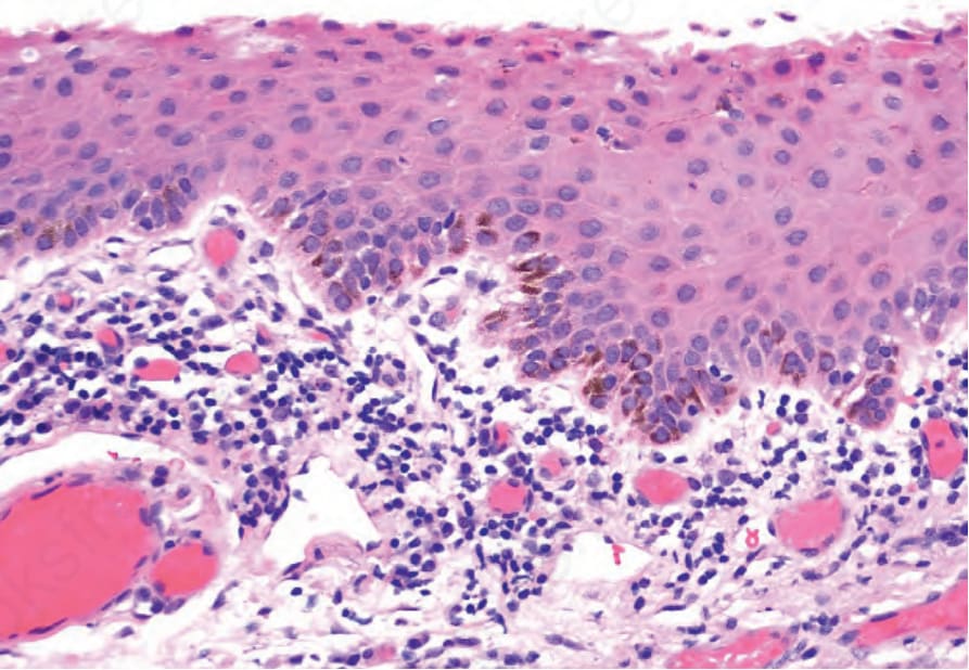
Fig. 27.2 Normal limbus: histology shows nonkeratinizing squamous epithelium and palisades of Vogt with melanin pigmentation of the basal epithelium and scattered intraepithelial dendritic melanocytes.
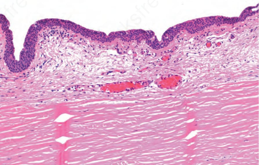
Fig. 27.3 Normal bulbar conjunctiva: histology shows squamous mucosa with scattered goblet cells and loose connective tissue in the substantia propria.
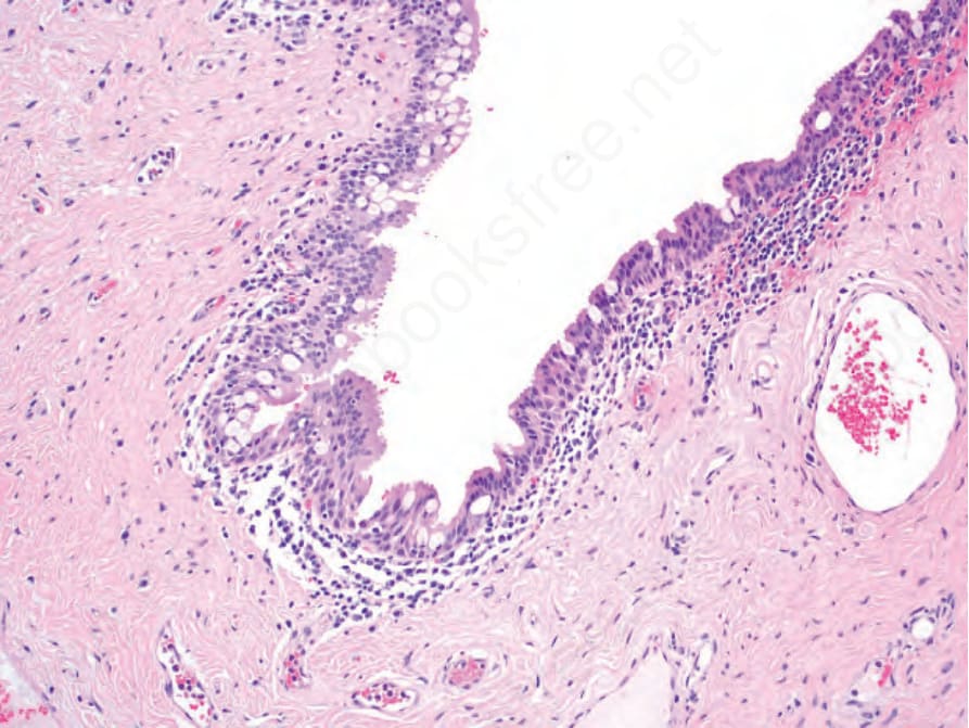
Fig. 27.4 Normal fornix: histology shows pseudostratified columnar epithelium with numerous goblet cells and lymphocytes and plasma cells in the substantia propria.