基底-黑色素細胞腫瘤 (Basomelanocytic Tumor)
臨床特徵 (Clinical Features)
- 基底-黑色素細胞腫瘤 (basomelanocytic tumor) 為一種極為罕見的雙相 (biphasic) 腫瘤,與前述的真皮鱗狀-黑色素細胞腫瘤 (dermal squamomelanocytic tumor) 相似,或可能彼此相關。
- 然而,basomelanocytic tumor 是 basaloid carcinoma 與 melanoma 的組合。誠如所預期,腫瘤中的黑色素細胞 (melanocytic) 成分似乎是較具侵襲性的成分,且曾有轉移並導致致命結局的報告。
組織病理特徵 (Histopathology)
- 在這種混合性腫瘤中,於少數已報導的病例裡,basaloid 成分可呈現 basaloid squamous cell carcinoma 的外觀,或更接近 basal cell carcinoma 的型態 (Fig. 26.128)。
- 與所謂的碰撞瘤 (collision tumors) 不同,雙相腫瘤顯示上皮成分與黑色素細胞成分之間有緊密的關聯。表皮內 (intraepidermal) 成分並非總是存在。
- 已有報導描述 melanoma 轉移至 basal cell carcinoma,或 melanoma in situ 對其進行群落化 (colonization) 的病例,作為 basomelanocytic tumor 的擬態 (mimics)。
- melanoma 成分以傳統黑色素細胞標記如 S100 protein、MART-1 與 HMB-45 染色呈陽性 (Fig. 26.129)。包含 AE1/AE3 在內的 cytokeratins 可標記 basaloid 成分,Ber-EP4 亦可呈陽性 (Fig. 26.130)。在 melanoma 成分中可見特徵性的拷貝數變異 (copy number alterations)。
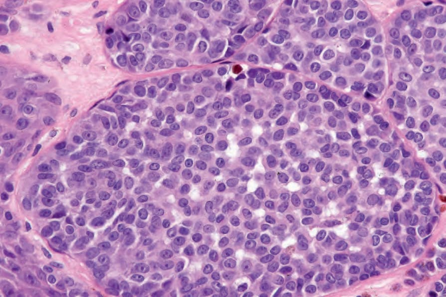
圖 26-122:兒童黑色素瘤 (childhood melanoma):膨脹性結節 (expansile nodules) 的存在是重要的診斷線索。圖片承蒙 M. Little, MD, University College Hospital, Galway, Ireland 提供。
Fig. 26.122 Childhood melanoma: the presence of expansile nodules is an important diagnostic clue. By courtesy of M. Little, MD, University College Hospital, Galway, Ireland.
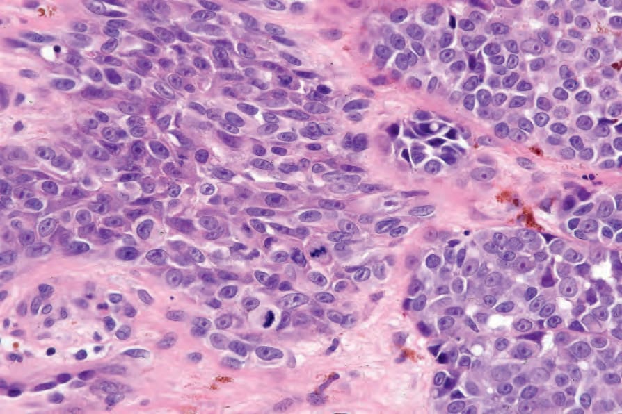
圖 26-123:兒童黑色素瘤 (childhood melanoma):此視野中可見多個真皮內有絲分裂 (dermal mitoses)。圖片承蒙 M. Little, MD, University College Hospital, Galway, Ireland 提供。
Fig. 26.123 Childhood melanoma: multiple dermal mitoses are present in this field. By courtesy of M. Little, MD, University College Hospital, Galway, Ireland.
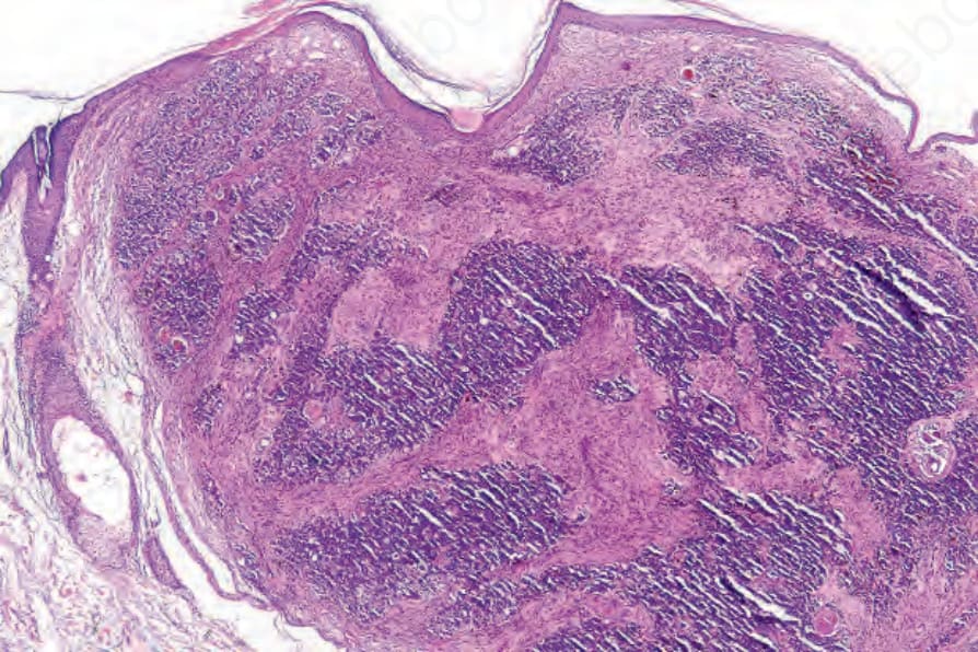
圖 26-124:真皮鱗狀-黑色素細胞腫瘤 (dermal squamomelanocytic tumor):一個顏面部腫瘤的低倍視野。圖片承蒙 S. Poole, MD, Beth Israel Deaconess Medical Center, Boston, USA 提供。
Fig. 26.124 Dermal squamomelanocytic tumor: low-power view of a facial tumor. By courtesy of S. Poole, MD, Beth Israel Deaconess Medical Center, Boston, USA.
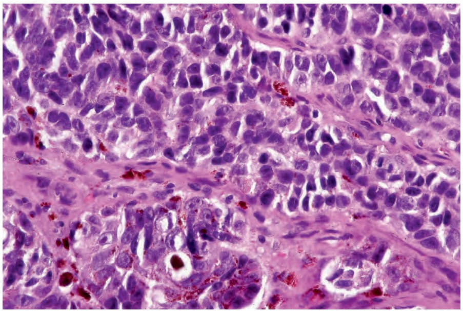
圖 26-125:真皮鱗狀增生性腫瘤 (dermal squamoproliferative tumor):此視野中,腫瘤由色素含量不一、核呈不規則深染 (irregular hyperchromatic nuclei) 的細胞所組成。圖片承蒙 S. Poole, MD, Beth Israel Deaconess Medical Center, Boston, USA 提供。
Fig. 26.125 Dermal squamoproliferative tumor: in this field, the tumor is composed of variably pigmented cells with irregular hyperchromatic nuclei. By courtesy of S. Poole, MD, Beth Israel Deaconess Medical Center, Boston, USA.
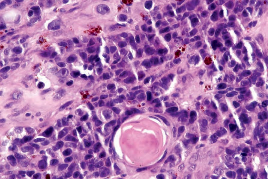
圖 26-126:真皮鱗狀增生性腫瘤 (dermal squamoproliferative tumor):可見局灶性鱗狀分化 (focal squamous differentiation)。圖片承蒙 S. Poole, MD, Beth Israel Deaconess Medical Center, Boston, USA 提供。
Fig. 26.126 Dermal squamoproliferative tumor: focal squamous differentiation is evident. By courtesy of S. Poole, MD, Beth Israel Deaconess Medical Center, Boston, USA.
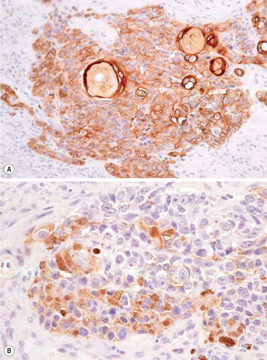
圖 26-127:真皮鱗狀增生性腫瘤 (dermal squamoproliferative tumor):(A) 鱗狀成分以 keratin 免疫組織化學染色凸顯;(B) S100 protein。圖片承蒙 S. Poole, MD, Beth Israel Deaconess Medical Center, Boston, USA 提供。
Fig. 26.127 Dermal squamoproliferative tumor: (A) the squamous component is highlighted with keratin immunohistochemistry; (B) S100 protein. By courtesy of S. Poole, MD, Beth Israel Deaconess Medical Center, Boston, USA.

圖 26-128:基底-黑色素細胞腫瘤 (basomelanocytic tumor):環繞著未分化腫瘤細胞的是一排顯著的嗜鹼性細胞柵欄狀排列 (palisade),為 basal cell carcinoma 的典型表現。圖片承蒙 L. Erickson, MD, Mayo Clinic, Rochester, Minnesota 提供。
Fig. 26.128 Basomelanocytic tumor: surrounding undifferentiated tumor cells is a striking palisade of basophilic cells typical of basal cell carcinoma. By courtesy of L. Erickson, MD, Mayo Clinic, Rochester, Minnesota.
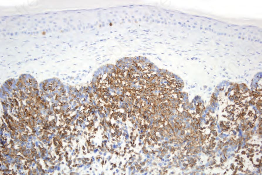
圖 26-129:基底-黑色素細胞腫瘤 (basomelanocytic tumor):內側細胞表現 HMB-45。Mart-1 亦呈陽性。圖片承蒙 L. Erickson, MD, Mayo Clinic, Rochester, Minnesota 提供。
Fig. 26.129 Basomelanocytic tumor: the inner cells express HMB-45. Mart-1 was also positive. By courtesy of L. Erickson, MD, Mayo Clinic, Rochester, Minnesota.
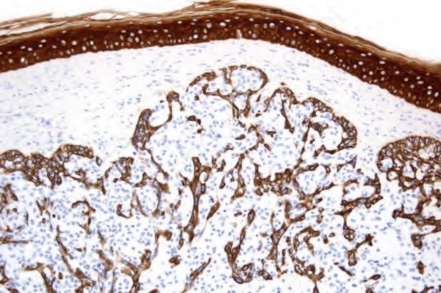
圖 26-130:基底-黑色素細胞腫瘤 (basomelanocytic tumor):背景的 basaloid 細胞族群表現 keratin。圖片承蒙 L. Erickson, MD, Mayo Clinic, Rochester, Minnesota 提供。
Fig. 26.130 Basomelanocytic tumor: the background population of basaloid cells expresses keratin. By courtesy of L. Erickson, MD, Mayo Clinic, Rochester, Minnesota.