Basomelanocytic tumor
Basomelanocytic tumor
Clinical features Basomelanocytic tumor is a very rare biphasic neoplasm similar, or perhaps related, to the dermal squamomelanocytic tumor described above.1
1352 Melanoma
However, basomelanocytic tumor is a combination of basaloid carcinoma and melanoma.2–4 As would be expected, the melanocytic component of the tumor appears to be the more aggressive component and has been reported to metastasize with fatal outcome.3
Histologic features In this combined tumor, the basaloid component in the few described cases can take the appearance of a basaloid squamous cell carcinoma or be more basal cell carcinoma-like (Fig. 26.128). Unlike so-called collision tumors, biphasic tumors show an intimate association of the epithelial and melanocytic components. An intraepidermal component is not always present. Cases of melanoma metastatic to a basal cell carcinoma or colonization by melanoma in situ have been described as mimics of basomelanocytic tumor.5,6
The melanoma component stains with traditional melanocytic markers such as S100 protein, MART-1, and HMB-45 (Fig. 26.129). Cytokeratins, including AE1/AE3, mark the basaloid component, and Ber-EP4 can also be positive (Fig. 26.130).3,4 Characteristic copy number alterations can be seen in the melanoma component.1
1353 Lentiginous melanoma
A
B
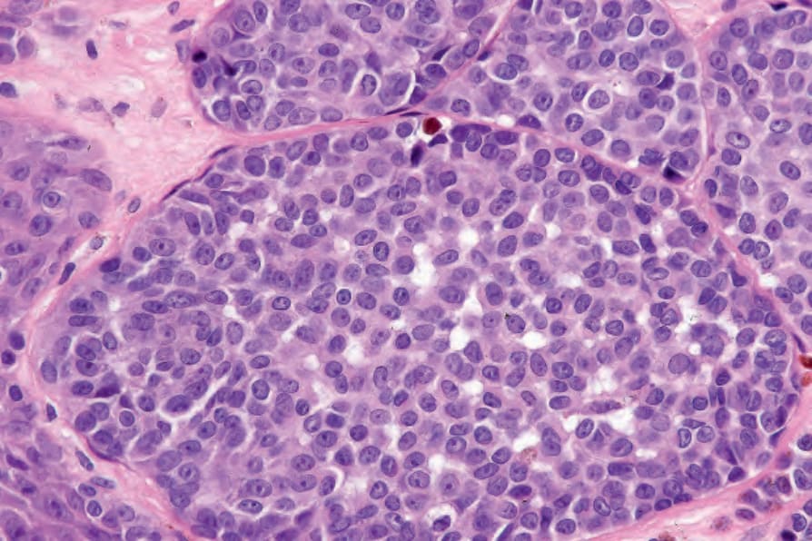
Fig. 26.122 Childhood melanoma: the presence of expansile nodules is an important diagnostic clue. By courtesy of M. Little, MD, University College Hospital, Galway, Ireland.
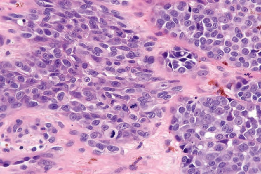
Fig. 26.123 Childhood melanoma: multiple dermal mitoses are present in this field. By courtesy of M. Little, MD, University College Hospital, Galway, Ireland.
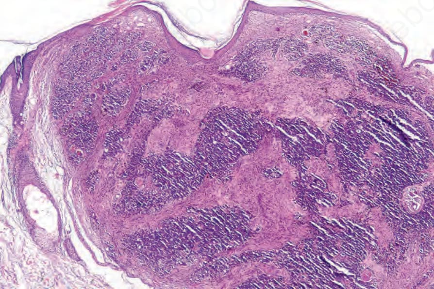
Fig. 26.124 Dermal squamomelanocytic tumor: low-power view of a facial tumor. By courtesy of S. Poole, MD, Beth Israel Deaconess Medical Center, Boston, USA.
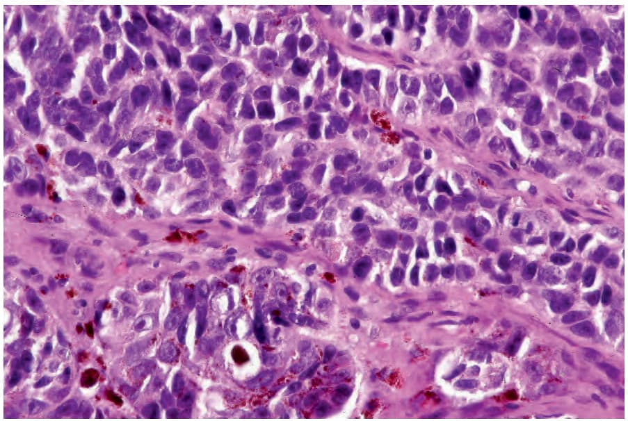
Fig. 26.125 Dermal squamoproliferative tumor: in this field, the tumor is composed of variably pigmented cells with irregular hyperchromatic nuclei. By courtesy of S. Poole, MD, Beth Israel Deaconess Medical Center, Boston, USA.
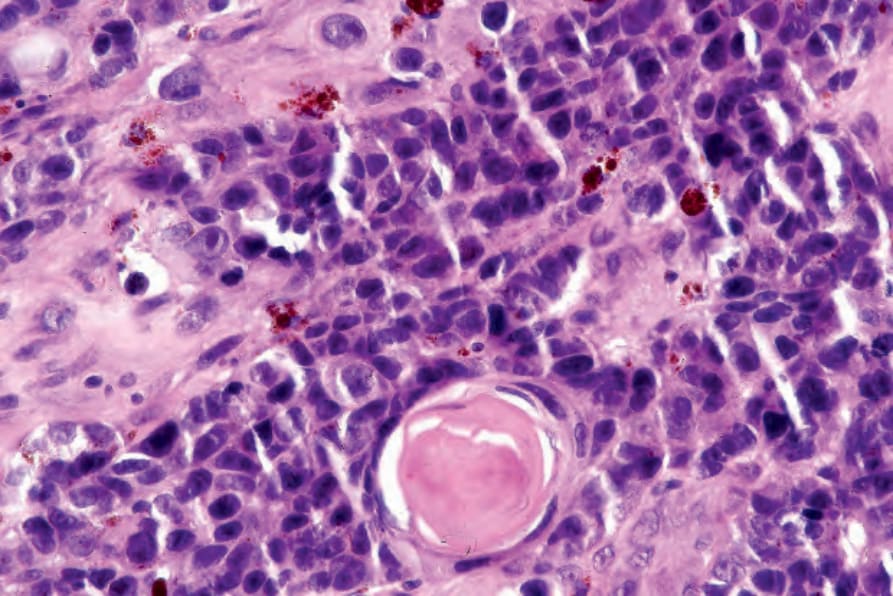
Fig. 26.126 Dermal squamoproliferative tumor: focal squamous differentiation is evident. By courtesy of S. Poole, MD, Beth Israel Deaconess Medical Center, Boston, USA.
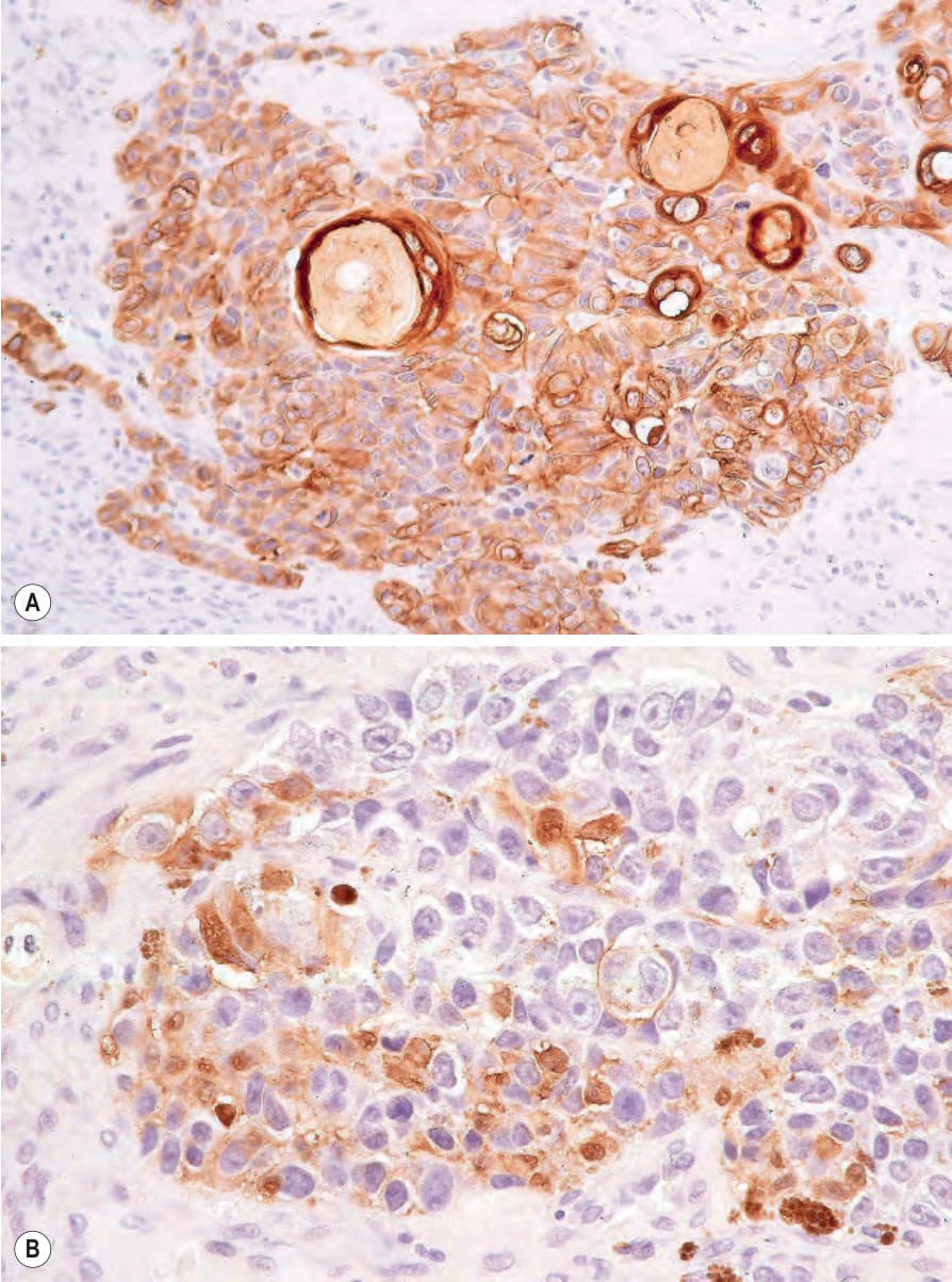
Fig. 26.127 Dermal squamoproliferative tumor: (A) the squamous component is highlighted with keratin immunohistochemistry; (B) S100 protein. By courtesy of S. Poole, MD, Beth Israel Deaconess Medical Center, Boston, USA.

Fig. 26.128 Basomelanocytic tumor: surrounding undifferentiated tumor cells is a striking palisade of basophilic cells typical of basal cell carcinoma. By courtesy of L. Erickson, MD, Mayo Clinic, Rochester, Minnesota.
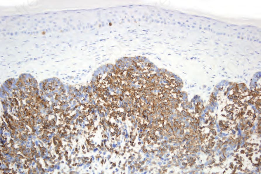
Fig. 26.129 Basomelanocytic tumor: the inner cells express HMB-45. Mart-1 was also positive. By courtesy of L. Erickson, MD, Mayo Clinic, Rochester, Minnesota.
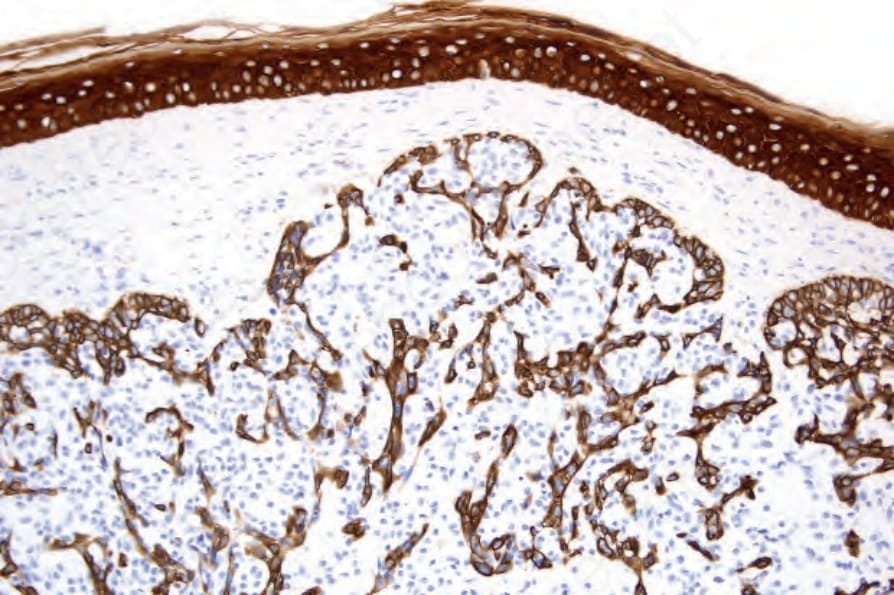
Fig. 26.130 Basomelanocytic tumor: the background population of basaloid cells expresses keratin. By courtesy of L. Erickson, MD, Mayo Clinic, Rochester, Minnesota.