臨床特徵 (Clinical Features)
- 在病理文獻中,真皮鱗狀黑色素細胞腫瘤 (dermal squamomelanocytic tumor) 的報告極為罕見。
- 患者表現為棕色或紫黑色的顏面結節 (facial nodule),直徑可達 1.0 cm。
- 文獻記載的年齡範圍為 32–94 歲,已報告的病例少於十五例,兩性分布相當。
- 曾有一例記載有前驅病灶為惡性小痣 (lentigo maligna)。
- 相對較短的追蹤資料(平均 2.7 年)顯示無復發或轉移之證據。
- 近期報告的一例「轉移性」病例,據稱顯示對前哨淋巴結 (sentinel lymph node) 的微轉移 (micrometastasis),但其外觀更符合包膜痣 (capsular nevus) 的特徵。
組織病理特徵 (Histopathology)
- 雖然可能可辨識出前驅病灶或源自表皮 (epidermis) 的來源,腫瘤也可能表現為一個與表皮無任何連結、獨立存在的上真皮結節 (upper dermal nodule)(Fig. 26.124)。
- 亦曾描述過僅局限於表皮 (epidermis) 的病例。
- 腫瘤由界線分明的小葉狀腫瘤 (lobular tumor) 構成,係由緊密混合的惡性鱗狀上皮 (malignant squamous epithelium) 與通常較不明顯的一群惡性黑色素細胞 (malignant melanocytes) 所組成(Figs 26.125 與 26.126)。
- 亦曾描述過基質分化 (matrical differentiation)。
- 此兩群細胞在形態學上彼此不同,而此區別可藉由 S100 protein 或其他黑色素細胞標記 (melanocytic markers) 以及 cytokeratin 免疫組織化學染色而更加凸顯(Fig. 26.127)。
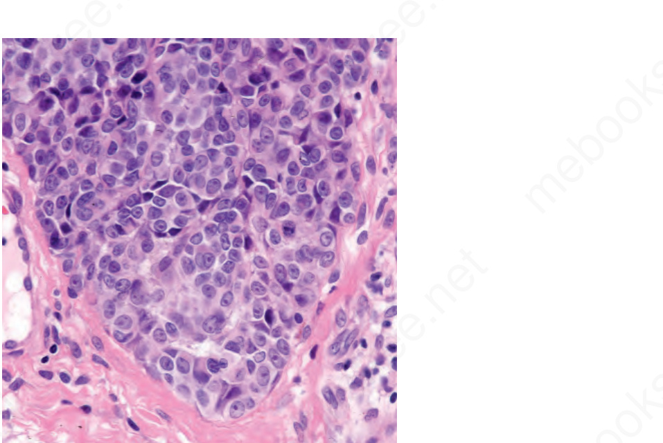
圖 26-121:兒童期黑色素瘤 (childhood melanoma):隨深度增加並無成熟現象 (maturation)。注意其界線分明的下緣。By courtesy of M. Little, MD, University College Hospital, Galway, Ireland.
Fig. 26.121 Childhood melanoma: there is no maturation with depth. Note the sharply delineated lower border. By courtesy of M. Little, MD, University College Hospital, Galway, Ireland.
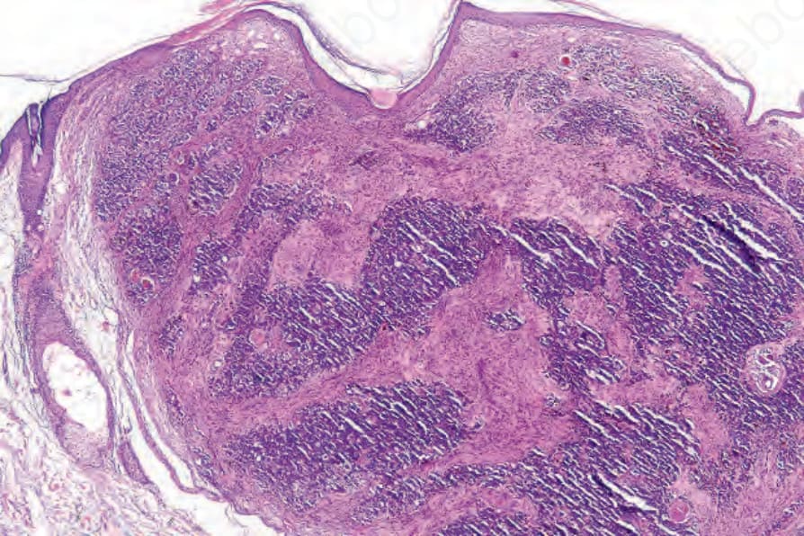
圖 26-124:真皮鱗狀黑色素細胞腫瘤 (dermal squamomelanocytic tumor):一顆顏面腫瘤的低倍視野。By courtesy of S. Poole, MD, Beth Israel Deaconess Medical Center, Boston, USA.
Fig. 26.124 Dermal squamomelanocytic tumor: low-power view of a facial tumor. By courtesy of S. Poole, MD, Beth Israel Deaconess Medical Center, Boston, USA.
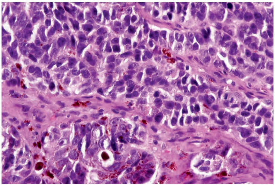
圖 26-125:真皮鱗狀增生性腫瘤 (dermal squamoproliferative tumor):在此視野中,腫瘤由色素深淺不一、具不規則深染細胞核 (irregular hyperchromatic nuclei) 的細胞所組成。By courtesy of S. Poole, MD, Beth Israel Deaconess Medical Center, Boston, USA.
Fig. 26.125 Dermal squamoproliferative tumor: in this field, the tumor is composed of variably pigmented cells with irregular hyperchromatic nuclei. By courtesy of S. Poole, MD, Beth Israel Deaconess Medical Center, Boston, USA.
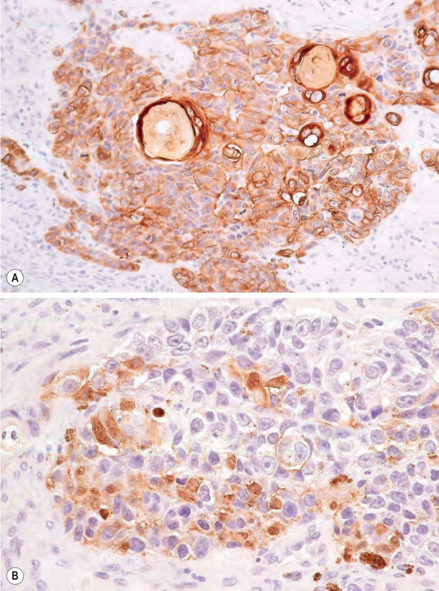
圖 26-127:真皮鱗狀增生性腫瘤 (dermal squamoproliferative tumor):(A) 鱗狀成分以 keratin 免疫組織化學染色凸顯;(B) S100 protein。By courtesy of S. Poole, MD, Beth Israel Deaconess Medical Center, Boston, USA.
Fig. 26.127 Dermal squamoproliferative tumor: (A) the squamous component is highlighted with keratin immunohistochemistry; (B) S100 protein. By courtesy of S. Poole, MD, Beth Israel Deaconess Medical Center, Boston, USA.