Dermal squamomelanocytic tumor
Dermal squamomelanocytic tumor
Clinical features Reports of this tumor are exceedingly rare in the pathology literature.1–6 Patients present with brown or purple-black facial nodules measuring up to 1.0 cm in diameter. The age range documented is 32–94 years and there have been less than 15 cases reported with equal sex distribution.5 A precursor lentigo maligna has been documented in one case.1 The relatively short follow-up information (mean 2.7 years) has shown no evidence of recurrence or metastasis.2 A recently reported ‘metastatic’ case purportedly showing micrometastasis to a sentinel lymph node has an appearance much more characteristic of a capsular nevus.6,7
Histologic features Although a precursor lesion or origin from the epidermis may be identifiable, the tumor may present as an upper dermal nodule independent of any epidermal connection (Fig. 26.124).2 A case limited to the epidermis has also been described.3 It consists of a sharply delineated, lobular tumor composed of intimately admixed malignant squamous epithelium and a usually less prominent population of malignant melanocytes (Figs 26.125 and
26.126).2 Matrical differentiation has also been described.5 The two populations are distinctive morphologically, but the distinction is highlighted by S100 protein or other melanocytic markers and cytokeratin immunohistochemistry (Fig. 26.127).2
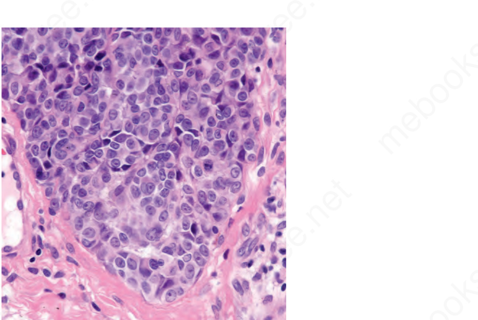
Fig. 26.121 Childhood melanoma: there is no maturation with depth. Note the sharply delineated lower border. By courtesy of M. Little, MD, University College Hospital, Galway, Ireland.
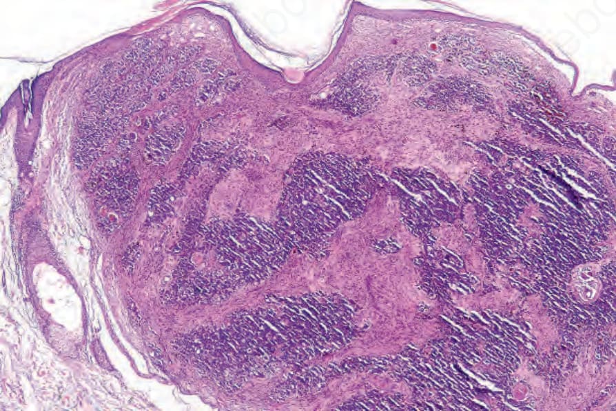
Fig. 26.124 Dermal squamomelanocytic tumor: low-power view of a facial tumor. By courtesy of S. Poole, MD, Beth Israel Deaconess Medical Center, Boston, USA.
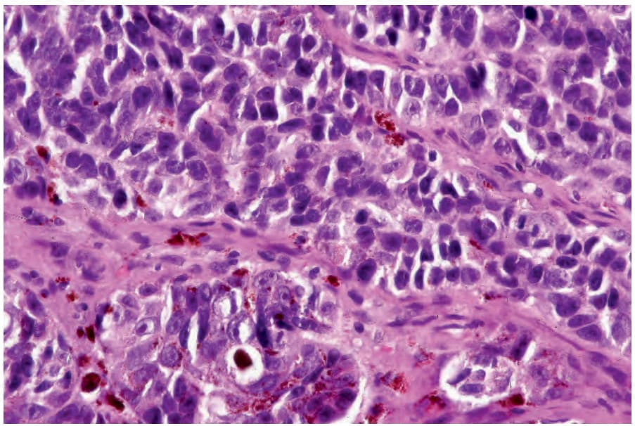
Fig. 26.125 Dermal squamoproliferative tumor: in this field, the tumor is composed of variably pigmented cells with irregular hyperchromatic nuclei. By courtesy of S. Poole, MD, Beth Israel Deaconess Medical Center, Boston, USA.
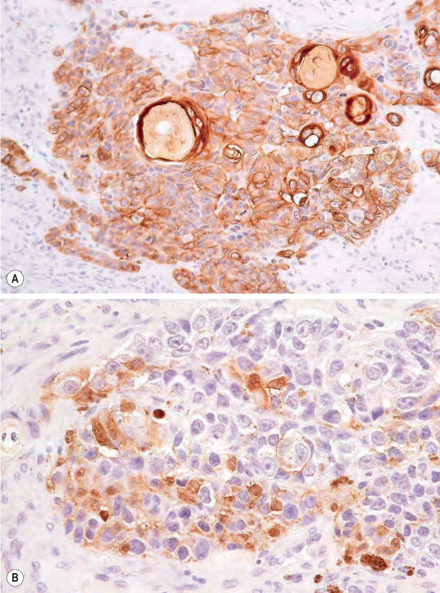
Fig. 26.127 Dermal squamoproliferative tumor: (A) the squamous component is highlighted with keratin immunohistochemistry; (B) S100 protein. By courtesy of S. Poole, MD, Beth Israel Deaconess Medical Center, Boston, USA.