肢端雀斑樣黑色素瘤 (Acral Lentiginous Melanoma)
組織病理特徵 (Histopathology)
- 在肢端雀斑樣黑色素瘤 (acral lentiginous melanoma) 放射狀生長期 (radial growth phase) 的早期階段,變化可能相當細微,僅由不規則的表皮增生 (irregular epidermal hyperplasia) 及散在、位於基底層的非典型黑色素細胞 (atypical melanocytes) 所構成。166–170
- 已建立的病灶顯示棘層肥厚 (acanthosis),伴隨表皮突 (epidermal ridges) 明顯延長,以及明顯的黑色素細胞非典型性 (melanocytic atypia)(Figs 26.33 and 26.34)。
- 表皮下層被大量非典型黑色素細胞所浸潤,其特徵為細胞核多形性 (nuclear pleomorphism) 與深染 (hyperchromatism),並呈現一種細胞質固定皺縮假象 (cytoplasmic fixation retraction artifact)。
- 核仁 (nucleoli) 明顯,可辨識出有絲分裂象 (mitotic figures)。
- 雖然最常見的是梭形 (spindled) 型態,但有時也可見上皮樣 (epithelioid) 與巨細胞 (giant cells)。
- 也可偵測到散在的接合處巢 (junctional nests) 病灶,通常位於表皮突的尖端 (Fig. 26.35)。
- 常見有濃密的帶狀慢性發炎細胞浸潤 (bandlike chronic inflammatory cell infiltrate)。
- 侵襲性腫瘤 (invasive tumor) 通常為梭形細胞型,並可能引發促結締組織增生反應 (desmoplastic reaction)(Fig. 26.36)。
- 沿汗腺上皮 (sweat gland epithelium) 的深部延伸很常見,部分病例中可見嗜神經性 (neurotropism)。
- 受侵犯汗腺的橫切面 (cross-sectioning) 可能導致誤判為侵襲。
- 偶爾,肢端腫瘤可能顯示表淺擴散性原位 (superficial spreading in situ) 成分,或代表缺乏放射狀生長期的新生結節型黑色素瘤 (de novo nodular melanoma)。
細胞型態 (Cell types)
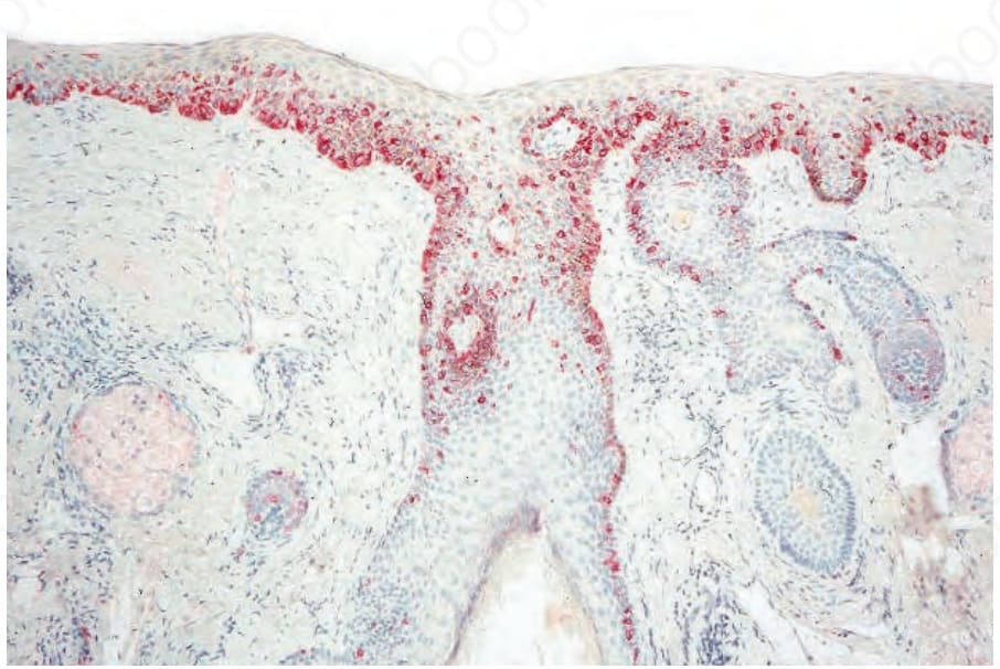
圖 26-26:惡性小痣 (lentigo maligna):偶爾難以區分日光性角化症 (actinic keratosis) 與 lentigo maligna。在此類病例中,使用紅色顯色劑 (red chromogen)(此例為 alkaline phosphatase)的免疫組織化學 (immunohistochemistry) 可使區分變得容易。
Fig. 26.26 Lentigo maligna: occasionally it is difficult to distinguish between actinic keratosis and lentigo maligna. In such cases, immunohistochemistry using a red chromogen (in this case alkaline phosphatase) can make the distinction easy.
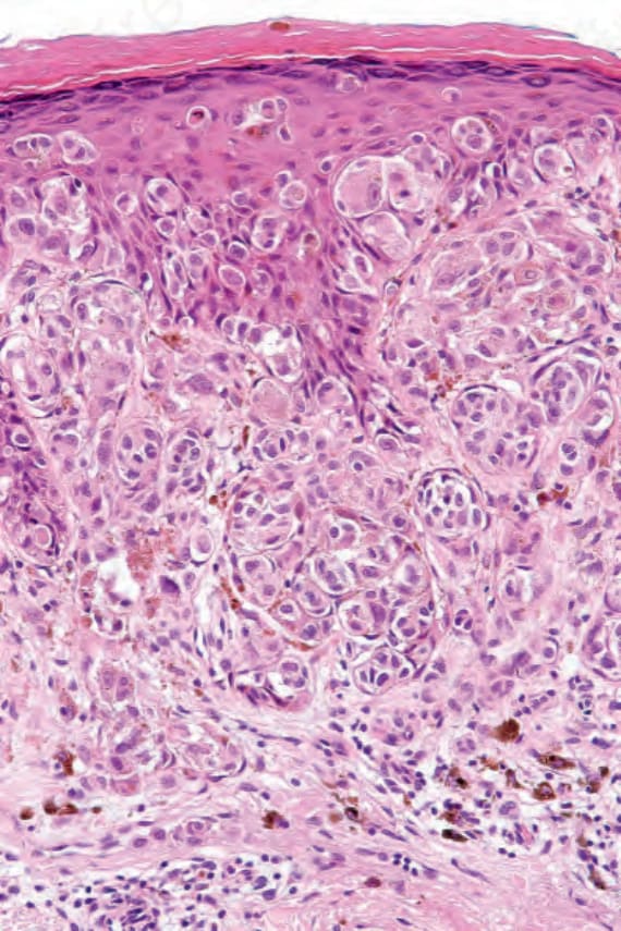
圖 26-29:表淺擴散性黑色素瘤 (superficial spreading melanoma):侵襲性腫瘤通常為上皮樣 (epithelioid) 型,如本視野所示。注意豐富的細胞質、細胞核多形性 (nuclear pleomorphism) 及明顯的核仁 (prominent nucleoli)。
Fig. 26.29 Superficial spreading melanoma: invasive tumor is usually of the epithelioid type as shown in this field. Note the abundant cytoplasm, nuclear pleomorphism, and prominent nucleoli.
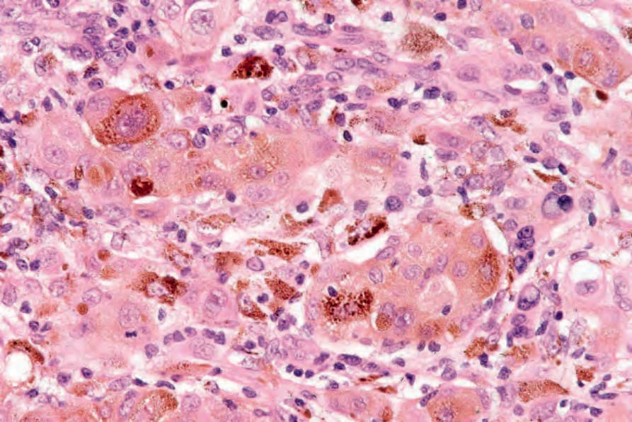
圖 26-30:表淺擴散性黑色素瘤 (superficial spreading melanoma):此例顯示濃重的黑色素沉著 (melanin pigmentation)。
Fig. 26.30 Superficial spreading melanoma: in this example there is heavy melanin pigmentation.
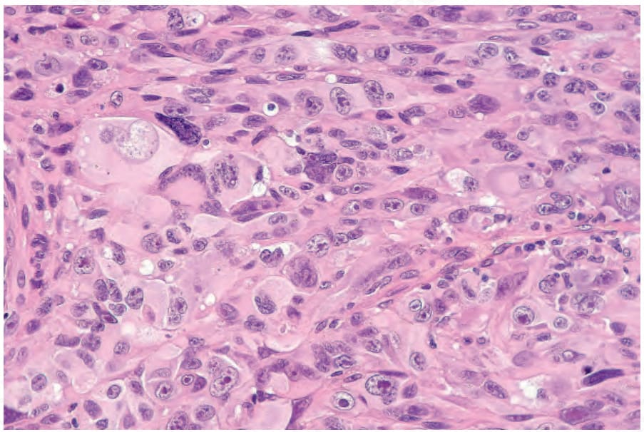
圖 26-31:表淺擴散性黑色素瘤 (superficial spreading melanoma):無色素性腫瘤 (amelanotic tumors) 的診斷,尤其在如本例般高度多形性時,若無明顯的接合處 (junctional) 成分,則往往須仰賴免疫組織化學 (immunohistochemistry)。
Fig. 26.31 Superficial spreading melanoma: diagnosis of amelanotic tumors, particularly when very pleomorphic as in this example, often depends upon immunohistochemistry if a junctional component is not evident.
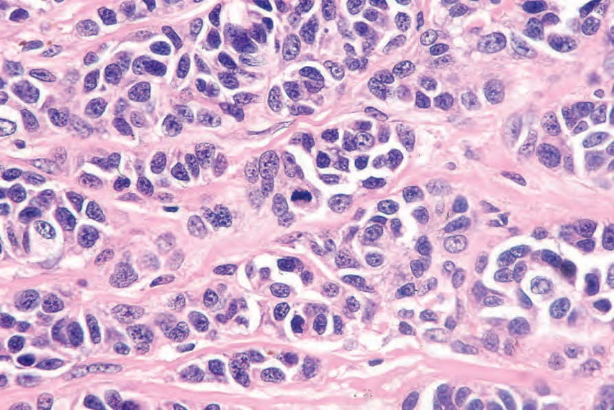
圖 26-32:表淺擴散性黑色素瘤 (superficial spreading melanoma):此例於視野中央顯示一個有絲分裂象 (mitotic figure)。
Fig. 26.32 Superficial spreading melanoma: this example shows a mitotic figure in the center of the field.
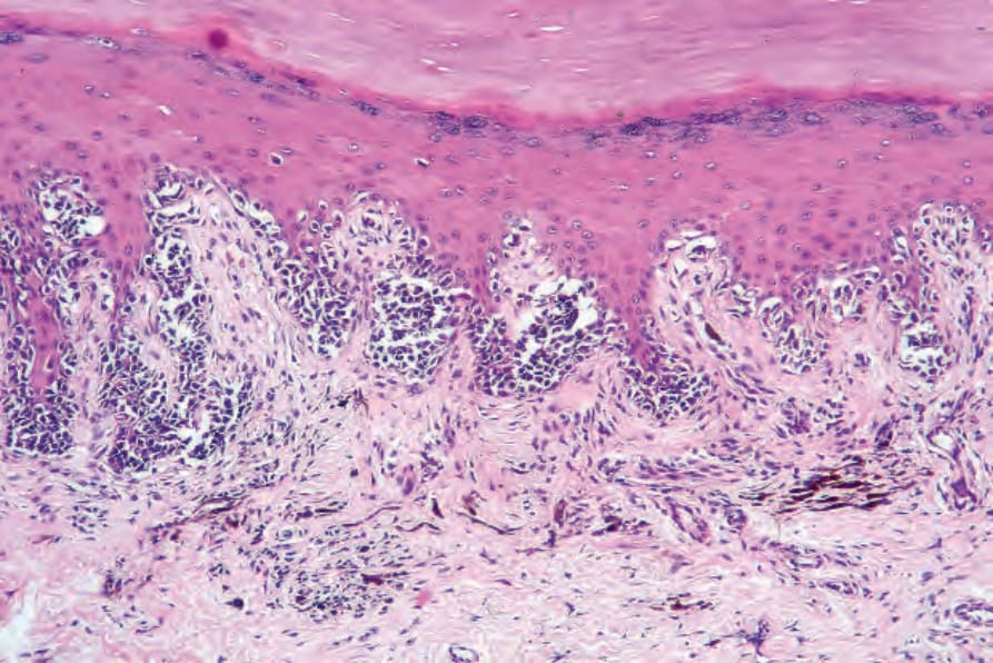
圖 26-33:肢端雀斑樣黑色素瘤 (acral lentiginous melanoma):在此原位 (in situ) 病灶中,有不規則棘層肥厚 (irregular acanthosis)、顆粒層增厚 (hypergranulosis) 與角化過度 (hyperkeratosis)。腫瘤細胞深染 (hyperchromatic),呈雀斑樣 (lentiginous) 及巢狀 (nested) 分布。真皮有瘢痕形成,並有明顯的噬黑色素細胞 (melanophages) 與慢性發炎細胞。
Fig. 26.33 Acral lentiginous melanoma: in this in situ lesion, there is irregular acanthosis, hypergranulosis, and hyperkeratosis. Tumor cells are hyperchromatic and distributed in a lentiginous and nested pattern. The dermis is scarred and there are conspicuous melanophages and chronic inflammatory cells.
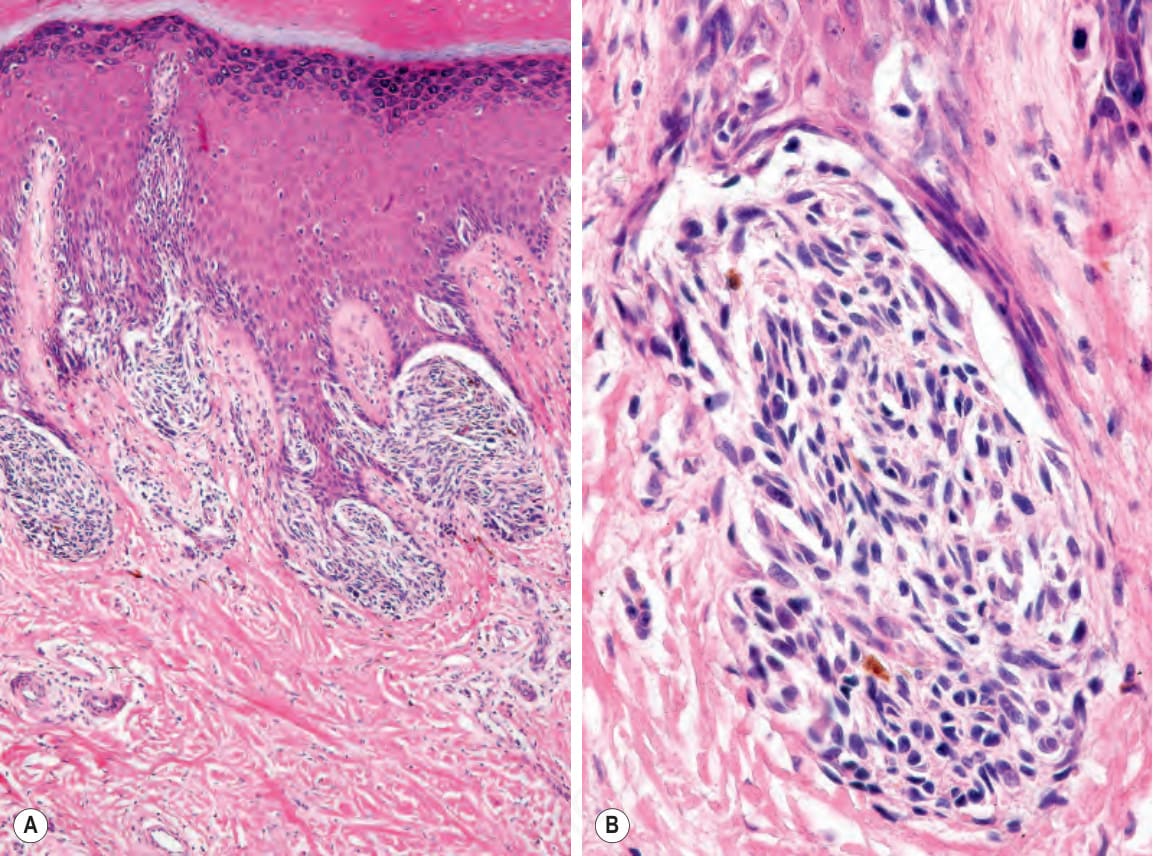
圖 26-35:(A, B) 肢端雀斑樣黑色素瘤 (acral lentiginous melanoma):表皮突 (rete) 尖端可見大型接合處巢 (junctional nests)。
Fig. 26.35 (A, B) Acral lentiginous melanoma: large junctional nests are present at the tips of the rete.
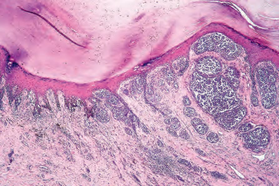
圖 26-36:肢端雀斑樣黑色素瘤 (acral lentiginous melanoma):此例中,侵襲性成分為混合性上皮樣 (epithelioid)、梭形細胞 (spindled cell) 與促結締組織增生型 (desmoplastic)。
Fig. 26.36 Acral lentiginous melanoma: in this example, the invasive component is mixed epithelioid, spindled cell, and desmoplastic.