黑色素細胞的惡性轉化 (Malignant Transformation of Melanocytes)
由黑色素細胞 (melanocyte) → 黑色素細胞痣 (melanocytic nevus) → 異型痣 (dysplastic nevus) → 原位黑色素瘤 (melanoma in situ),最終演進至侵襲性黑色素瘤 (invasive melanoma) 的這一進程,一直作為黑色素瘤發生 (melanoma genesis) 的合理模型。雖然這種逐步進展確實發生於某些黑色素瘤,但有一部分黑色素瘤的發生並不伴隨相關的痣或原位成分(例如 primary dermal melanoma、blue nevus-like melanoma)。黑色素瘤形成所必需的基因改變數目,會因個體本身的遺傳易感性 (genetic susceptibility),以及所發展出之基因改變的順序與組合而有所不同。使用既定基因元件 (defined genetic elements) 的體外研究指出,在移植於小鼠的人類皮膚上,僅需三種不同的突變即足以誘發類黑色素瘤病灶 (melanoma-like lesions)。12 與實驗情境不同,真實癌症中的基因改變通常是依序產生的。驅動增殖的基因突變,如 BRAF、NRAS 與 GNAQ/11,會驅動黑色素細胞的克隆性擴增 (clonal expansion)。若此類起始突變發生於 senescence 路徑完整的細胞中,失調的生長會觸發具保護性的腫瘤抑制檢查點 (tumor suppressor checkpoints) 以限制增殖,於是形成黑色素細胞痣。當這些誘發增殖的突變發生於帶有基因缺陷、且該缺陷已使此類檢查點機制失能的細胞中時,逐步進展模型中的某些階段可被跳過。此點可由帶有生殖系 CDKN2A 突變 (germline CDKN2A mutations) 的病人加以說明,這些病人在一條腫瘤抑制路徑上具有先天缺陷。與大多數無此突變的個體不同,這些個體會發展出大型非典型痣 (large atypical nevi),似乎跳過了第一個良性痣階段——在該階段中只存在單一致病突變(例如 BRAFV600E)。因此可以預期,若一個已帶有失活之腫瘤抑制路徑的細胞,隨後獲得一個誘發增殖的突變,便可能發生「de novo」黑色素瘤,跳過任何在表型上可見的前驅階段。35 基因異常的組合決定了惡性轉化的階段,其中某些組合比其他組合具有更強的轉化後果。例如,BRAF 與 BAP1 突變的組合似乎具有低惡性潛能,11 而將 GNAQ/11 與 BAP1 突變組合則會產生具有高轉移潛能 (high metastatic potential) 的腫瘤。36,37
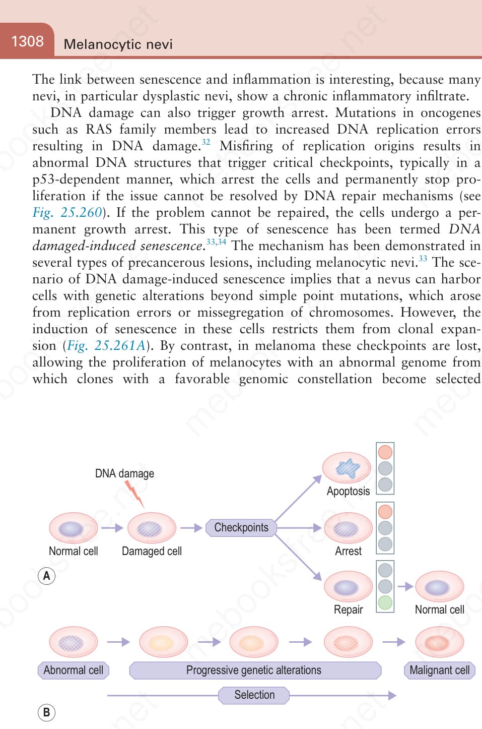
圖 25-261:限制基因改變細胞擴增的屏障 (A) 及其在癌症中的失效 (B):(A) 若 DNA 損傷的檢查點 (checkpoints) 完整,任何獲得 DNA 損傷的細胞都會被暫時性地停滯 (transiently arrested),以爭取時間修復該缺陷。若缺陷無法修復,細胞會被永久性停滯或導向死亡路徑 (death pathway)。(B) 若這些檢查點失效,帶有已獲得之基因改變的細胞便能克隆性擴增並獲得額外的基因改變。隨時間推移,帶有進一步促進生長與存活之突變的變異株 (variants) 將被選擇出來。
Fig. 25.261 Barriers that restrict expansion of genetically altered cells (A) and their failure in cancer (B): (A) If checkpoints of DNA damage are intact, any cells that acquire DNA damage are transiently arrested to give time to repair the defect. If the defect cannot be repaired, cells are permanently arrested or routed to a death pathway. (B) If these checkpoints fail, cells with acquired genetic alterations can clonally expand and acquire additional genetic alterations. Over time, variants with mutations that further promote growth and survival will be selected.
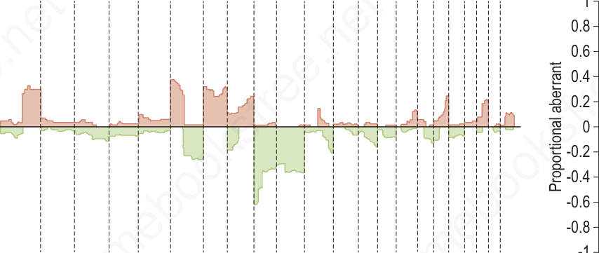
圖 25-262:黑色素瘤(惡性)與痣(良性)的比較基因組雜交 (comparative genomic hybridization):左側為黑色素瘤 (n = 133),伴隨多處成簇的拷貝數增益與缺失 (copy number gains and losses)。相對地,右側的痣 (n = 54) 則僅顯示極微小的變化,唯一例外是 Spitz 痣 (Spitz nevi) 中包含 HRAS 基因的 11p 拷貝數增加或擴增 (amplification)。這些差異可利用多重 FISH 檢測 (multiplexed FISH assays) 加以運用,以在困難病例中支持黑色素瘤或痣的診斷。
Fig. 25.262 Comparative genomic hybridization of melanoma (malignant) and nevi (benign): on the left, melanomas (n = 133) are associated with multiple copy number gains and losses that cluster. In contrast, the nevi (n = 54) on the right show minimal changes with the exception of the 11p copy number increase or amplification in Spitz nevi which includes the HRAS gene. These differences can be exploited using multiplexed FISH assays to support the diagnosis of melanoma or nevus in challenging cases.