Epithelioid blue nevus
臨床特徵 (Clinical Features)
- 上皮樣藍痣 (epithelioid blue nevus) 是藍痣 (blue nevus) 的一種罕見變異型,最常見於罹患 Carney complex 的病人。少數類似腫瘤在無 Carney complex 的情況下出現也有記載。上皮樣藍痣亦曾於巨大型先天性黑色素細胞痣 (giant congenital melanocytic nevus) 的情境中被描述。
- Carney complex 是一種體染色體顯性 (autosomal dominant) 疾病,病人會出現多種病灶,包括皮膚雀斑樣痣 (cutaneous lentigines) 與藍痣、皮膚、乳腺與心臟黏液瘤 (myxomas)、因原發性色素性結節性腎上腺增生 (primary pigmented nodular adrenal hyperplasia) 所致的 Cushing syndrome、因腦下垂體腺瘤 (pituitary adenoma) 所致的肢端肥大症 (acromegaly),以及因大細胞鈣化型 Sertoli 細胞瘤 (large cell calcifying Sertoli cell tumor) 所致的性早熟 (sexual precocity)。惡性黑色素性 schwann 細胞腫瘤 (malignant melanotic schwannian tumors,先前稱為 psammomatous melanotic schwannoma) 也是其特徵之一。
- 上皮樣藍痣表現為藍色至黑色或紫色、通常呈圓頂狀的病灶,最常見於四肢與軀幹,典型者直徑可達 1.0 cm。曾有黏膜 (口腔與生殖器) 受侵犯的描述。有時可見多發性病灶。亦曾報告一例廣泛型先天性變異,出現於一名具有超過 1000 個上皮樣藍痣的嬰兒。散發性病灶在形態上相似。它們在生物學上為良性。
組織病理特徵 (Histopathology)
- 上皮樣藍痣為位於真皮內、邊界不清、常呈圓頂狀、卵圓形至球形或楔形的腫塊,有時延伸至皮下脂肪 (Fig. 25.235)。偶爾可見合併性病灶 (combined lesions)。
- 它由以下兩種細胞混合組成:大小不一、重度色素沉著的球狀細胞 (globular cells),具有小的泡狀核 (vesicular nuclei) 與明顯的嗜伊紅性核仁 (eosinophilic nucleoli);以及僅有輕度色素的胞質、泡狀核與單一大型嗜伊紅性核仁的多角形細胞 (polygonal cells) (Fig. 25.236)。
- 這些細胞以間質性分布,呈單一散在、短列狀,有時於真皮膠原束之間呈束狀 (fascicles)。皮膚附屬器 (adnexae) 典型受侵犯。有時可見稀疏分布的有絲分裂象 (mitotic figures)。背景中有較少數的紡錘狀與樹突狀細胞 (spindled and dendritic cells)。纖維化 (fibrosis) 不是此病灶的特徵。
- 部分作者將上皮樣藍痣 (包括散發性以及在 Carney complex 情境下者) 與色素合成型 (動物型) 黑色素瘤 (pigment synthesizing (animal-type) melanoma) 視為同一臨床與病理譜系的一部分,並在此情境下使用色素性上皮樣黑色素細胞瘤 (pigmented epithelioid melanocytoma) 一詞。被歸類為 pigmented epithelioid melanocytoma 的病灶似乎具有低度惡性潛能,常伴有區域淋巴結轉移 (高達 60%)、罕見遠端轉移,以及良好的長期臨床病程。然而,pigmented epithelioid melanocytoma 一詞仍有爭議,其他作者建議使用色素合成型黑色素瘤 (pigment synthesizing melanoma) 一詞來指稱此類病灶。仍需進一步的長期研究。
- 散發性上皮樣藍痣在形態上完全相同。上皮樣合併痣 (epithelioid combined nevus) 與促結締組織增生型 Spitz (desmoplastic Spitz)、深部穿透型 (deep penetrating) 或一般型痣 (banal nevus) 的特徵相關。
免疫組化與特殊染色 (Immunohistochemistry & Special Stains)
- 球狀細胞表現 CD68 與 CD163;上皮樣型表現 S100 protein 與 HMB-45,但不表現 CD68。
鑑別診斷 (Differential Diagnosis)
- 上皮樣藍痣應與色素合成型 (動物型、馬型) 黑色素瘤 (pigment synthesizing (animal-type, equine) melanoma) 區別。細胞學異型性 (cytological atypia)、有絲分裂活性 (mitotic activity) 與表皮受侵犯 (epidermal involvement) 較支持後者的診斷。
Epithelioid and fusiform blue nevus of chronically sun-damaged skin
1299 真皮黑色素細胞病灶 (dermal melanocytoses)
組織病理特徵 (Histopathology)
- 在淺層,毛囊神經嵴錯構瘤 (pilar neurocristic hamartoma) 可顯示一般型真皮內痣細胞 (banal intradermal nevus cells) 的聚集以及類似普通藍痣 (common blue nevus-like) 的特徵。然而在網狀真皮 (reticular dermis) 中,色素性紡錘狀細胞 (pigmented spindled cells) 圍繞著毛囊的下段以及鄰近的小汗腺 (eccrine sweat glands) (pilar neurocristic hamartoma) (Figs 25.237 與 25.238)。毛囊數目可能減少或顯得發育不良 (dystrophic)。毛囊間真皮 (interfollicular dermis) 內含有散在的重度色素沉著紡錘狀細胞與樹突狀細胞,令人聯想到於類似神經纖維瘤的非色素性紡錘狀細胞 (neurofibroma-like nonpigmented spindled cells) 背景中出現的蒙古斑 (Mongolian blue spot),後者含有邊界清晰的 Schwann cell nodules,有時伴有類似 Meissner 觸覺小體 (Meissner tactoid body-like) 的結構。曾有花環狀巨細胞 (floret-like giant cells) 的描述。無細胞學異型性,且無有絲分裂。其上覆的表皮可能色素過度沉著並顯示
臨床特徵 (Clinical Features)
- 此近期描述的疾病實體代表藍痣的一種亞型,好發於頭頸部與四肢的日光損傷皮膚 (sun-damaged skin)。上皮樣與紡錘狀藍痣 (epithelioid and fusiform blue nevus) 呈女性優勢 (約 1.7 : 1),最常見於第七個十年的人生 (40 至 84 歲;平均 63 歲),表現為單發、雜色的斑疹或丘疹,直徑可達 1 cm。上皮樣與紡錘狀藍痣與 Carney complex 無關。
- 此病灶完全為良性,完整切除後尚未有復發的報告。然而,由於它出現於日光損傷皮膚,加上一部分病灶具有輕度細胞多形性 (cellular pleomorphism)、核異型性 (nuclear atypia) 與罕見的有絲分裂活性,上皮樣與紡錘狀藍痣可能被誤認為黑色素瘤 (melanoma)。
組織病理特徵 (Histopathology)
- 此黑色素細胞增生典型地以淺層真皮為中心,由具有豐富充滿黑色素胞質的上皮樣與紡錘狀黑色素細胞 (epithelioid and fusiform melanocytes) 呈叢狀生長 (plexiform growth) 所組成。第二個黑色素細胞成分,即傳統型藍痣 (conventional blue nevus) 也可被辨識。日光彈力組織變性束 (solar elastotic bundles) 特徵性地混雜於上皮樣/紡錘狀黑色素細胞增生之內。未見黑色素細胞有顯著異型性,且通常無有絲分裂活性。上皮樣成分的成熟 (maturation) 典型地保留。然而,在一部分這類增生中,可觀察到局部中度至高度核異型性,伴隨核增大、深染 (hyperchromasia)、明顯核仁,以及偶發的有絲分裂活性 (少於 1 個有絲分裂/mm²)。
鑑別診斷 (Differential Diagnosis)
- 叢狀生長型態 (plexiform growth pattern)、缺乏融合性高度異型性、低有絲分裂活性,以及無異型性有絲分裂 (atypical mitoses),應有助於與黑色素瘤區別。
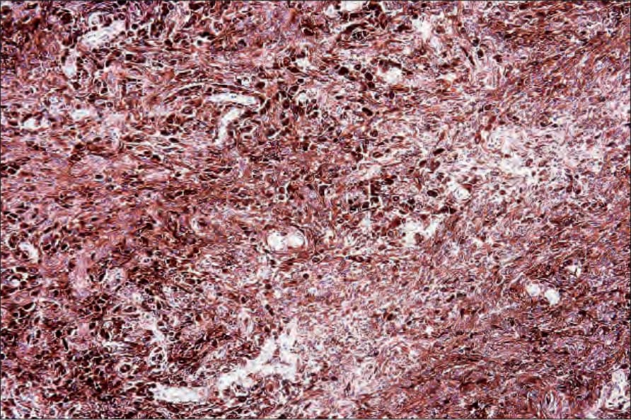
圖 25-235:上皮樣藍痣 (epithelioid blue nevus):重度色素沉著的上皮樣細胞散布於樹突狀細胞與噬黑色素細胞 (melanophages) 之間。承蒙 C.D.M. Fletcher, MD, Brigham and Women’s Hospital and Harvard Medical School, Boston, USA 提供。
Fig. 25.235 Epithelioid blue nevus: heavily pigmented epithelioid cells are dispersed among dendritic cells and melanophages. By courtesy of C.D.M. Fletcher, MD, Brigham and Women’s Hospital and Harvard Medical School, Boston, USA.

圖 25-236:上皮樣藍痣 (epithelioid blue nevus):上皮樣細胞呈重度色素沉著,並具有泡狀核 (vesicular nuclei) 與明顯核仁 (prominent nucleoli)。承蒙 C.D.M. Fletcher, MD, Brigham and Women’s Hospital and Harvard Medical School, Boston, USA 提供。
Fig. 25.236 Epithelioid blue nevus: the epithelioid cells are heavily pigmented and have vesicular nuclei with prominent nucleoli. By courtesy of C.D.M. Fletcher, MD, Brigham and Women’s Hospital and Harvard Medical School, Boston, USA.
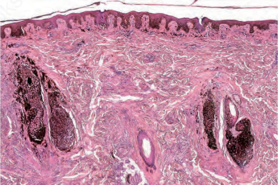
圖 25-237:毛囊神經嵴錯構瘤 (pilar neurocristic hamartoma):此例顯示一個明顯以毛囊為中心 (folliculocentric) 的病灶。汗腺 (sweat glands) 亦受侵犯。
Fig. 25.237 Pilar neurocristic hamartoma: this example shows a strikingly folliculocentric lesion. The sweat glands were also involved.