色素減退型普通藍痣 (Hypopigmented Common Blue Nevus)
臨床特徵 (Clinical Features)
- 色素減退型普通藍痣 (hypopigmented common blue nevus;amelanotic blue nevus) 是 common blue nevus 一個罕見且近期才被描述的變異型,發病年齡與 common blue nevus 相近,最常見於四肢與臀部。
- 由於缺乏或僅有極少量黑色素 (melanin) 色素,臨床上通常被認為是一個良性的尋常痣 (banal nevus) 或皮膚纖維瘤 (dermatofibroma;fibrous histiocytoma)。
組織病理特徵 (Histopathology)
- 與其較典型的對應病灶相同,hypopigmented variant 表現為一個界限不清、呈浸潤性的真皮內腫瘤 (intradermal tumor)。
- 病灶由雙極 (bipolar) 與樹突狀 (dendritic) melanocytes 組成,散布於緻密的膠原基質 (collagenous stroma) 中(Figs 25.233 與 25.234)。
- 有時也可見席紋狀 (storiform) 分布。
- 細胞具有不明顯的嗜伊紅性細胞質 (eosinophilic cytoplasm) 與梭形、深染的細胞核 (fusiform hyperchromatic nuclei),核內常含有小核仁 (small nucleoli)。
- 偶可見核內細胞質假包涵體 (intranuclear cytoplasmic pseudoinclusions)。
- 例外情況下可見具多空泡細胞質 (multivacuolated cytoplasm) 與扇貝狀細胞核 (scalloped nuclei)、形似皮脂腺細胞 (sebocytes) 的 melanocytes。
- 不見有絲分裂 (mitoses)。
- 雖然在極小比例的細胞中可見黑色素 (melanin) 色素,但多數情況下並無色素;然而,黑色素偶爾在病灶邊緣相對明顯。
免疫組化與特殊染色 (Immunohistochemistry & Special Stains)
- 針對 S100 protein 或 HMB-45 的免疫組化染色,有助於顯示此痣性細胞族群 (nevoid population) 的雙極與樹突狀特性。
1298 Melanocytic nevi
鑑別診斷 (Differential Diagnosis)
- Hypopigmented common blue nevus 與 sclerosing (common) blue nevus(見前文)有相當程度的重疊,且兩者很可能是相同的病變。
- 最常見的是與 dermatofibroma (fibrous histiocytoma) 混淆。事實上,兩者在組織學上可能有相當程度的重疊;在那些看不到任何色素的病例中,可能需要以 S100 protein 免疫組化染色來建立正確的診斷。
- 偶爾,硬化 (sclerosis) 呈現同心圓狀 (concentric) 或層狀 (laminated) 分布,類似所謂的席紋狀膠原瘤 (storiform collagenoma)。此時可能同樣需要免疫組化來作出區分。
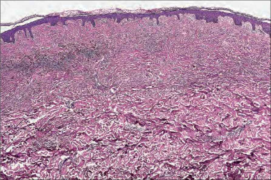
圖 25-233:色素減退型藍痣 (hypopigmented blue nevus):視野左側可見殘餘色素沉著 (residual pigmentation);右側則為一個細胞稀疏的硬化性成分 (paucicellular sclerosing component)。
Fig. 25.233 Hypopigmented blue nevus: residual pigmentation is seen on the left side of the field. On the right, there is a paucicellular sclerosing component.
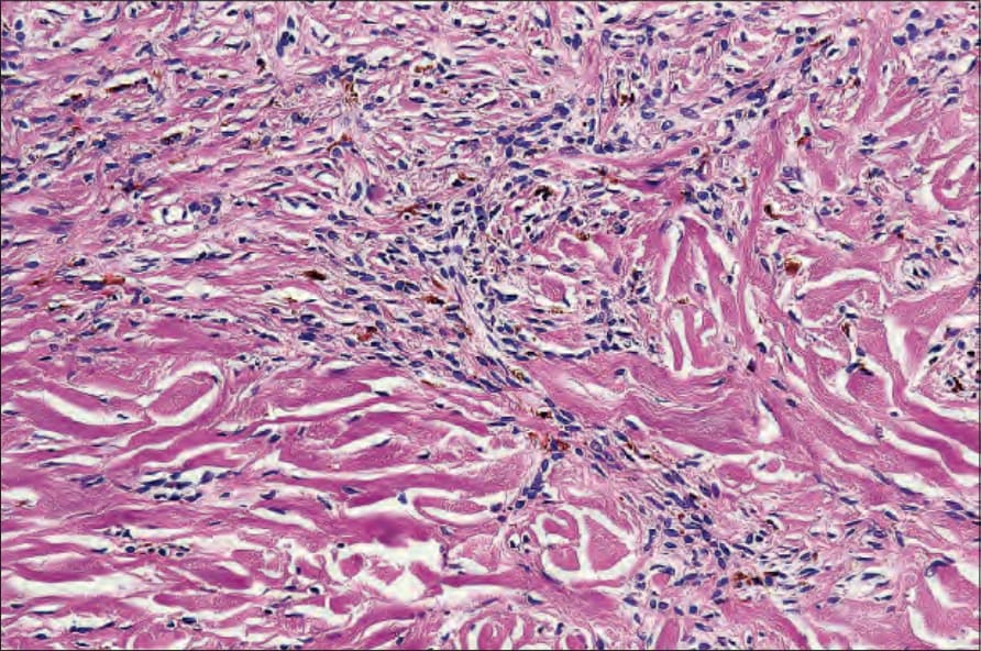
圖 25-234:色素減退型藍痣 (hypopigmented blue nevus):可見殘餘色素沉著 (residual pigmentation)。注意無色素的梭形細胞 (nonpigmented spindle cells)。
Fig. 25.234 Hypopigmented blue nevus: residual pigmentation is present. Note the nonpigmented spindle cells.
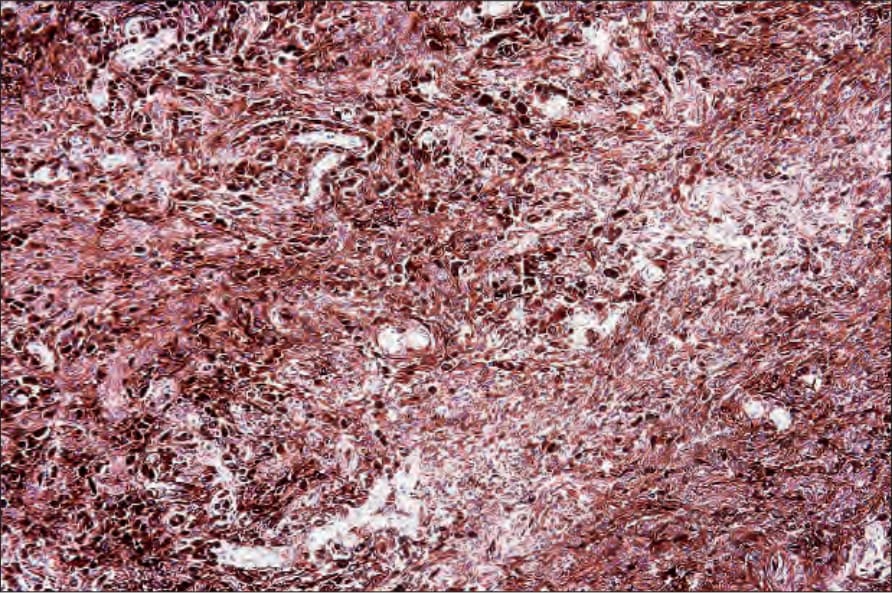
圖 25-235:上皮樣藍痣 (epithelioid blue nevus):濃染的上皮樣細胞 (heavily pigmented epithelioid cells) 散布於樹突狀細胞 (dendritic cells) 與噬黑色素細胞 (melanophages) 之間。承蒙 C.D.M. Fletcher, MD, Brigham and Women’s Hospital and Harvard Medical School, Boston, USA 提供。
Fig. 25.235 Epithelioid blue nevus: heavily pigmented epithelioid cells are dispersed among dendritic cells and melanophages. By courtesy of C.D.M. Fletcher, MD, Brigham and Women’s Hospital and Harvard Medical School, Boston, USA.