Hypopigmented common blue nevus
Hypopigmented common blue nevus
Clinical features Hypopigmented common blue nevus (amelanotic blue nevus) is a rare and recently described variant of common blue nevus, which presents at a similar age and is seen most often on the extremities and buttocks.1–4 Due to the lack or paucity of melanin pigment, it is usually thought to represent a banal nevus or a dermatofibroma (fibrous histiocytoma) clinically.
Histologic features As with its more typical counterpart, the hypopigmented variant presents as an ill-defined infiltrating intradermal tumor. It is composed of bipolar and dendritic melanocytes dispersed in a dense collagenous stroma (Figs 25.233 and 25.234).1 A storiform distribution may also sometimes be seen. The cells have indistinct eosinophilic cytoplasm and fusiform hyperchromatic nuclei often containing small nucleoli. Intranuclear cytoplasmic pseudoinclusions are occasionally evident. Melanocytes with multivacuolated cytoplasm and scalloped nuclei resembling sebocytes can exceptionally be seen.5 Mitoses are not present. Although melanin pigment may be seen in a very small proportion of cells, it is mostly absent. Occasionally, however, it is relatively conspicuous at the edge of the lesion.2
Immunohistochemistry for S100 protein or HMB-45 is of value in demonstrating the bipolar and dendritic nature of the nevoid population.1
1298 Melanocytic nevi
Differential diagnosis Hypopigmented common blue nevus shows considerable overlap and is probably identical to sclerosing (common) blue nevus (see above).
Most commonly, it is confused with dermatofibroma (fibrous histiocytoma). There may, in fact, be considerable histologic overlap, and in those examples where no pigment is visible, immunohistochemical staining with S100 protein may be necessary to establish the correct diagnosis.
Occasionally, the sclerosis adopts a concentric or laminated distribution resembling so-called storiform collagenoma.3 Immunohistochemistry may again be necessary to afford the distinction.
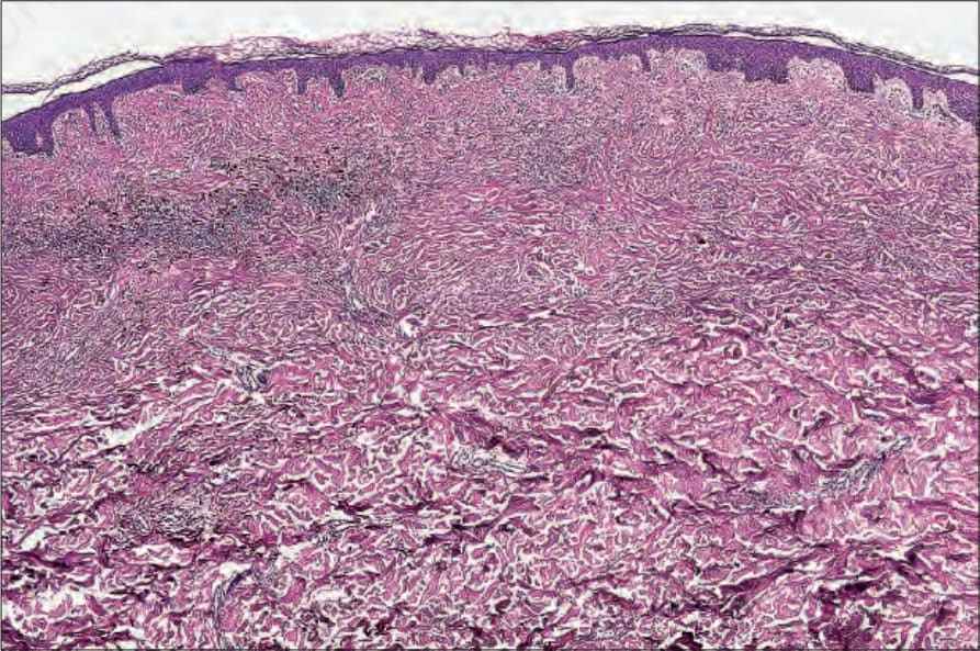
Fig. 25.233 Hypopigmented blue nevus: residual pigmentation is seen on the left side of the field. On the right, there is a paucicellular sclerosing component.
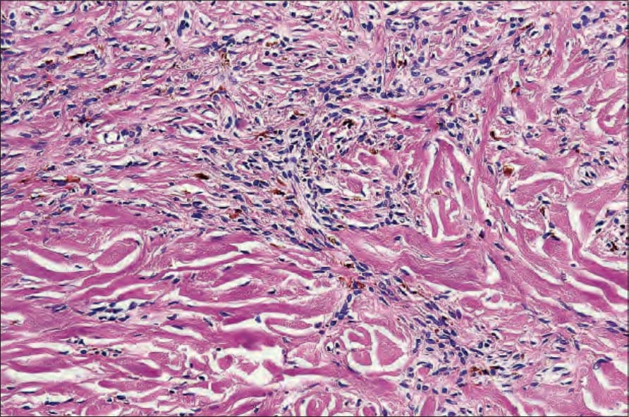
Fig. 25.234 Hypopigmented blue nevus: residual pigmentation is present. Note the nonpigmented spindle cells.
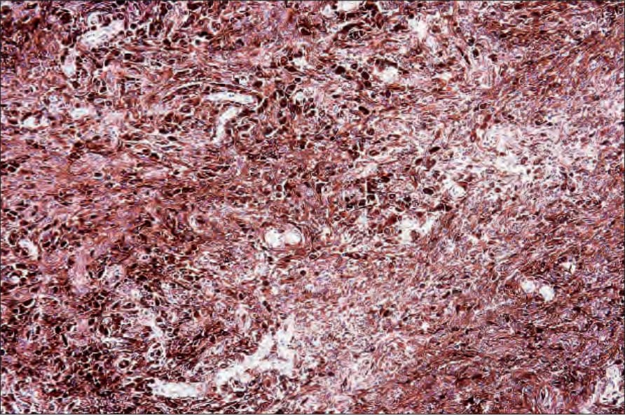
Fig. 25.235 Epithelioid blue nevus: heavily pigmented epithelioid cells are dispersed among dendritic cells and melanophages. By courtesy of C.D.M. Fletcher, MD, Brigham and Women’s Hospital and Harvard Medical School, Boston, USA.