疾病定義與分類
- 深部穿透性痣 (deep penetrating nevus) 是一種特殊的黑色素細胞痣 (melanocytic nevus),最初由 Seab 等人於 1989 年描述。Barnhill 等人其後報告的叢狀梭形細胞痣 (plexiform spindle cell nevus) 在組織學特徵上基本相同。
- 雖然「deep penetrating nevus」一詞意指病變延伸至深層真皮或皮下組織,但真正定義此病變的,其實是其生長型態與細胞形態的組合。
- 深部穿透性痣的一種表淺變異型於 1994 年被報告,命名為帶有局灶上皮樣成分的黑色素細胞痣 (melanocytic nevus with focal epithelioid component),亦稱選殖性痣 (clonal nevus)。此變異型與 deep penetrating nevus 共享相似的年齡、解剖分布與細胞學特徵,但缺乏黑色素細胞的深部延伸。
- 深部穿透性痣難以歸類。在某些病例中,它似乎代表一種自成一格 (sui generis) 的獨立病變;在另一些情況下,它則代表混合性痣 (combined nevus) 的一種變異型。相當比例的病例中存在樹突狀細胞 (dendritic cells),此點提示許多病例或許更適合歸入藍痣 (blue nevus) 光譜之內。
臨床特徵 (Clinical Features)
- 深部穿透性痣並不常見,其重要性在於臨床與組織學上皆可能被誤認為黑色素瘤 (melanoma)。
- 發病年齡範圍相當廣 (0–77 歲),但報告中年齡超過 50 歲者不足 5%。病人最常見於第二或第三個十年,表現為單發、邊界清楚、通常直徑小於 1 cm、圓頂狀的藍色或黑色丘疹或結節。
- 色素沉著可呈多樣性,從淺棕色到黑色不等,但多數病變色素濃重。尤其當其為混合性黑色素細胞痣 (combined melanocytic nevus) 的一部分時,病變可能呈現不對稱且色素分布不均,因而在臨床上引起對黑色素瘤的懷疑。
- 顏面、上軀幹或四肢近端尤其好發 (Fig. 25.161)。近期報告了一例發生於足底的深部穿透性痣。亦有一例獨特的多發性深部穿透性痣於耳前皮膚呈線狀分布的病例被發表。
- 深部穿透性痣顯示輕微的女性優勢 (1.3 : 1)。
組織病理特徵 (Histopathology)
- 深部穿透性痣在掃描放大倍率下的一個特徵性形態,是其對稱、楔形、邊界銳利的構型,寬基部位於最上方、與表面上皮平行 (Fig. 25.162)。深部漸尖的成分通常延伸至下方網狀真皮,甚至皮下脂肪。沿皮膚附屬器或神經血管束 (neurovascular bundles) 的一處或多處此類延伸常可見到。
- 60% 至 85% 的深部穿透性痣中可見交界成分 (junctional component),通常侷限於少數小巢。黑色素細胞向上延伸及佩吉特樣播散 (pagetoid spread) 並非此病變的特徵。然而在三分之一至三分之二的病例中,深部穿透性痣呈現為混合性病變,以平凡 (banal) 或 Spitz 特徵作為表淺成分 (Figs 25.163 與 25.164)。
- 乳頭層真皮常未受侵犯,尤其在僅具深部穿透性痣成分的病變中。真皮成分通常界線銳利,沿神經血管束與附屬器結構分布,常呈現束狀 (fascicular) 或叢狀 (plexiform) 輪廓 (Fig. 25.165),故有其別名叢狀梭形細胞痣 (plexiform spindle cell nevus)。
- 真皮黑色素細胞成分由疏鬆的細胞巢與垂直走向的上皮樣及梭形黑色素細胞束所構成。上皮樣黑色素細胞在病變較表淺處較常見,且常出現透明細胞 (clear cells) 之病灶 (Figs 25.166 與 25.167)。梭形黑色素細胞一般在較深處佔優勢 (Fig. 25.168)。可見細胞巢的融合,尤其在上方真皮與病變中央區域,但此現象一般為局灶性。病變周邊與基底處可出現黑色素細胞的失黏附 (discohesion)。
- 神經周圍延伸 (perineural extension) 與立毛肌 (arrector pili muscle) 浸潤常見。看不到黑色素細胞明顯的成熟現象 (maturation)。輕度核多形性 (nuclear pleomorphism) 常見,核深染 (nuclear hyperchromatism) 也可能是一項特徵。中度核多形性亦可顯現,但通常為局灶且隨機分布,且通常不伴隨有絲分裂活性的增加。在個別的黑色素細胞巢或束內可見細胞核大小的差異。核仁為小至中等大小、嗜伊紅性。
- 然而,有絲分裂 (mitoses) 非常稀少,或更常見的是完全沒有 (Fig. 25.169)。有絲分裂數目範圍為 0 至 1.2/mm²。但出現多於偶見的有絲分裂則是令人憂慮的特徵,應提高對非典型深部穿透性痣 (atypical deep penetrating nevus,見下文) 或具深部穿透性痣特徵之黑色素瘤的懷疑。深部穿透性痣中找不到非典型有絲分裂。核內假包涵體 (intranuclear pseudoinclusions) 常可見到。黑色素細胞的細胞質呈淡粉紅色至雙嗜性 (amphophilic)。偶見的樹突狀黑色素細胞 (dendritic melanocytes) 常存在。
- 一種多變的反應性慢性發炎細胞浸潤伴隨此痣。通常基質反應極微。黑色素噬細胞 (melanophages) 是深部穿透性痣中恆定的發現。它們可以稀少且局灶,或豐富且散布於整個病變之中。它們通常環繞個別的黑色素細胞巢與束,並在病變周邊處尤為顯著。
致病機轉/分子 (Pathogenesis / Molecular)
- 黑色素細胞一致呈 S100 與 HMB-45 陽性。增殖細胞核抗原 (proliferating cell nuclear antigen, PCNA) 通常由少於 5% 的細胞表現。
- 雖然在 32 例深部穿透性痣中的 2 例檢測到 HRAS 突變,但其中無一顯示 GNAQ 或 GNA11 突變 (此兩者常見於藍痣),提示其可能與類 Spitz 黑色素細胞增生 (spitzoid melanocytic proliferations) 有關。
- 近期已證實這些病變是由 MAP-kinase 路徑與 beta catenin 訊息傳導的合併活化所致,且 B-catenin 的表現在整個腫瘤中皆保留。
鑑別診斷 (Differential Diagnosis)
- 主要鑑別診斷包括黑色素瘤 (melanoma),後者偶可表現為深部穿透性生長型態。鑑別要點包括:缺乏非典型交界黑色素細胞成分、真皮黑色素細胞呈非隨機性細胞學異型性、更嚴重的多形性、核仁顯著、過多的有絲分裂 (包括異常型態),以及成熟受損 (Figs 25.170–25.173)。
- 已有人提出非典型 (或高度惡性度) 深部穿透性痣的概念,適用於符合下列一項或多項組織學特徵的增生:病變直徑大於 5 mm、不對稱、邊界不清、侵犯皮下組織、細胞密度增加、結節狀或片狀生長、中度至重度細胞學異型性,以及有絲分裂活性大於 2/mm²。關於非典型深部穿透性痣的有限資料提示,其中一部分病變可能發展出淋巴結沉積 (lymph-node deposits),然而其結局未必不良,與所謂的非典型 Spitz 腫瘤 (atypical Spitz tumors) 相類比。此外,非典型深部穿透性痣以 FISH 或 CGH 檢測時,一般缺乏黑色素瘤典型的拷貝數異常 (copy number aberrations)。
- 應與深部穿透性痣區分的良性黑色素細胞病變包括普通型藍痣 (common blue nevi) 與細胞性藍痣 (cellular blue nevi)。普通型藍痣由位於硬化性基質內、帶色素的梭形與樹突狀黑色素細胞構成;細胞性藍痣則由色素稀少、具卵圓形細胞核、核仁不明顯及透明細胞質的黑色素細胞之細胞巢與束所組成。
治療與預後 (Treatment & Prognosis)
- 深部穿透性痣是一種良性的黑色素細胞增生。局部復發極不常見,且通常與不完全或邊緣性切除有關。
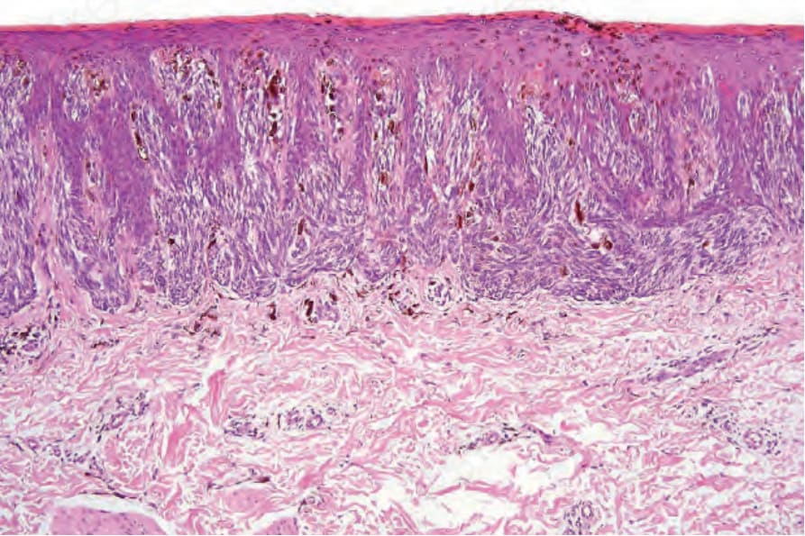
圖 25-155:Reed 色素性梭形細胞瘤 (pigmented spindle cell tumor of Reed):真皮乳頭被梭形細胞的交界細胞巢撐開。上方表皮內偶有痣細胞 (nevus cells) 存在,尤其在病變中央,並不少見;在病變整體外觀的脈絡下審視,不應構成警示來源。
Fig. 25.155 Pigmented spindle cell tumor of Reed: the dermal papillae are distended by junctional nests of spindle cells. The presence of occasional nevus cells in the upper epidermis, particularly in the center of the lesion, is not uncommon and when taken in the context of the overall appearances of the lesion should not be a source of alarm.
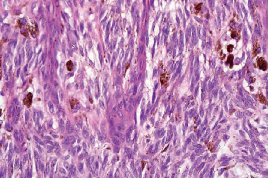
圖 25-156:Reed 色素性梭形細胞瘤 (pigmented spindle cell tumor of Reed):痣細胞具有漸尖的囊泡狀細胞核與小核仁。注意缺乏多形性。也可見到色素濃重的黑色素噬細胞 (melanophages)。
Fig. 25.156 Pigmented spindle cell tumor of Reed: the nevus cells have tapered vesicular nuclei with small nucleoli. Note the lack of pleomorphism. Heavily pigmented melanophages are also evident.
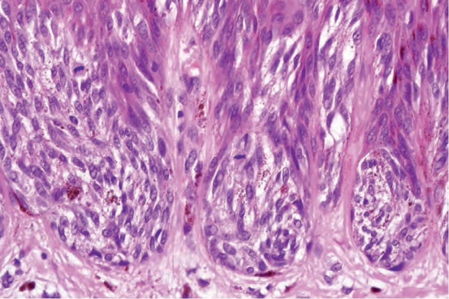
圖 25-157:Reed 色素性梭形細胞瘤 (pigmented spindle cell tumor of Reed):如本視野所見的交界處有絲分裂象 (junctional mitotic figures) 並不少見,除非數量眾多或型態異常,否則不必然是令人擔憂的來源。
Fig. 25.157 Pigmented spindle cell tumor of Reed: junctional mitotic figures, as seen in this field, are not uncommon and should not necessarily be a source of concern unless numerous or atypical.
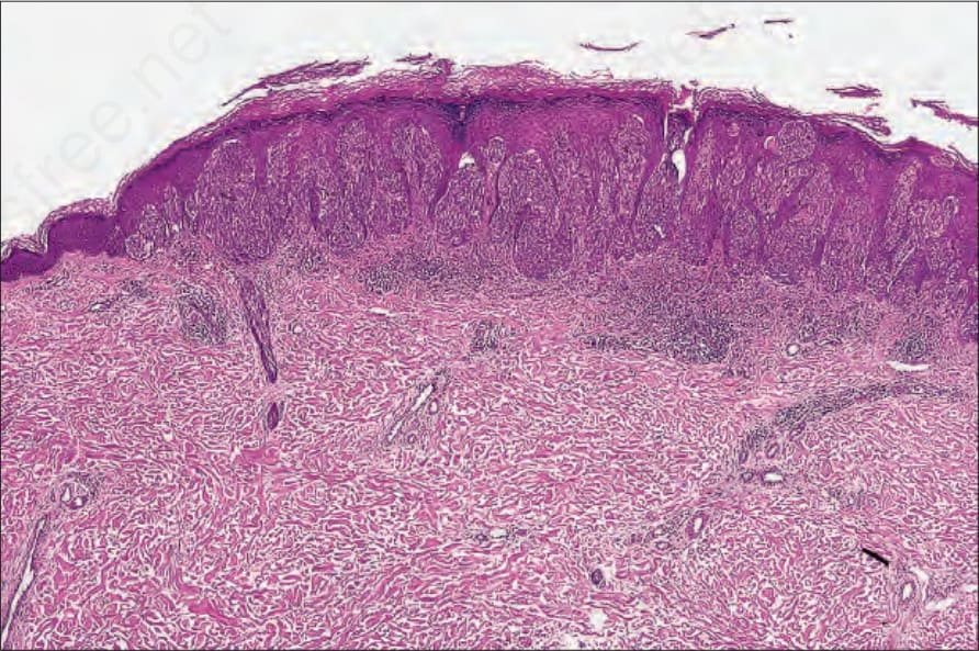
圖 25-158:Reed 梭形細胞瘤 (spindle cell tumor of Reed):一例無黑色素 (amelanotic) 病例的低倍視野。與 Spitz 痣 (Spitz nevus) 有明顯重疊。
Fig. 25.158 Spindle cell tumor of Reed: low-power view of an amelanotic example. There is obvious overlap with Spitz nevus.
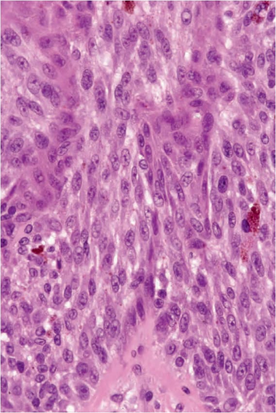
圖 25-159:Reed 梭形細胞瘤 (spindle cell tumor of Reed):仔細搜尋通常可發現小灶的黑色素 (melanin pigment)。細胞核非常均一。
Fig. 25.159 Spindle cell tumor of Reed: careful search usually reveals small foci of melanin pigment. The nuclei are very uniform.
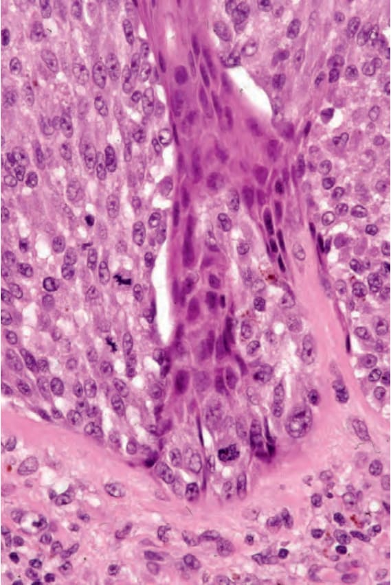
圖 25-160:Reed 梭形細胞瘤 (spindle cell tumor of Reed):注意其有絲分裂象 (mitotic figures)。
Fig. 25.160 Spindle cell tumor of Reed: note the mitotic figures.
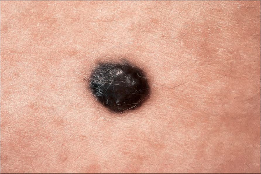
圖 25-161:深部穿透性痣 (deep penetrating nevus):此痣色素濃重且邊界銳利。承蒙倫敦皮膚病學研究所 (Institute of Dermatology, London, UK) 惠予提供。
Fig. 25.161 Deep penetrating nevus: the nevus is intensely pigmented and sharply circumscribed. By courtesy of the Institute of Dermatology, London, UK.
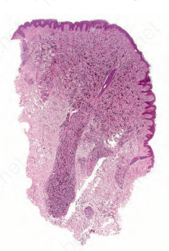
圖 25-162:深部穿透性痣 (deep penetrating nevus):此痣呈楔形,寬基部位於最上方,並有一深部穿透的下緣。
Fig. 25.162 Deep penetrating nevus: the nevus is wedge shaped with the broad base uppermost and a deeply penetrating lower border.
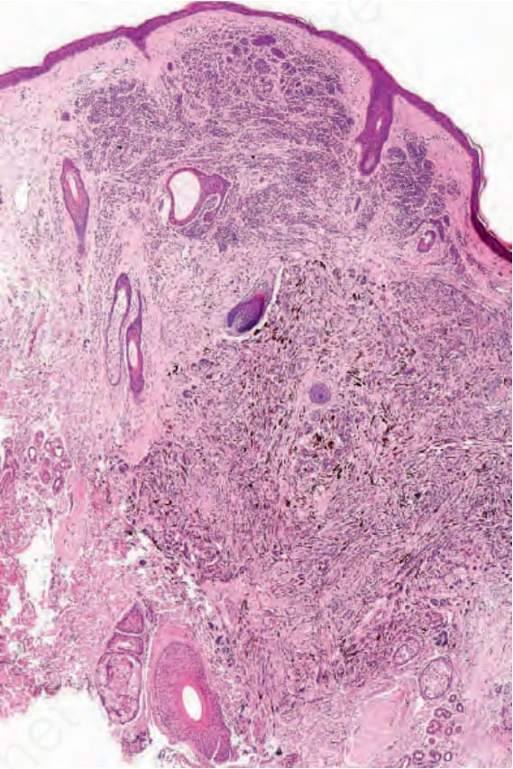
圖 25-163:深部穿透性痣 (deep penetrating nevus):此例為混合性變異型,平凡 (banal) 的真皮成分覆蓋於深部穿透性痣之上。
Fig. 25.163 Deep penetrating nevus: this example is a combined variant with a banal dermal component overlying the deep penetrating nevus.
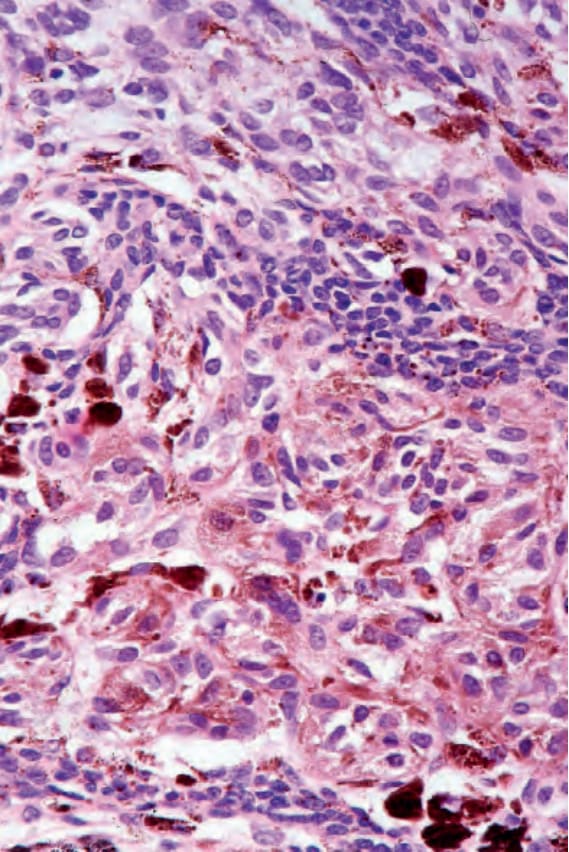
圖 25-164:深部穿透性痣 (deep penetrating nevus):Fig. 25.163 的高倍視野。
Fig. 25.164 Deep penetrating nevus: high-power view of Fig. 25.163.
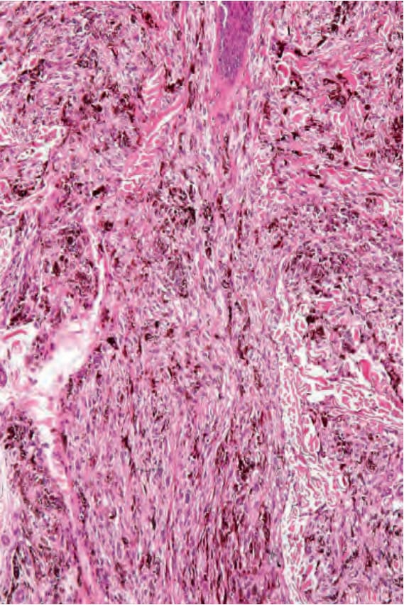
圖 25-165:深部穿透性痣 (deep penetrating nevus):深部延伸通常沿毛囊 (hair follicles) 與神經血管束 (neurovascular bundles) 分布。
Fig. 25.165 Deep penetrating nevus: the deep extension typically follows the hair follicles and neurovascular bundles.
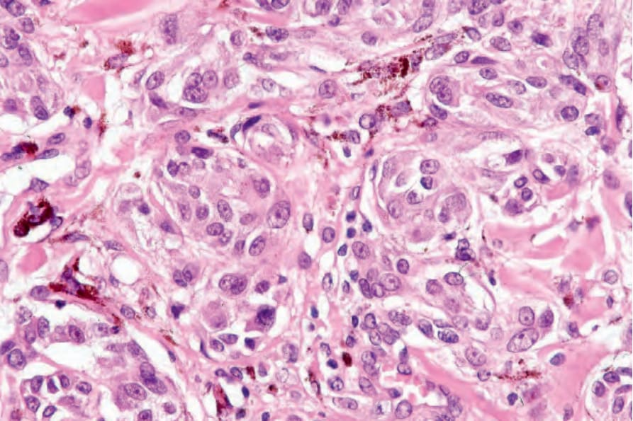
圖 25-166:深部穿透性痣 (deep penetrating nevus):在表淺處,此痣由上皮樣黑色素細胞 (epithelioid melanocytes) 構成,其細胞質淡染或細微帶色素,並具囊泡狀細胞核,常帶有小的嗜伊紅性核仁。
Fig. 25.166 Deep penetrating nevus: superficially, the nevus consists of epithelioid melanocytes with pale-staining or finely pigmented cytoplasm and vesicular nuclei, often with small eosinophilic nucleoli.
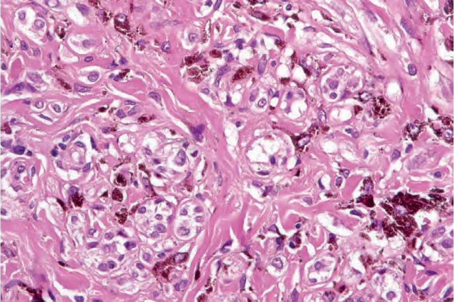
圖 25-167:深部穿透性痣 (deep penetrating nevus):透明細胞 (clear cells) 常存在。
Fig. 25.167 Deep penetrating nevus: clear cells are commonly present.
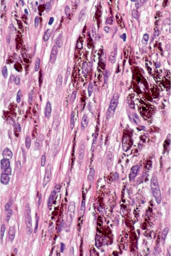
圖 25-168:深部穿透性痣 (deep penetrating nevus):在較深處,梭形細胞型態佔優勢,且色素濃重的黑色素噬細胞 (melanophages) 常很顯眼。
Fig. 25.168 Deep penetrating nevus: in the deeper reaches, spindled cell forms predominate and heavily pigmented melanophages are often conspicuous.
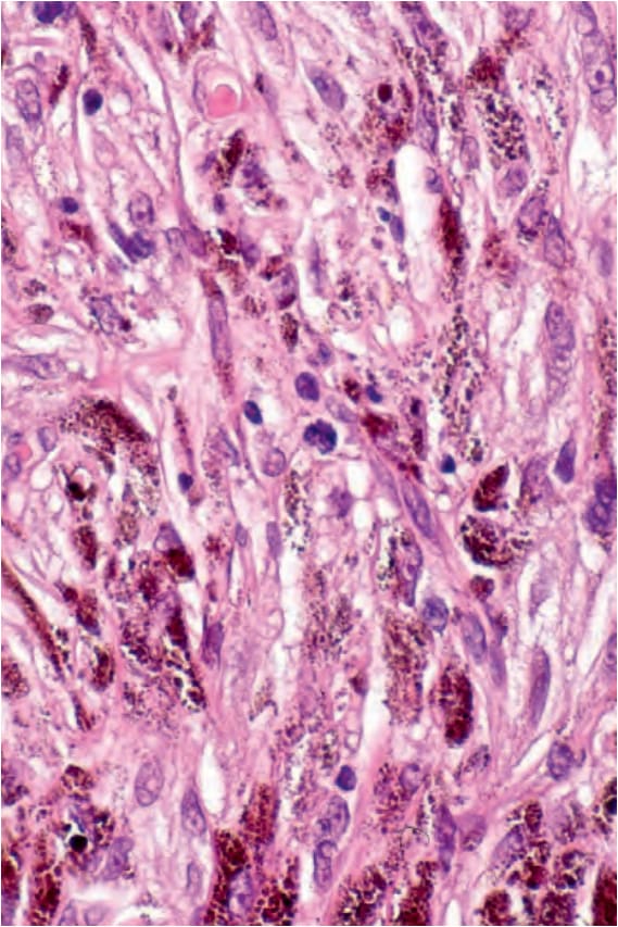
圖 25-169:深部穿透性痣 (deep penetrating nevus):可發現非常偶見的有絲分裂 (mitoses),但它們從不眾多或異常。
Fig. 25.169 Deep penetrating nevus: very occasional mitoses may be found but they are never numerous or abnormal.
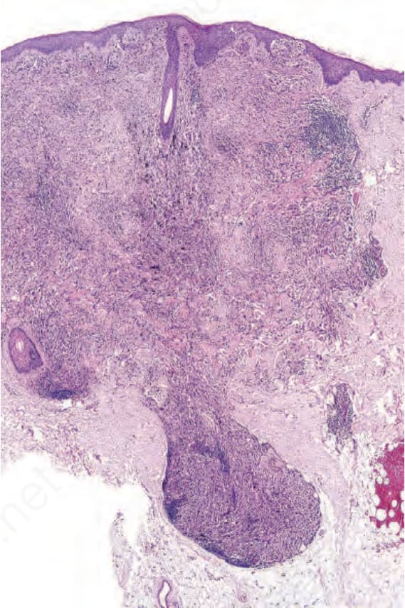
圖 25-170:具深部穿透性生長型態的黑色素瘤 (melanoma with deep penetrating growth pattern):在低倍放大下,此病變顯示深部穿透性生長型態。
Fig. 25.170 Melanoma with deep penetrating growth pattern: at low-power magnification, this lesion shows a deep penetrating growth pattern.
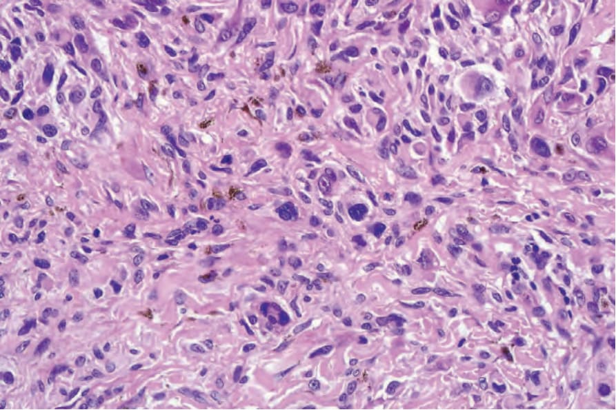
圖 25-171:具深部穿透性生長型態的黑色素瘤 (melanoma with deep penetrating growth pattern):然而在高倍放大下,可見明顯的核多形性 (nuclear pleomorphism) 與核深染 (hyperchromatism)。
Fig. 25.171 Melanoma with deep penetrating growth pattern: at high-power magnification, however, there is marked nuclear pleomorphism and hyperchromatism.