色素性上皮樣細胞痣 (Pigmented Epithelioid Cell Nevus)
Pigmented epithelioid cell nevus
臨床特徵 (Clinical Features)
此 Spitz nevus 的少見變異型在文獻中所受關注甚少。其表現為兒童或……(出現於兒童或)一個小而深色素的丘疹。
Pagetoid Spitz nevus 是 Spitz nevus 中相對罕見的變異型。它在女性中遠為常見,對四肢有顯著好發傾向(僅有少數病例報告發生於四肢以外),典型表現為直徑小於 6 mm 的淡棕至深棕色斑 (macule)。1–6 雖然 Busam 與 Barnhill 的開創性論文指出其僅發生於兒童與青少年,1 但近期一項針對 pagetoid Spitz nevi 的大型研究顯示其年齡分布範圍遠為廣泛(從 15 歲至 57 歲;平均年齡 34 歲)。6 Pagetoid Spitz nevus 通常表現為單發性病灶。1–6 然而,亦曾有報告描述單一病人身上發生多發性 pagetoid Spitz nevi 的例外案例,該病例最初被誤診為多發性原位黑色素瘤 (multiple melanoma in situ)。7
組織病理特徵 (Histopathology)
在組織學上,Spitz nevus 於其演化的早期階段,其特徵為一種完全位於表皮內 (intraepidermal) 的黑色素細胞增生,此增生可……(在組織學上可與)原位黑色素瘤 (in situ melanoma) 相混淆。
在表皮內 Spitz nevus 的 pagetoid 變異型中,數目增多、一致、增大的上皮樣黑色素細胞 (epithelioid melanocytes) 帶有豐富的嗜伊紅性細胞質,表現為一個小而界限分明且對稱的病灶,通常伴隨輕至中度的棘層肥厚 (acanthosis)(圖 25.136–25.138)。1 典型上,每個痣細胞 (nevus cell) 都藉由一個發育良好的回縮假象 (retraction artifact) 與相鄰細胞分隔開來。表皮內擴散 (intraepidermal spread) 通常局限於表皮的下半部,但亦可發生全層侵犯 (full-thickness involvement)。痣細胞為卵圓形或多角形,具有毛玻璃樣細胞質 (ground-glass cytoplasm) 以及圓形至卵圓形、一致的囊泡狀核 (vesicular nuclei),核內含有明顯的嗜伊紅性核仁。1 腫瘤細胞呈現有稜角的細胞質輪廓 (angulated cytoplasmic outline)。通常可見凋亡形態 (apoptotic forms),但不見有絲分裂 (mitoses)。在某些病灶中,可能有伴隨的接合部巢狀成分 (junctional nested component),一般僅代表此增生的次要成分(少於整體黑色素細胞群的 30%)。6 表淺真皮內可見血管周圍淋巴組織球性浸潤 (perivascular lymphohistiocytic infiltrate),常伴隨噬黑色素細胞 (melanophages)。6
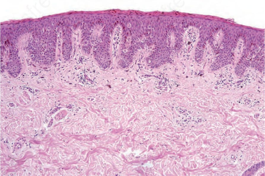
圖 25.136:Pagetoid Spitz nevus:低倍視野顯示一個大致位於基底部的上皮樣痣細胞群 (epithelioid nevus population)。此切片取自一名 5 歲兒童。
Fig. 25.136 Pagetoid Spitz nevus: low-power view showing a largely basally located epithelioid nevus population. This biopsy was taken from a 5-year-old child.
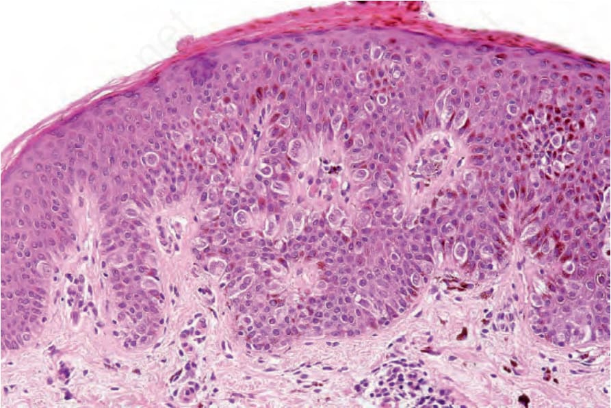
圖 25.137:Pagetoid Spitz nevus:可見中央的 pagetoid 擴散 (central pagetoid spread)。
Fig. 25.137 Pagetoid Spitz nevus: there is central pagetoid spread.
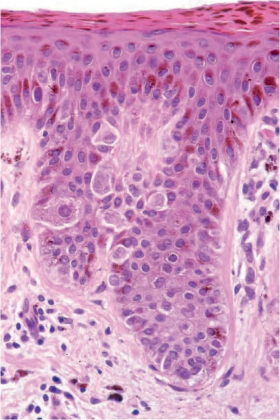
圖 25.138:Pagetoid Spitz nevus:痣細胞具有豐富的嗜伊紅性細胞質以及一致的囊泡狀核 (vesicular nuclei),核內含明顯核仁。
Fig. 25.138 Pagetoid Spitz nevus: the nevus cells have abundant eosinophilic cytoplasm and uniform vesicular nuclei with prominent nucleoli.