Pigmented epithelioid cell nevus
Pigmented epithelioid cell nevus
Clinical features This uncommon variant of Spitz nevus has received scant attention in the literature. It presents as a small, darkly pigmented papule in children or
Clinical features Pagetoid Spitz nevus is a relatively rare variant of Spitz nevus. It much more common in females, shows striking predilection for extremities (with only a handful of the cases reported outside extremities), and typically presents as a light to dark brown macule measuring less than 6 mm in diameter.1–6 Although a seminal paper by Busam and Barnhill suggested an exclusive occurrence in children and adolescents,1 a recent large study on pagetoid Spitz nevi demonstrated much broader age distribution (from 15 to 57 years; average age, 34 years).6 Pagetoid Spitz nevus usually presents as a solitary lesion.1–6 Nevertheless, an exceptional example of multiple pagetoid Spitz nevi occurring in a single patient, initially misdiagnosed as multiple melanoma in situ, has also been reported.7
Histologic features Histologically, Spitz nevus in its early stages of evolution is characterized by a wholly intraepidermal melanocytic proliferation which can be
1271 Desmoplastic nevus
re-excision to ensure complete removal is advised.4 In addition, fluorescence in situ hybridization assay can be performed to detect aberrations on chromosome 6 (6p25, 6q23, and Cep6) and chromosome 11 (11q13), their presence being suggestive of malignant melanocytic proliferations.8,9
confused histologically with in situ melanoma. In the pagetoid variant of intraepidermal Spitz nevus, increased numbers of uniform, enlarged, epithelioid melanocytes with abundant eosinophilic cytoplasm present as a small circumscribed and symmetrical lesion usually associated with mild to moderate acanthosis (Figs 25.136–25.138).1 Typically, each nevus cell is separated from adjacent cells by a well-developed retraction artifact. Intraepidermal spread is usually limited to the lower half of the epidermis, but full-thickness involvement can occur. The nevus cells are oval or polygonal with ground-glass cytoplasm and round to oval uniform vesicular nuclei containing prominent eosinophilic nucleoli.1 Tumor cells display an angulated cytoplasmic outline. Apoptotic forms are usually present, but mitoses are not seen. In some lesions, there may be an associated junctional nested component, generally representing a minor component of the proliferation (less than 30% of the total melanocytic population).6 A perivascular lymphohistiocytic infiltrate is present in the superficial dermis, frequently associated with melanophages.6
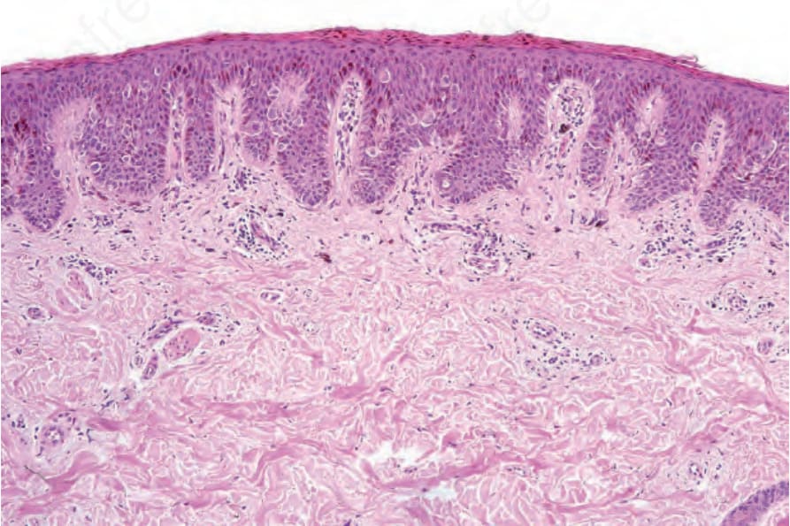
Fig. 25.136 Pagetoid Spitz nevus: low-power view showing a largely basally located epithelioid nevus population. This biopsy was taken from a 5-year-old child.
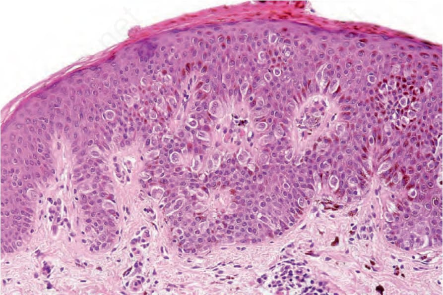
Fig. 25.137 Pagetoid Spitz nevus: there is central pagetoid spread.
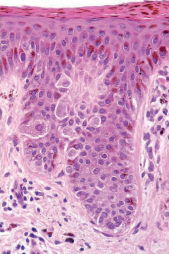
Fig. 25.138 Pagetoid Spitz nevus: the nevus cells have abundant eosinophilic cytoplasm and uniform vesicular nuclei with prominent nucleoli.