氣球細胞痣 (Balloon Cell Nevus)
臨床特徵 (Clinical Features)
- 氣球細胞痣 (balloon cell nevus) 為黑色素細胞痣 (melanocytic nevus) 的一種罕見變異型,咸認其代表一種退化性變化 (degenerative change),係因黑色素前驅物 (melanin precursors) 堆積於前黑色素小體 (premelanosomes),導致細胞質出現空泡化 (vacuolation) 所致。
- 此現象出現於不到 2% 的一般痣 (common nevi) 中。
- 氣球細胞痣雖可發生於任何年齡,但約 80% 在生命的前三個十年內被診斷。
- 男女發生率相等,好發於頭頸部 (head and neck),其次為軀幹與四肢。
- 此病灶並無特別具鑑別性的臨床特徵,通常表現為平滑、圓頂狀 (dome-shaped) 的紅色或棕色丘疹 (papule) 或結節 (nodule)。
- 氣球細胞痣亦曾在黏膜部位 (mucosal sites)、結膜 (conjunctiva) 與虹膜 (iris) 被報告過。
- 在皮膚鏡 (dermoscopy) 下,氣球細胞巢 (balloon cell nests) 對應於痣內白色與黃色的小球/團塊 (white and yellow globules/clods)。
組織病理特徵 (Histopathology)
- 此痣可為複合型 (compound) 或皮內型 (intradermal),且依定義必須顯示氣球細胞 (balloon cells) 佔優勢(大於 50%)(此為原發現象,primary phenomenon),因為偶見的典型黑色素細胞痣也可能含有少數散在的氣球細胞(此為次發現象,secondary phenomenon)(Fig. 25.95)。
- 後者(氣球細胞)呈圓形或卵圓形、大小不一,具有豐富的泡沫狀 (foamy)、淡染 (pale-staining) 或透明的細胞質。
- 細胞含有一個位於中央、相當深染 (hyperchromatic) 或具空泡的細胞核,並有顯著的核仁 (nucleolus)(Fig. 25.96)。
- 受影響的細胞常與脂肪細胞 (adipocytes) 有顯著相似性。
- 可能出現多核巨細胞 (multinucleate giant cells) 此一特徵。
- 無有絲分裂活性 (mitotic activity)。
- 色素含量不定,從含有豐富黑色素的病灶,到完全無黑色素 (amelanotic) 的例子皆有。
- 在痣的周邊有時可見較明顯的黑色素細胞特徵 (melanocytic features)(Fig. 25.97)。
- 氣球細胞變化 (balloon cell change) 亦曾在發生於大型先天性黑色素細胞痣 (large congenital melanocytic nevus) 背景中的增生性結節 (proliferative nodules) 內被報告。
鑑別診斷 (Differential Diagnosis)
- 氣球細胞變化 (balloon cell change) 亦曾被記載於 Spitz nevi、dysplastic nevi、cellular blue nevi、combined nevi 與 melanoma(包括轉移性腫瘤)中。
- 後者(melanoma)可藉由核多形性 (nuclear pleomorphism)、核仁明顯 (nucleolar prominence) 與有絲分裂活性 (mitotic activity) 的存在而加以區分。
- 須注意,balloon cell melanoma 偶爾可呈現出具欺騙性的溫和外觀(類似於 nevoid melanoma),因此氣球細胞痣應始終在多個切面層次 (multiple levels) 仔細審視,以排除 melanoma。
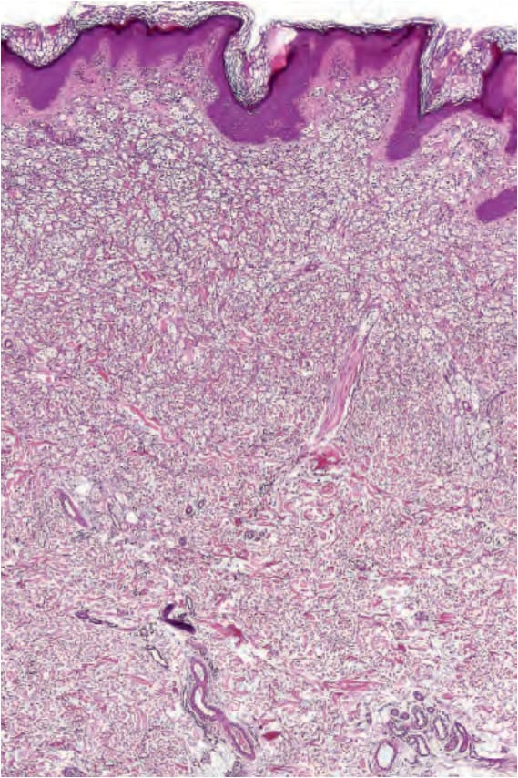
圖 25-95:氣球細胞痣 (balloon cell nevus):掃描視野顯示一個由淡染細胞 (pale-staining cells) 組成的真皮結節 (dermal nodule)。
Fig. 25.95 Balloon cell nevus: scanning view showing a dermal nodule composed of palestaining cells.
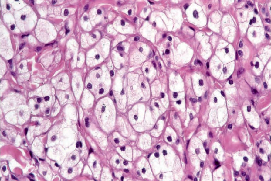
圖 25-96:氣球細胞痣 (balloon cell nevus):痣細胞具有淡染、略呈泡沫狀的細胞質 (pale, slightly foamy cytoplasm) 與位於中央的深染細胞核 (central hyperchromatic nuclei)。
Fig. 25.96 Balloon cell nevus: the nevus cells have pale, slightly foamy cytoplasm and central hyperchromatic nuclei.
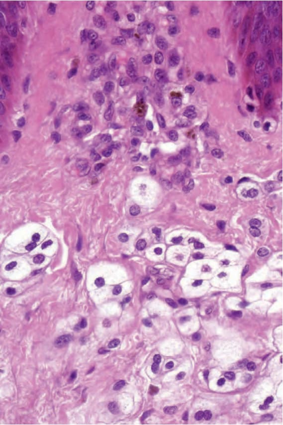
圖 25-97:氣球細胞痣 (balloon cell nevus):藉由辨識較典型的痣細胞 (typical nevus cells) 有助於診斷。
Fig. 25.97 Balloon cell nevus: diagnosis is facilitated by the identification of more typical nevus cells.