Balloon cell nevus
Balloon cell nevus
Clinical features Balloon cell nevus is a rare variant of melanocytic nevus and is believed to represent a degenerative change due to accumulation of melanin precursors in premelanosomes with resultant vacuolation of the cytoplasm.1–5 This phenomenon is present in less than 2% of common nevi.2 Although balloon cell nevus may present at any age, about 80% are diagnosed in the first three decades of life. The incidence is equal in men and women, and there is a predilection for the head and neck, followed by trunk and extremities. The lesion displays no particular distinguishing clinical features, and usually presents as a smooth, dome-shaped, red or brown papule or nodule.2 Balloon cell nevi have also been reported at mucosal sites, conjunctiva, and iris.5–11
On dermoscopy, balloon cell nests correspond to areas of white and yellow globules/clods within the nevus.12,13
Histologic features The nevus may be compound or intradermal and, by definition, must show a predominance (greater than 50%) of balloon cells (primary phenomenon), since the occasional typical melanocytic nevus may contain a few scattered balloon cells (secondary phenomenon) (Fig. 25.95).1,2 The latter are round or oval and variably sized, with abundant foamy pale-staining or clear cytoplasm. They contain a central rather hyperchromatic or vesicular nucleus with a conspicuous nucleolus (Fig. 25.96). Not uncommonly, affected cells display a striking resemblance to adipocytes. Multinucleate giant cells may be a feature. Mitotic activity is absent. Pigmentation is variable, ranging from lesions with abundant melanin to completely amelanotic examples. More obvious melanocytic features are sometimes evident at the periphery of the nevus (Fig. 25.97).
Balloon cell change has also been reported in proliferative nodules developing in the background of a large congenital melanocytic nevus.14
Differential diagnosis Balloon cell change has also been documented in Spitz nevi, dysplastic nevi, cellular blue nevi, combined nevi, and melanoma (including metastatic tumors).15–18 The last may be distinguished by the presence of nuclear pleomorphism, nucleolar prominence, and mitotic activity.
It should be noted that balloon cell melanoma can, on occasion, appear deceptively bland (similar to nevoid melanoma) and, as a result, balloon cell nevi should always be carefully scrutinized at multiple levels to exclude a melanoma.
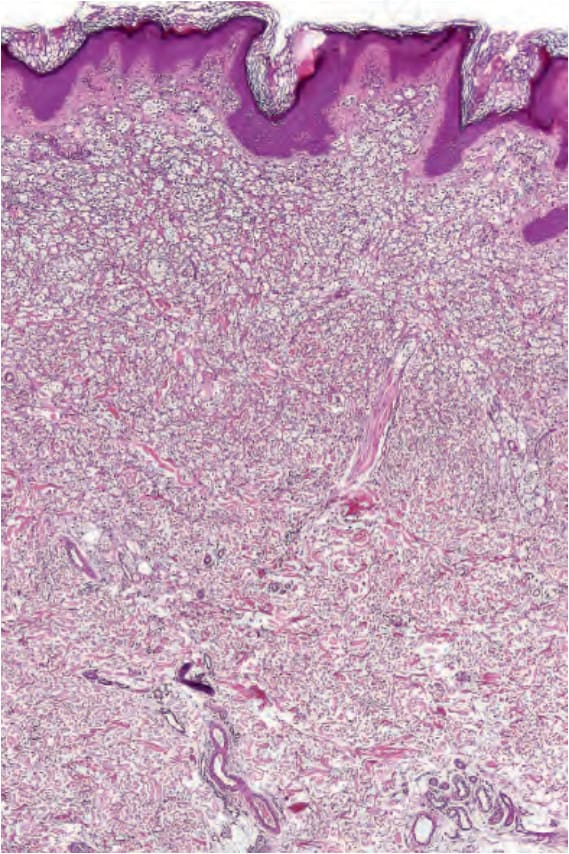
Fig. 25.95 Balloon cell nevus: scanning view showing a dermal nodule composed of palestaining cells.
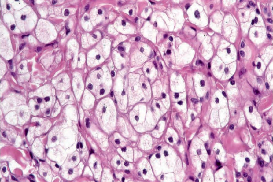
Fig. 25.96 Balloon cell nevus: the nevus cells have pale, slightly foamy cytoplasm and central hyperchromatic nuclei.
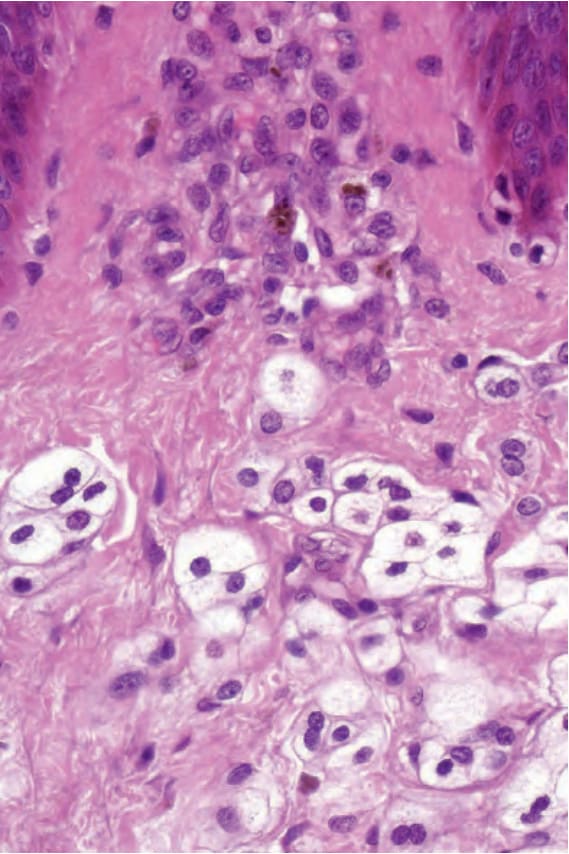
Fig. 25.97 Balloon cell nevus: diagnosis is facilitated by the identification of more typical nevus cells.