下肢/踝部痣 (Nevi of the Lower Leg/Ankle)
下肢/踝部痣 (nevi of the lower leg/ankle)
已有文獻報導下肢/踝部的非典型痣 (atypical nevi of the lower leg/ankle)。這類病灶一般為小型病灶,大小介於 2 至 4 mm(平均直徑 3 mm),且呈現女性好發,比例為 4:1。
組織病理特徵 (Histopathology)
下肢/踝部的 atypical nevi 可為交界型 (junctional) 或複合型 (compound)。病灶可呈不對稱、缺乏側向界限分明 (lateral circumscription),並顯示單一細胞增生 (single cell proliferation),尤其是在側緣部位。雖然可見局灶性向上遷移至表皮下層 (focal upward migration into the lower layers of the epidermis),但一般不會見到 pagetoid 擴散至表皮上層。細胞學異型性 (cytological atypia) 通常為輕度至中度。
真皮成分若存在,則薄且不顯著。可見輕度、非劇烈 (nonbrisk) 的發炎細胞浸潤。通常不會發現真皮纖維化 (dermal fibrosis)。
鑑別診斷 (Differential Diagnosis)
與 dysplastic nevus 的區別在於缺乏典型出現於 dysplastic nevi 的間質反應 (stromal response),包括板層狀或同心圓狀纖維化 (lamellar or concentric fibrosis) 與血管增生 (vascular proliferation)。然而,有人提出至少部分下肢/踝部的 atypical nevi 實際上可能代表早期的 dysplastic nevus。
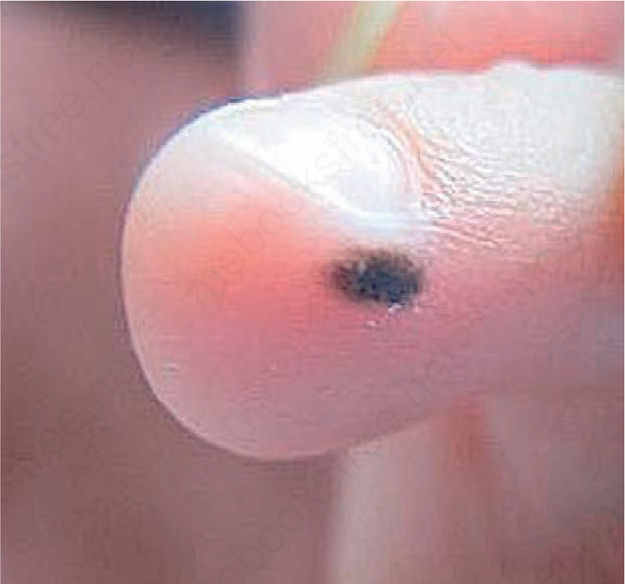
圖 25-80:肢端痣 (acral nevus):注意其不規則輪廓 (irregular outline)。Courtesy of Yi-Guo Feng, Xi’an, China。
Fig. 25.80 Acral nevus: Note irregular outline. Courtesy of Yi-Guo Feng, Xi’an, China.