Nevi of the lower leg/ankle
Nevi of the lower leg/ankle
Atypical nevi of the lower leg/ankle have been reported.1 They are generally small lesions, measuring between 2 and 4 mm (mean diameter, 3 mm), and show female predominance with a ratio of 4 : 1.
Histologic features Atypical nevi of the lower leg/ankle can be junctional or compound. The lesions may be asymmetrical, lack lateral circumscription, and display single cell proliferation, especially at the lateral aspects.1 Although focal upward migration into the lower layers of the epidermis may be seen, pagetoid spread into the upper layers is generally not seen. Cytological atypia is usually mild to moderate in degree.
The dermal component, when present, is thin and unremarkable. A mild nonbrisk inflammatory cell infiltrate can be seen. Usually, no dermal fibrosis is identified.
Differential diagnosis Distinction from dysplastic nevus is made by the absence of the stromal response, typically seen in dysplastic nevi, including lamellar or concentric fibrosis and vascular proliferation. However, it has been suggested that at least some atypical nevi of the lower leg/ankle might actually represent an early dysplastic nevus.
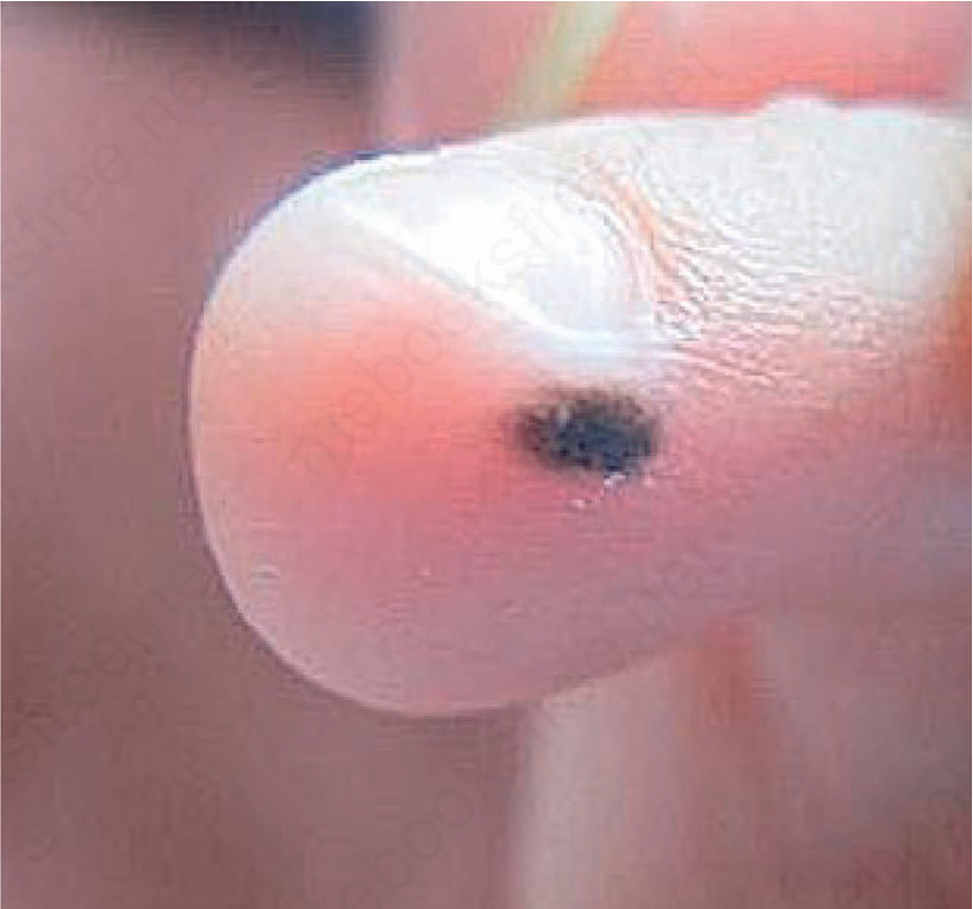
Fig. 25.80 Acral nevus: Note irregular outline. Courtesy of Yi-Guo Feng, Xi’an, China.