頭皮痣 (Nevi on the Scalp)
臨床特徵 (Clinical Features)
- 頭皮痣 (nevi on the scalp) 最常發生於枕部 (occipital region),其次為左頂部 (left parietal region)、右頂部 (right parietal region) 及額部 (frontal region)(Fig. 25.65)。
- 其數目與全身痣的總數相關,最常見於四十多歲(第四個十年),平均年齡 35 歲。頭皮痣以男性為多。
- 約 10% 的頭皮痣會表現出令人困擾的組織學特徵。此類痣通常見於青少年與年輕成人,其組織學特徵與發生於乳線 (mammary line)、生殖器部位及屈側部位 (flexural sites) 的病灶相似。
組織病理特徵 (Histopathology)
- 在此部位,非典型黑色素細胞痣 (atypical melanocytic nevi) 通常可辨識出兩種主要的形態學型態:大巢狀型態 (large nested pattern) 與類似分化不良痣 (dysplastic nevus) 的型態。
- 一般而言,頭皮上的 atypical nevi 以不對稱 (asymmetry) 及橫向界限不清 (poor lateral circumscription) 為特徵(Figs 25.66–25.70)。
- 巢狀增生型態 (nested pattern of proliferation) 由位於表皮突 (rete ridges) 尖端與側面、並沿真皮–表皮交界 (dermal–epidermal junction) 隨機散布的大型黑色素細胞巢 (large nests of melanocytes) 所組成。此外,黑色素細胞巢的形狀有所變化,常見奇形怪狀的形態 (bizarre forms) 以及巢內腫瘤細胞的失黏附 (discohesion)。可見到皮膚附屬器 (skin adnexa) 的侵犯,且可能相當明顯。
- 沿真皮–表皮交界的局灶性雀斑樣增生 (focal lentiginous proliferation) 經常出現。
- 黑色素細胞異型性 (melanocytic atypia) 通常輕微(雖然偶有嚴重的細胞學異型性出現)且為隨機分布,由染色質深染的細胞核 (hyperchromatic nuclei) 與不明顯的核仁 (indistinct nucleoli) 所構成。在病灶中央部位有時可見到單個黑色素細胞的向上遷移 (upward migration of isolated melanocytes)。
- 類分化不良痣型態 (dysplastic nevus-like pattern) 的特徵為表皮突的橋接 (bridging of the rete ridges)、交界成分 (junctional component) 延伸超過真皮成分 (dermal component),以及乳頭真皮纖維增生 (papillary dermal fibroplasia)。真皮黑色素細胞成分通常無明顯異常,但偶可見到淺層的有絲分裂活性 (superficial mitotic activity)。
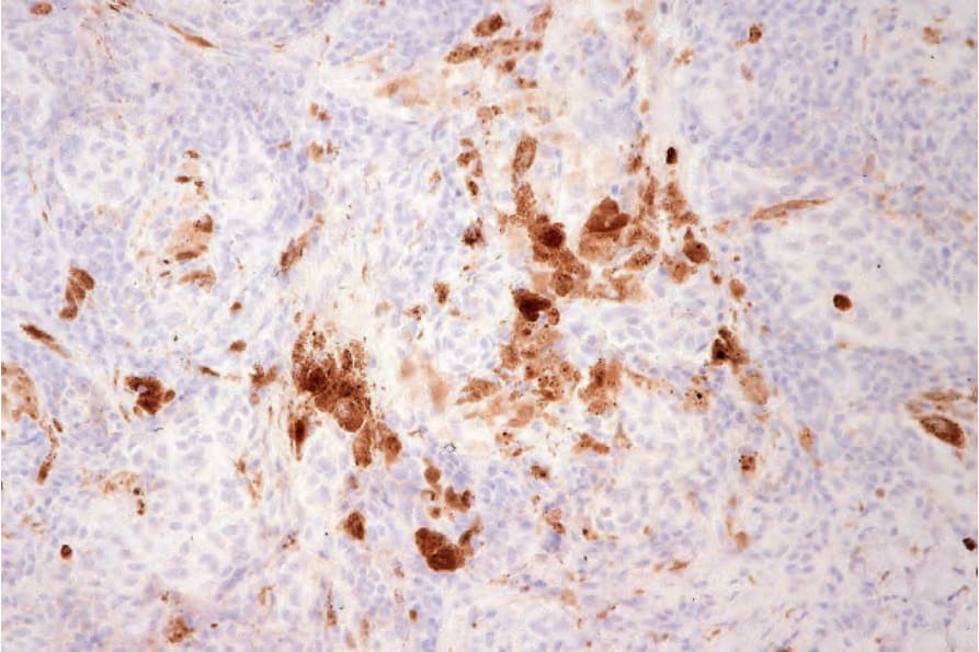
圖 25-64:純系性痣 (clonal nevus):痣細胞不表現 MIB-1。承蒙 W. Grayson 醫師提供,National Health Laboratory Service, Johannesburg, South Africa。
Fig. 25.64 Clonal nevus: the nevus cells do not express MIB-1. By courtesy of W. Grayson, MD, National Health Laboratory Service, Johannesburg, South Africa.
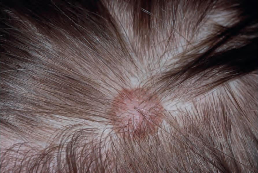
圖 25-65:頭皮痣 (scalp nevus):此部位的病灶有時會表現出細胞學特徵,可能令不警覺者誤判為黑色素瘤 (melanoma)。注意中央的蒼白區 (central pallor) 伴隨周邊的色素加深邊緣 (rim of hyperpigmentation)。
Fig. 25.65 Scalp nevus: lesions at this site may on occasions show cytological features that can raise concern for a diagnosis of melanoma by the unwary. Note the central pallor with a rim of hyperpigmentation.
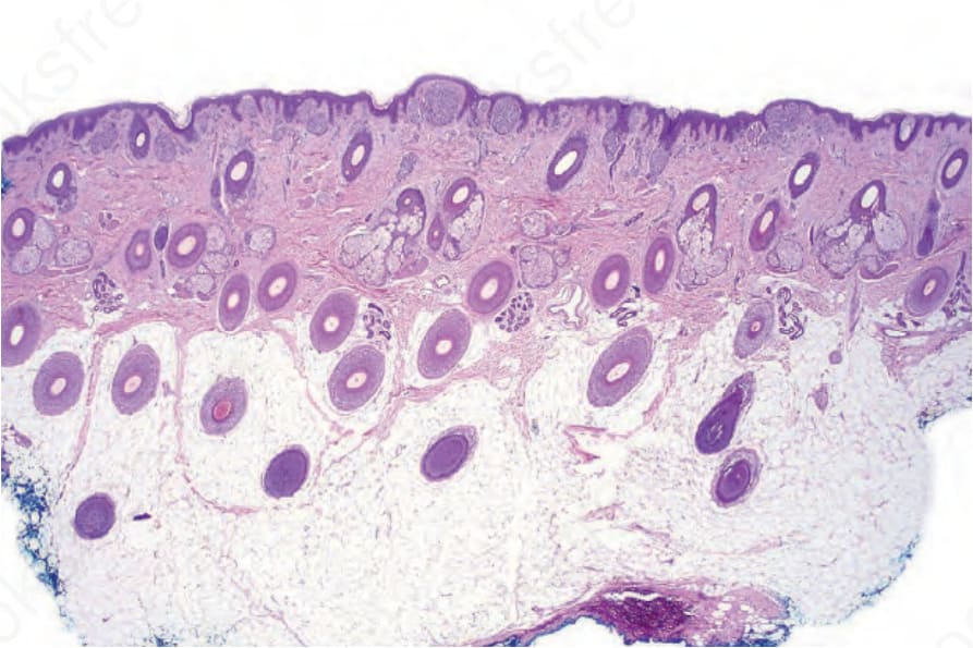
圖 25-66:頭皮痣 (scalp nevus):即使在低倍放大下,也可辨識出大型膨脹性的交界巢 (large expansile junctional nests)。
Fig. 25.66 Scalp nevus: even at low-power magnification, large expansile junctional nests can be appreciated.
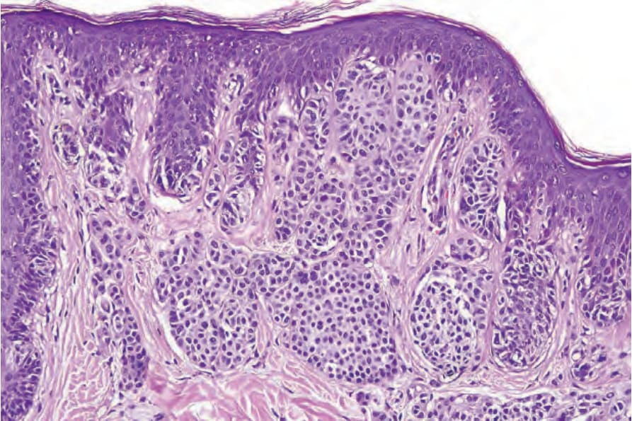
圖 25-67:頭皮痣 (scalp nevus):真皮巢 (dermal nests) 大於交界巢 (junctional ones),此特徵總是令人擔憂。
Fig. 25.67 Scalp nevus: the dermal nests are larger than the junctional ones, a feature which always results in concern.
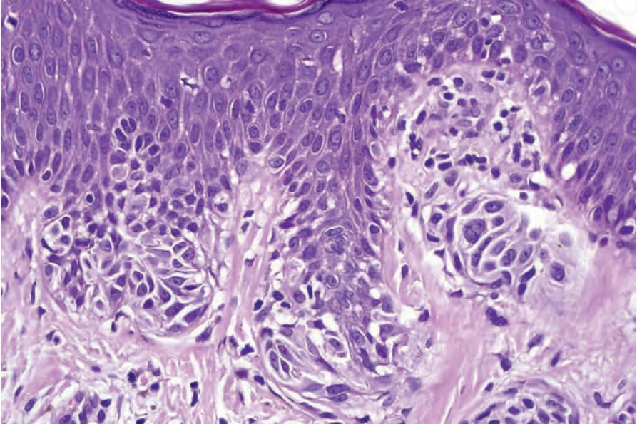
圖 25-68:頭皮痣 (scalp nevus):在此視野中,交界痣細胞 (junctional nevus cells) 顯示中度至重度的細胞學異型性 (cytological atypia)。
Fig. 25.68 Scalp nevus: in this field, the junctional nevus cells show moderate to severe cytological atypia.
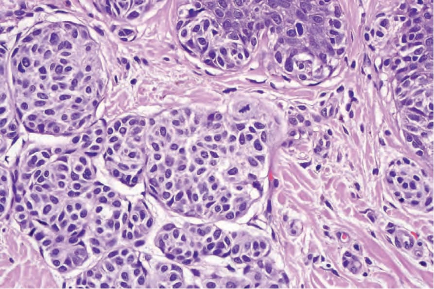
圖 25-69:頭皮痣 (scalp nevus):注意淺層真皮成分中的有絲分裂象 (mitotic figure)。
Fig. 25.69 Scalp nevus: note the mitotic figure in the superficial dermal component.
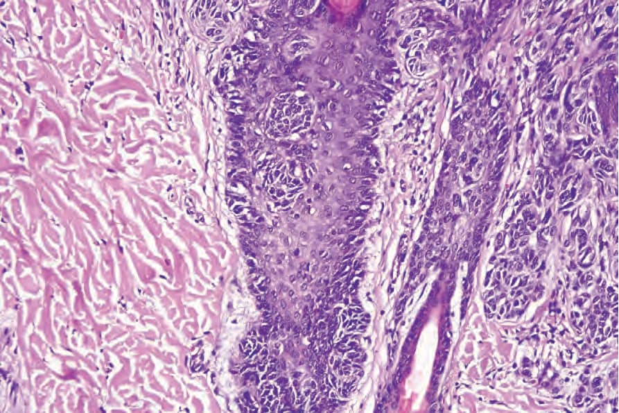
圖 25-70:頭皮痣 (scalp nevus):可見毛囊侵犯 (follicular involvement),此為這些病灶具有顯著復發率 (significant recurrence rate) 的原因。
Fig. 25.70 Scalp nevus: there is follicular involvement accounting for the significant recurrence rate for these lesions.
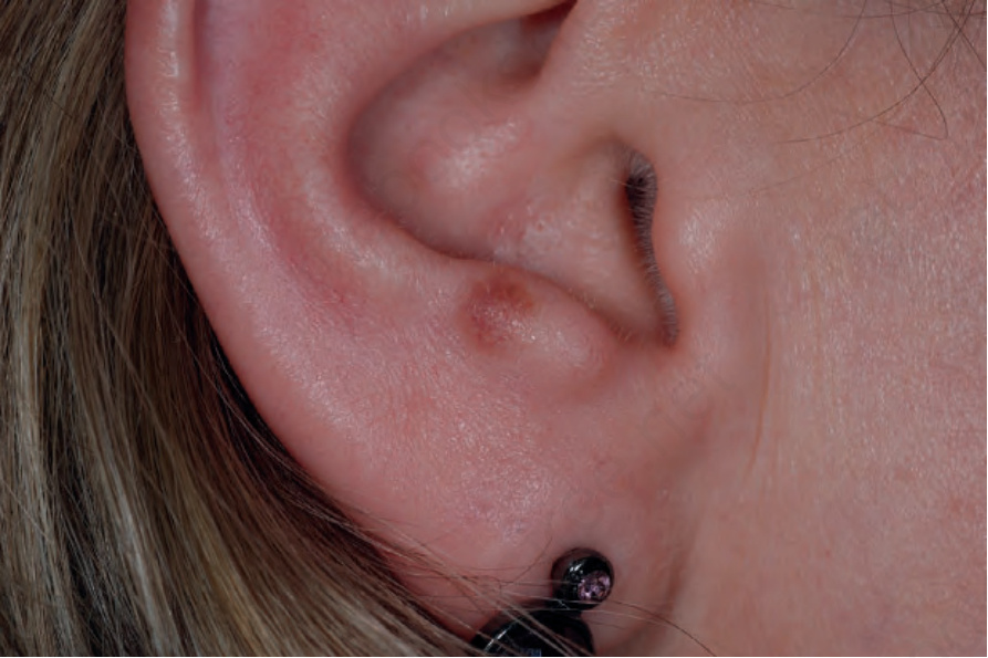
圖 25-71:耳部痣 (nevus of ear):注意界限不清的色素性斑狀病灶 (ill-defined pigmented, macular lesion)。
Fig. 25.71 Nevus of ear: note the ill-defined pigmented, macular lesion.
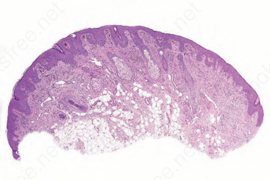
圖 25-72:耳部痣 (nevus of ear):以掃描式視野 (scanning view) 觀察一個主要為交界型 (largely junctional) 的病灶。
Fig. 25.72 Nevus of ear: scanning view of a largely junctional lesion.