假黑色素細胞巢 (Pseudomelanocytic nests)
假黑色素細胞巢 (Pseudomelanocytic nests)
臨床特徵 (Clinical features)
-
假黑色素細胞巢 (pseudomelanocytic nests) 是表皮內非黑色素細胞 (nonmelanocytic) 之聚集團塊或細胞碎片,在組織學檢查時模擬黑色素細胞增生 (melanocytic proliferation)。這些細胞巢通常發生於苔癬樣 (lichenoid) 的背景中。
-
雖然這些病灶確實偶爾會發生惡性轉化 (malignant transformation),但此類事件甚為罕見;因此,對於其他方面典型的黑色素細胞痣 (melanocytic nevi),並無理由進行廣泛的預防性切除 (Figs 25.23 and 25.24)。據估計,任一顆痣演變為黑色素瘤 (melanoma) 的可能性約為 1/100 000。後續死亡率約為每 1/500 000 顆原始痣。然而,痣的盛行率在流行病學上具重大意義,痣數目增多與後續發生 melanoma(淺表擴散型 superficial spreading 及結節型 nodular 亞型)的風險增高相關。惡性小痣黑色素瘤 (lentigo maligna melanoma) 並非源自既存的痣,亦與其盛行率無關。在這些病灶處於發育早期的幼童與青少年中,色素增多是可預期的。然而在成人,交界活性 (junctional activity) 的證據則須謹慎看待;正是那些色素逐漸明顯增加、或在較年長年齡層中重新發生 (de novo) 的痣,常被切除以供組織學評估。
黑色素細胞痣 (Melanocytic nevus)
臨床特徵 (Clinical features)
-
黑色素細胞痣 (melanocytic nevi) 最早出現於兒童早期,並在第二與第三個十年期間數目增多。在男性,頭部、頸部與軀幹特別容易受侵犯,而在女性則上肢與下肢較常受累。後天性黑色素細胞痣偶可見呈聚集 (agminate,成群) 分布的型態。Melanocytic nevi 在中年期退化,且多數在老年人中已完全消退。它們在膚色蒼白、淺色眼睛的個體中較為常見。在亞洲人 (Asians) 與非裔加勒比海人 (Afro-Caribbeans) 中,melanocytic nevi 之發生則少得多。在這些種族中,肢端 (acral) 部位特別容易受侵犯。深棕色或黑色頭髮與痣數目增多相關,而紅色頭髮似乎具保護作用。Melanocytic nevi 之發育與生命前二十年內的日曬程度相關。間歇性強烈日曬比慢性曝曬更為重要。事實上,慢性日曬與低痣數相關(亦即其似乎具保護作用)。痣數目增多者,常見於日曬後傾向曬傷 (sunburn) 而非曬黑 (tan) 的個體,並與雀斑 (freckling) 程度相關。由於這些因素在 melanoma 的病因學中亦屬重要,故早年高度的雀斑與痣可能具預測價值。
-
Melanocytic nevi 依其演化階段呈現多樣的特徵。交界痣 (junctional nevi) 通常為斑狀 (macular) 或略隆起,直徑可達 0.5 cm,顏色由淺至深棕色 (Figs 25.25–25.27)。它們界限分明、邊緣規則,且通常色素均勻,但有時中央區域較深。典型情況下,病灶表面可清楚辨識皮膚紋路 (skin lines)。
-
複合痣 (compound nevus) 隆起,有時呈圓頂狀 (dome-shaped) 或疣狀 (warty),且常仍深度色素沉著 (Figs 25.28 and 25.29)。偶爾其表面突出粗糙的毛髮;拔除這些毛髮可能使毛囊的真皮成分受創,導致肉芽腫性發炎 (granulomatous inflammation),這可能使病人與臨床醫師對惡性轉化之可能性產生疑慮。
-
皮內痣 (intradermal nevus) 常無色素,可表現為圓頂狀結節 (dome-shaped nodule)、乳頭瘤狀 (papillomatous) 病灶,或有蒂的皮膚贅瘤 (pedunculated skin tag) (Figs 25.30 and 25.31)。
-
痣在懷孕或口服避孕藥 (oral contraceptive pill) 的影響下可能變得色素更深。然而,並無證據顯示懷孕會以任何顯著方式刺激其發育或改變既存痣的生物學潛能。
組織學特徵 (Histologic features)
-
關於痣細胞 (nevocyte) 的本質及其與黑色素細胞 (melanocyte) 之區別,存在相當多的混淆。痣細胞 (nevocyte) 不過是已增殖以形成黑色素細胞痣的黑色素細胞。其電子顯微鏡下外觀與 melanocyte 相同,並具有相同的胞器與酵素系統;唯一的顯著差異在於,真皮成分缺乏樹突狀突起 (dendritic processes),且隨深度增加,黑色素合成 (melanin synthesis) 被中止。
-
在發育的最早期階段,黑色素細胞的交界巢 (junctional nests) 出現於表皮下部(受基底膜 basement membrane 所限),通常位於有時加寬並延長的表皮突 (epidermal ridges) 之尖端,較少見於其側面(雀斑樣交界痣 lentiginous junctional nevus)(Figs 25.32 and 25.33)。黑色素細胞可為多角形 (polygonal) 與上皮樣 (epithelioid),或較罕見地呈紡錘狀 (spindled),胞質透明至淡染或輕度嗜伊紅 (eosinophilic),並含有均勻的圓形至卵圓形小核,核仁明顯(A型痣細胞 type A nevus cells)(Figs 25.34 and 25.35)。胞質典型含有稀疏、均勻分布的細緻黑色素顆粒。在良性交界痣中,生長朝向真皮侵犯。黑色素細胞任何朝表皮上層擴散 (pagetoid spread) 的傾向,提示惡性變化,應謹慎看待。然而,黑色素細胞單獨的向上移行 (upward migration) 本身並不能診斷為惡性,因為此特徵可見於發生於某些特殊部位的類痣樣病灶 (nevi-like lesions) 變異型(見下文)。表皮突之間的黑色素細胞有時可能顯得數目增多。依定義,交界痣完全位於表皮內 (intraepidermal);然而,在色素濃重的變異型中,黑色素典型存在於乳頭真皮 (papillary dermis) 內的巨噬細胞(噬黑色素細胞 melanophages)中(色素失禁 pigmentary incontinence)。
-
除了交界活性之外,複合痣 (compound nevi) 在乳頭真皮與淺層網狀真皮 (superficial reticular dermis) 內均顯示痣細胞 (nevus cells) 之細胞巢與細胞索 (strands) (Figs 25.36–25.39)。複合性平庸痣 (compound banal nevi) 通常界限相當分明且對稱。交界成分典型不會延伸超出真皮成分,亦即多數平庸痣 (banal nevi) 缺乏肩部 (shoulder)(與發育不良痣 dysplastic nevus 比較)。然而應注意,平庸性複合痣有時也可存在肩部,亦即肩部之存在本身並不使痣成為發育不良痣。Compound nevi 有時可伴有顯著的角化過度 (hyperkeratosis)、棘層肥厚 (acanthosis) 並形成角質假性囊腫 (keratinous pseudocyst),以及乳頭瘤病 (papillomatosis)(令人聯想到脂漏性角化症 seborrheic keratosis),造成疣狀的臨床外觀——即所謂的乳頭瘤狀(疣狀)黑色素細胞痣 (papillomatous (verrucous) melanocytic nevus) 或角化性黑色素細胞痣 (keratotic melanocytic nevus) (Fig. 25.40)。這些痣較常見於女性,且軀幹是最常受侵犯的部位。此變化可能與雌激素 (estrogens) 相關,因為痣細胞表現 pS2。
-
真皮病灶較淺表成分的細胞,可能保留與交界痣相似的細胞學特徵。較深部的細胞則小得多、胞質較少,並具有緻密、染色較深的核,類似淋巴球(B型痣細胞 type B nevus cells)(Figs 25.41–25.43)。有絲分裂活性 (mitotic activity) 偶可見於後天性黑色素細胞痣的真皮成分中(見鑑別診斷)。有絲分裂活性在懷孕期的黑色素細胞痣中亦可能增加。對其他方面平庸之複合性黑色素細胞痣的真皮有絲分裂進行分析,顯示其平均數為 0.024 個真皮有絲分裂/mm²。有絲分裂為正常且從不非典型,分布均勻,通常位於真皮上半部,且不呈群集出現。雖然孤立的規則有絲分裂亦可見於深部真皮黑色素細胞成分,但其頻率約比真皮上半部的有絲分裂少三倍。重要的是,具真皮有絲分裂的平庸痣在較年輕年齡層(例如 0 至 20 歲之間)顯著較常見。因此,在較年長病人中,若出現超過偶發的真皮有絲分裂,應謹慎看待。
-
真皮痣 (dermal nevus),即黑色素細胞痣的「末期 (end stage)」,特徵為色素逐漸減少並伴隨萎縮(所謂痣成熟 nevus maturation)(Fig. 25.44)。此通常伴隨疏鬆纖維組織 (loose fibrous tissue) 的堆積。罕見情況下,可能發展出緻密纖維化 (dense fibrosis),導致個別殘餘黑色素細胞分離(硬纖維性痣 desmoplastic nevus)(見下文)。在某些真皮痣中,可能出現令人擔憂的核多形性 (nuclear pleomorphism) 與核深染 (hyperchromatism)。然而後者顯得模糊塗抹 (smudged),且核缺乏核仁與有絲分裂活性(古老痣 ancient nevus、伴衰老非典型之痣 nevus with senescent atypia)。
-
在真皮痣中,黑色素細胞常發展出紡錘狀細胞與類許旺細胞 (Schwann cell-like) 的特徵(神經化 neurotization),例如纖維狀外觀伴淡嗜伊紅胞質與波浪狀核(C型痣細胞 type C nevus cells)(Fig. 25.45)。可能呈現膽鹼酯酶 (cholinesterase) 陽性,且常可發現類 Meissner 小體 (Meissner corpuscle-like) 結構 (Figs 25.46 and 25.47)。然而這些細胞仍真正屬黑色素細胞性:在超微結構上,它們含有黑色素體 (melanosomes)。此外,它們為 S100 與 dopa 陽性,且不顯示許旺細胞 (Schwann cell) 形態,亦不與抗髓鞘鹼性蛋白 (myelin basic protein) 抗體反應。偶爾,皮內痣可呈現真正的神經纖維瘤樣 (neurofibromatous) 外觀 (Fig. 25.48)。真皮痣其他「成熟」/衰老特徵包括巨細胞 (giant cell) 形成、黏液樣變性 (mucinous degeneration)、黃瘤化 (xanthomatization) 與脂肪堆積 (Figs 25.49–25.51)。更罕見的變化包括鈣化 (calcification) 與骨形成(通常基於退化的毛囊)(Fig. 25.52)。具骨形成的病灶稱為 Nanta 骨痣 (osteonevus of Nanta)。一個不罕見的觀察是假血管腔隙 (pseudovascular spaces) 之存在,賦予類血管瘤樣 (angiomatous-like) 外觀 (Figs 25.53 and 25.54)。
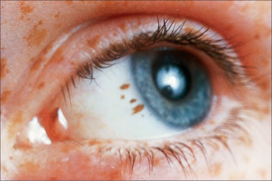
圖 25-22:結膜交界性黑色素細胞痣 (conjunctival junctional melanocytic nevus):呈均勻色素沉著與規則邊緣。承蒙已故 M. Beare, MD,Royal Victoria Hospital, Belfast, N. Ireland 提供。
Fig. 25.22 Conjunctival junctional melanocytic nevus: there is uniform pigmentation and a regular border. By courtesy of the late M. Beare, MD, Royal Victoria Hospital, Belfast, N. Ireland.
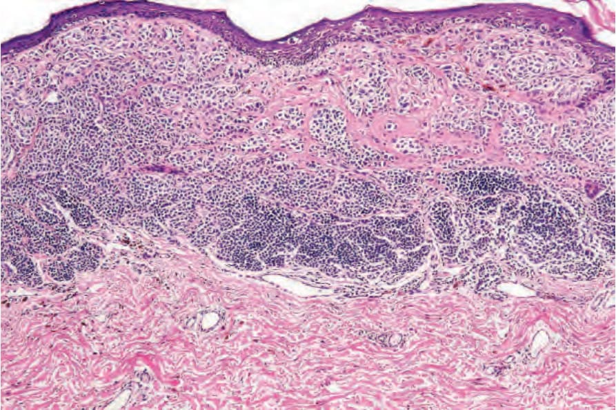
圖 25-23:黑色素瘤 (melanoma):此病灶發生於既存的複合性黑色素細胞痣 (compound melanocytic nevus) 內。殘餘的良性成分位於下方視野。比較 melanoma 的多形性與均勻一致的痣細胞群。
Fig. 25.23 Melanoma: this lesion has arisen within a pre-existent compound melanocytic nevus. The residual benign component is present in the lower field. Compare the pleomorphism of the melanoma with the uniform nevus population.
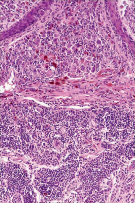
圖 25-24:黑色素瘤 (melanoma):Fig. 25.23 的高倍視野。
Fig. 25.24 Melanoma: high-power view of Fig. 25.23.
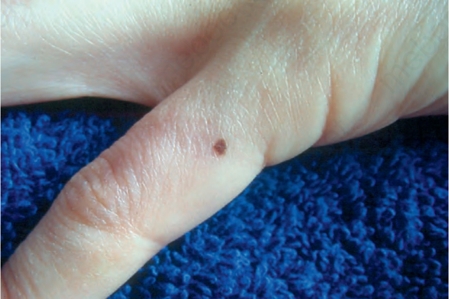
圖 25-25:交界性黑色素細胞痣 (junctional melanocytic nevus):病灶較小。注意其均勻著色。承蒙 M. Liang, MD,The Children’s Hospital, Boston, USA 提供。
Fig. 25.25 Junctional melanocytic nevus: the lesion is small. Note the uniform coloration. By courtesy of M. Liang, MD, The Children’s Hospital, Boston, USA.
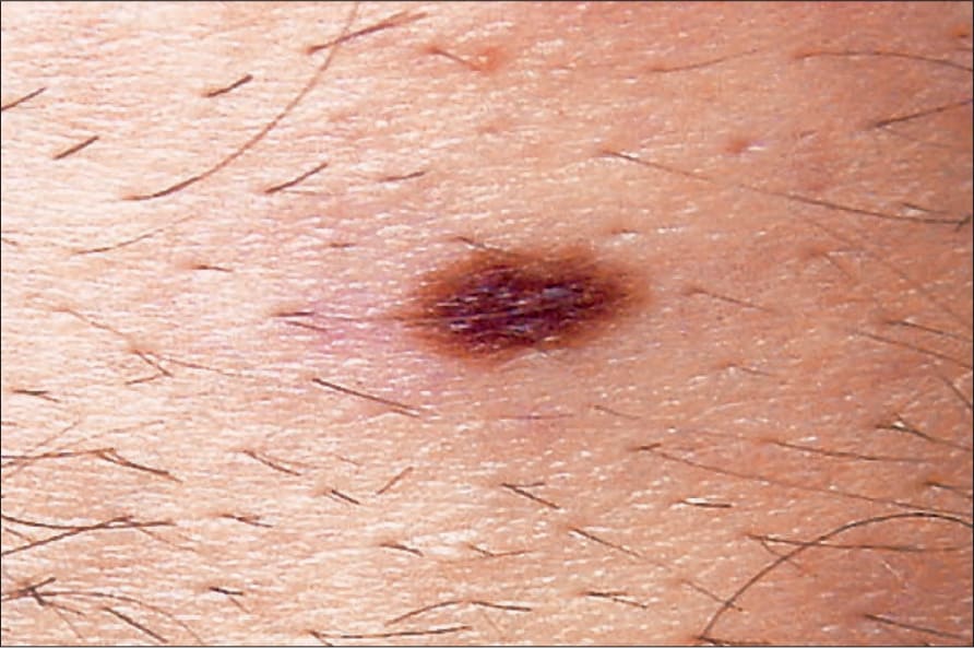
圖 25-26:交界性黑色素細胞痣 (junctional melanocytic nevus):平庸痣 (banal nevi) 典型界限分明。承蒙 M. Liang, MD,The Children’s Hospital, Boston, USA 提供。
Fig. 25.26 Junctional melanocytic nevus: banal nevi are typically sharply circumscribed. By courtesy of M. Liang, MD, The Children’s Hospital, Boston, USA.
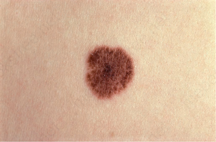
圖 25-27:交界痣 (junctional nevus):覆蓋於痣上方的皮膚紋路 (skin markings) 典型存在,與 melanoma 相反(melanoma 中皮膚紋路通常消失)。承蒙 Institute of Dermatology, London, UK 提供。
Fig. 25.27 Junctional nevus: the skin markings overlying the nevus are typically present, in contrast to melanoma when they are usually lost. By courtesy of the Institute of Dermatology, London, UK.
圖 25-28:複合痣 (compound nevus):此痣有中央隆起的圓頂狀成分。顏色均勻,邊緣界限分明。承蒙 Institute of Dermatology, London, UK 提供。
Fig. 25.28 Compound nevus: the nevus has a central raised dome-shaped component. Color is uniform, and the margin is sharply defined. By courtesy of the Institute of Dermatology, London, UK.
圖 25-29:複合痣 (compound nevus):此為色素濃重的例子。界限分明的邊緣是其良性本質的線索。承蒙 J. Dayrit, MD,Manila, The Philippines 提供。
Fig. 25.29 Compound nevus: this is a heavily pigmented example. The sharply defined border is a clue to its benign nature. By courtesy of J. Dayrit, MD, Manila, The Philippines.
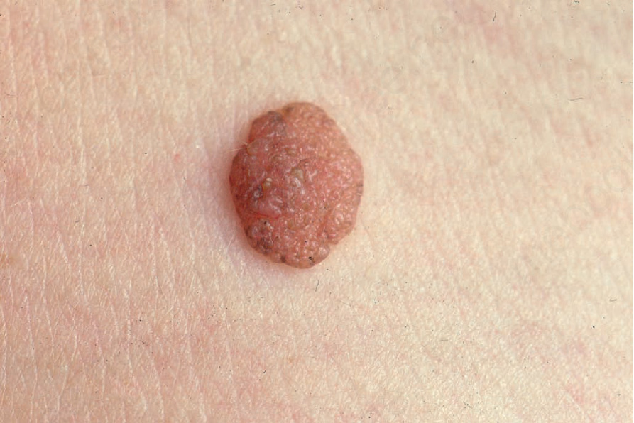
圖 25-30:真皮黑色素細胞痣 (dermal melanocytic nevus):表現為淡色、隆起的真皮疣狀結節是常見的發現。出自已故 N.P. Smith, MD 之收藏,the Institute of Dermatology, London, UK。
Fig. 25.30 Dermal melanocytic nevus: presentation as a pale, raised, dermal warty, nodule is a common finding. From the collection of the late N.P. Smith, MD, the Institute of Dermatology, London, UK.
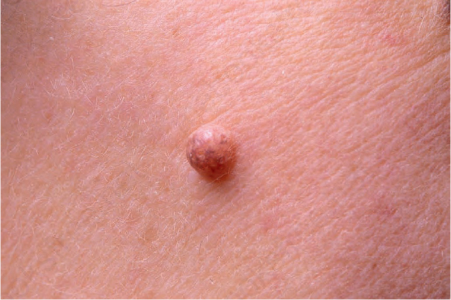
圖 25-31:真皮黑色素細胞痣 (dermal melanocytic nevus):此例呈圓頂狀外觀。有局部殘餘色素沉著。出自已故 N.P. Smith, MD 之收藏,the Institute of Dermatology, London, UK。
Fig. 25.31 Dermal melanocytic nevus: this example presents a dome-shaped appearance. There is focal residual pigmentation. From the collection of the late N.P. Smith, MD, the Institute of Dermatology, London, UK.
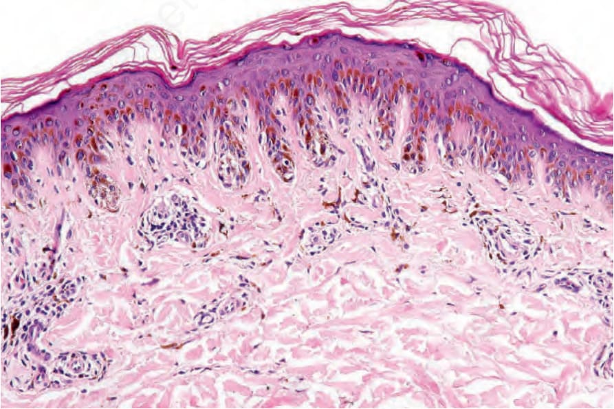
圖 25-32:雀斑樣交界痣 (lentiginous junctional nevus):注意表皮突 (rete ridges) 明顯延長。交界巢位於其尖端,為平庸痣 (banal nevi) 的特徵性位置。
Fig. 25.32 Lentiginous junctional nevus: note the marked elongation of the rete ridges. The junctional nests are present at their tips, a characteristic location in banal nevi.
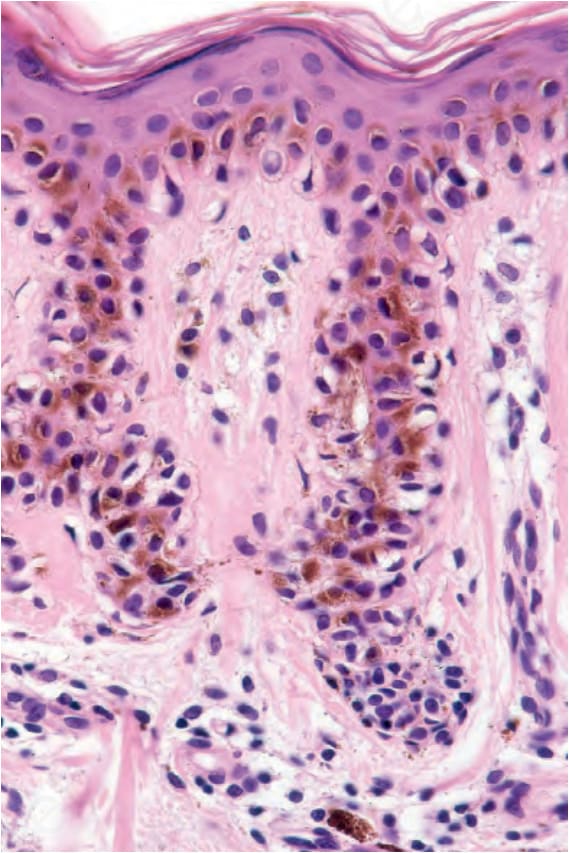
圖 25-33:雀斑樣交界痣 (lentiginous junctional nevus):在此視野中,痣細胞呈柵欄狀 (palisade) 分布,勾勒出表皮突 (rete) 的邊緣。注意色素失禁 (pigment incontinence)。
Fig. 25.33 Lentiginous junctional nevus: in this field, nevus cells are distributed as a palisade outlining the margins of the rete. Note the pigment incontinence.
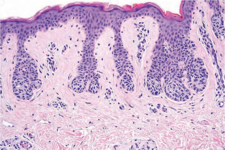
圖 25-34:交界痣 (junctional nevus):在此例中,黑色素細胞巢位於表皮突 (rete ridges) 的尖端(與發育不良痣 dysplastic nevi 比較,後者的細胞巢常見於 rete 側面並覆蓋於真皮乳頭尖端之上)。
Fig. 25.34 Junctional nevus: in this example, the nests of melanocytes are located at the tips of the rete ridges (compare with dysplastic nevi in which nests are often seen along the sides of the rete and overlying the tips of the dermal papillae).
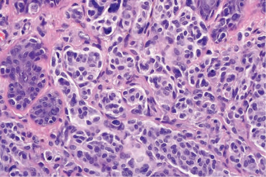
圖 25-35:A型痣細胞 (type A nevus cells):胞質淡染,核呈囊泡狀 (vesicular) 且均勻。核仁可見時典型為小型。
Fig. 25.35 Type A nevus cells: the cytoplasm is pale staining, and nuclei are vesicular and uniform. Nucleoli when visible are typically small.
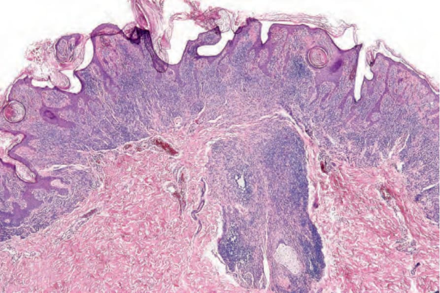
圖 25-36:複合痣 (compound nevus):如此視野所示之包繞毛囊 (ensheathing of the hair follicle) 更常為先天性病灶的特徵,但有時亦可見於後天性黑色素細胞痣。
Fig. 25.36 Compound nevus: ensheathing of the hair follicle as shown in this field is more often a feature of congenital lesions, but can sometimes be seen in acquired melanocytic nevi.
圖 25-37:複合痣 (compound nevus):病灶常具疣狀 (verrucous) 或疣樣 (warty) 外觀。
Fig. 25.37 Compound nevus: lesions often have a verrucous or warty appearance.
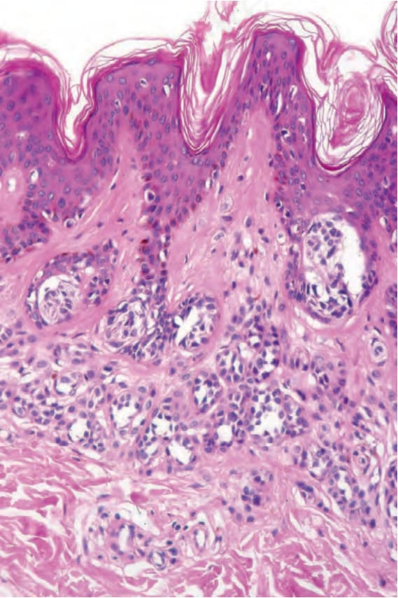
圖 25-38:複合痣 (compound nevus):交界成分與真皮成分皆存在。
Fig. 25.38 Compound nevus: both junctional and dermal components are present.
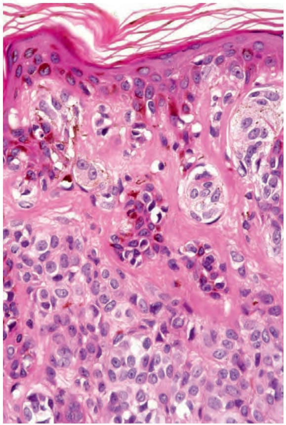
圖 25-39:複合痣 (compound nevus):A型痣細胞 (type A nevus cells) 的高倍視野。核均勻,許多含有小核仁。
Fig. 25.39 Compound nevus: high-power view of type A nevus cells. The nuclei are uniform and many contain small nucleoli.
圖 25-40:疣狀複合痣 (verrucous compound nevus):此變異型顯示明顯的乳頭瘤病 (papillomatosis) 與非常顯著的角化過度 (hyperkeratosis)。
Fig. 25.40 Verrucous compound nevus: this variant shows gross papillomatosis and very marked hyperkeratosis.
圖 25-41:真皮痣 (dermal nevus):在掃描放大倍率下,這些病灶典型具圓頂狀或疣狀型態。
Fig. 25.41 Dermal nevus: at scanning magnification, these lesions typically have a domeshaped or verrucous morphology.
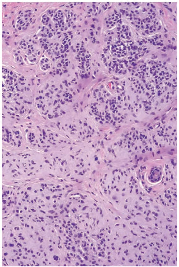
圖 25-42:真皮痣 (dermal nevus):染色深的淺表痣細胞為 B型細胞 (type B cells)。在較深處,可見紡錘狀型態,為成熟 (maturation) 的特徵。
Fig. 25.42 Dermal nevus: the darkly staining superficial nevus cells are type B cells. In the deeper reaches, spindled forms are evident, a feature of maturation.
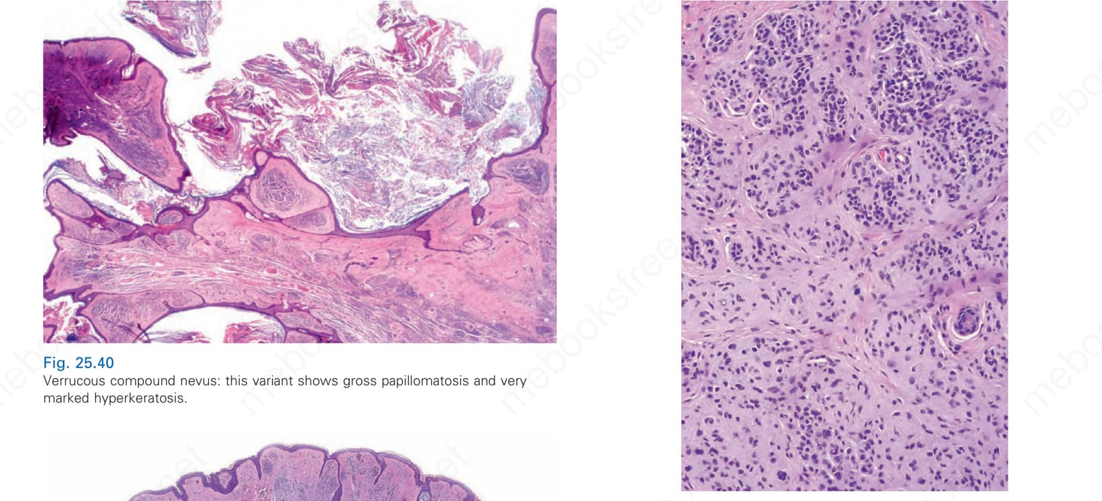
圖 25-43:真皮痣 (dermal nevus):B型細胞 (type B cells) 具均勻的核深染核,胞質少。
Fig. 25.43 Dermal nevus: type B cells have uniform hyperchromatic nuclei with little cytoplasm.
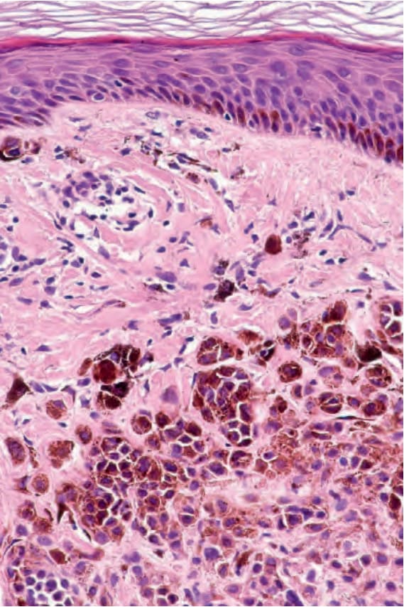
圖 25-44:真皮痣 (dermal nevus):黑色素 (melanin) 色素沉著典型隨深度減少。
Fig. 25.44 Dermal nevus: melanin pigmentation typically diminishes with depth.
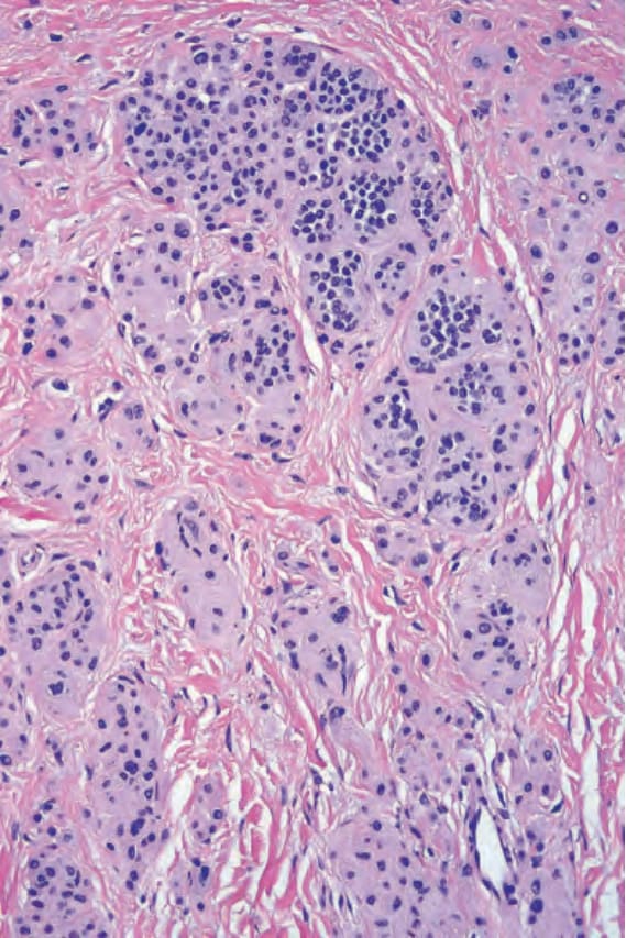
圖 25-45:真皮痣 (dermal nevus):此視野顯示成熟 (maturation),基部有紡錘狀的 C型細胞 (type C cells)。病灶頂部的核比基部的核大(成熟 maturation)。細胞巢存在時,亦隨深度縮小。
Fig. 25.45 Dermal nevus: this view shows maturation with spindle-shaped type C cells at the base. Nuclei at the top of the lesion are larger than those at the base (maturation). Nests, when present, also diminish in size with depth.
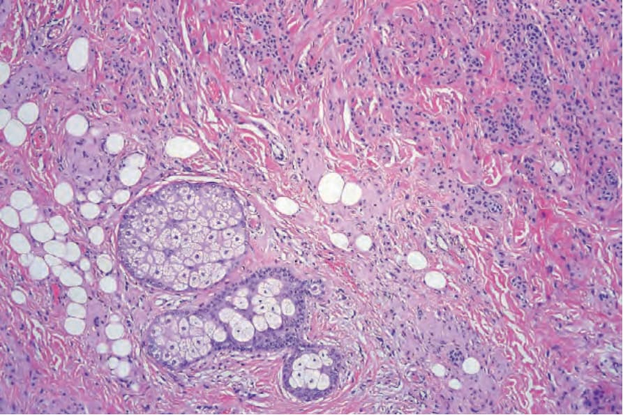
圖 25-46:真皮痣 (dermal nevus):神經化 (neurotization) 常伴隨典型呈層板狀的類 Meissner 小體 (Meissner corpuscle-like) 結構形成。
Fig. 25.46 Dermal nevus: neurotization is often accompanied by formation of typically lamellated, Meisner corpuscle-like structures.
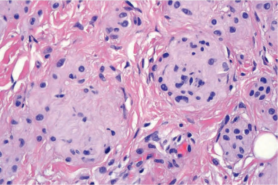
圖 25-47:真皮痣 (dermal nevus):類 Meissner 小體 (Meissner corpuscle-like) 結構的高倍視野。
Fig. 25.47 Dermal nevus: high-power view of Meisner corpuscle-like structure.
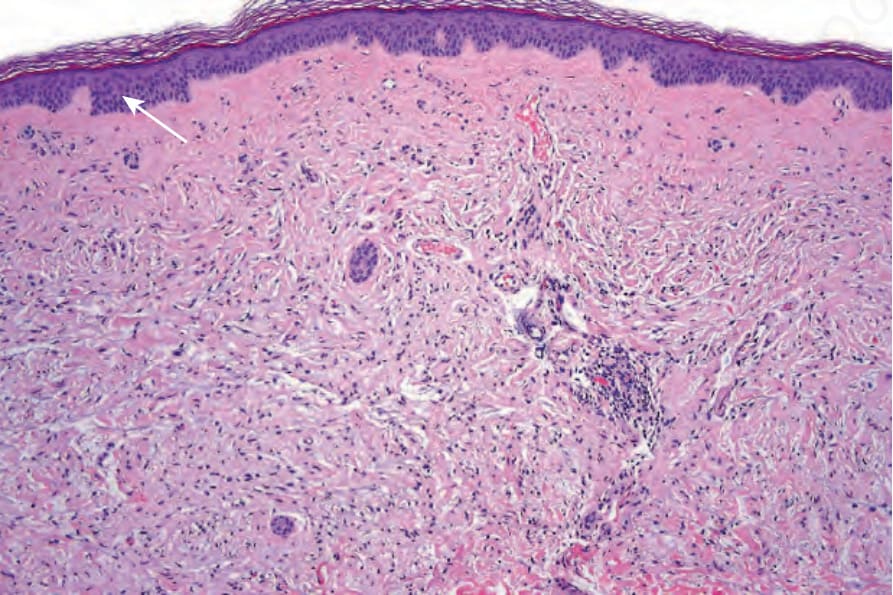
圖 25-48:真皮痣 (dermal nevus):此例已發生廣泛的神經化 (neurotization),使其與神經纖維瘤 (neurofibroma) 無法區別。視野左上角可見一兩個殘餘的痣細胞巢(箭頭所示)。
Fig. 25.48 Dermal nevus: this example has undergone extensive neurotization, making it indistinguishable from a neurofibroma. One or two residual nests of nevus cells are evident in the top-left corner of the field (arrowed).
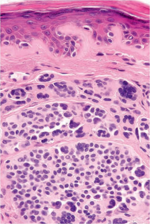
圖 25-49:真皮痣 (dermal nevus):常可發現多核巨細胞 (multinucleated giant cells),常伴模糊塗抹的染色質,且無臨床意義。
Fig. 25.49 Dermal nevus: multinucleated giant cells often with smudged chromatin are commonly found and are of no significance.
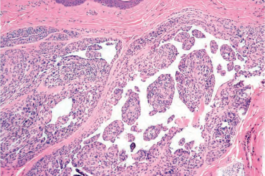
圖 25-50:真皮痣 (dermal nevus):極偶爾過量的基質黏液 (stromal mucin) 沉積會形成黏液池 (lakes)。在極端情況下,此有時稱為黏液樣痣 (myxoid nevus)。
Fig. 25.50 Dermal nevus: very occasionally excessive stromal mucin deposition results in lakes. In extreme cases, this is sometimes known as a myxoid nevus.
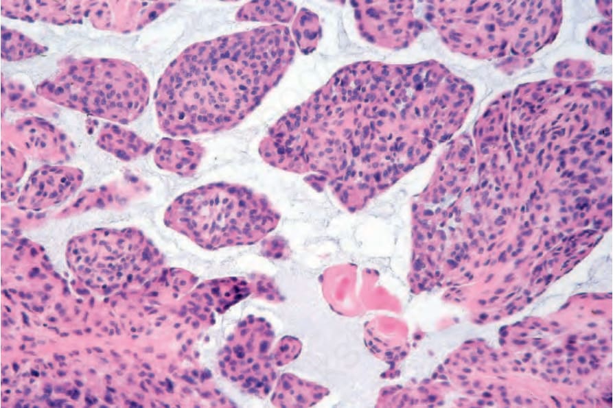
圖 25-51:真皮痣 (dermal nevus):Fig. 25.57 的高倍視野。注意細緻的黏液 (mucin) 縷絲。
Fig. 25.51 Dermal nevus: high-power view of Fig. 25.57. Note the delicate wisps of mucin.
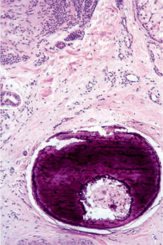
圖 25-52:真皮痣 (dermal nevus):局部鈣化 (calcification) 甚至骨形成並不罕見,通常代表被破壞的毛囊。
Fig. 25.52 Dermal nevus: focal calcification and even bone formation are not uncommon and usually represent a destroyed hair follicle.
圖 25-53:真皮痣 (dermal nevus):如此例所示之假血管腔隙 (pseudovascular spaces) 形成是常見的人工假象 (artifact)。
Fig. 25.53 Dermal nevus: the formation of pseudovascular spaces as shown in this example is a common artifact.
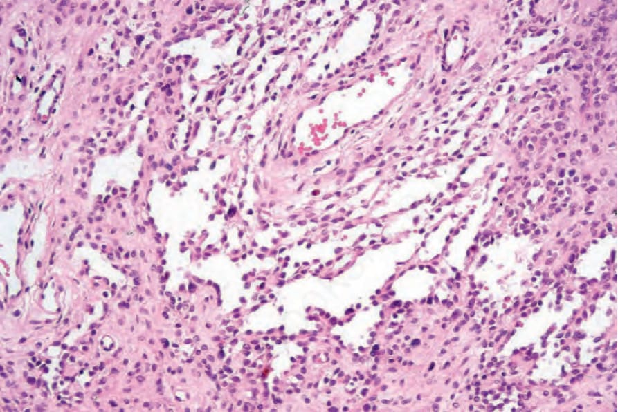
圖 25-54:真皮痣 (dermal nevus):假血管腔隙 (pseudovascular spaces) 由痣細胞 (nevus cells) 襯覆,且血管標記 (vascular markers) 為陰性。
Fig. 25.54 Dermal nevus: the pseudovascular spaces are lined by nevus cells and are negative for vascular markers.
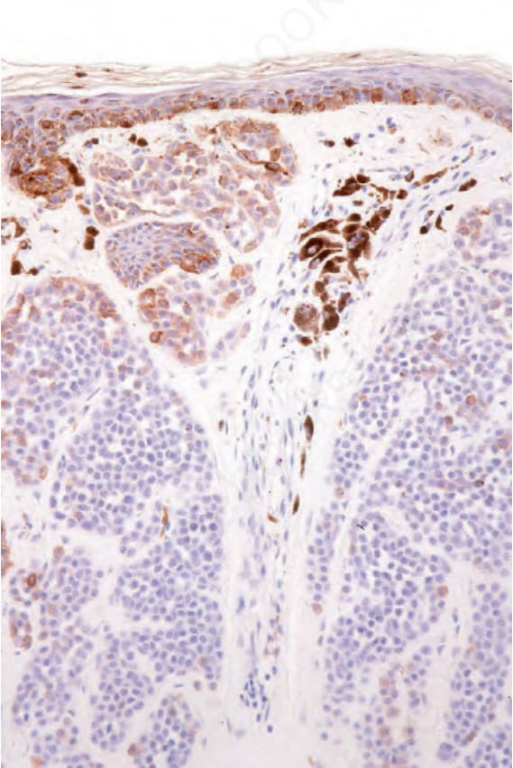
圖 25-58:真皮痣 (dermal nevus):淺表成分表現 HMB-45,但隨深度而消失。
Fig. 25.58 Dermal nevus: the superficial component expresses HMB-45 but this is lost with depth.
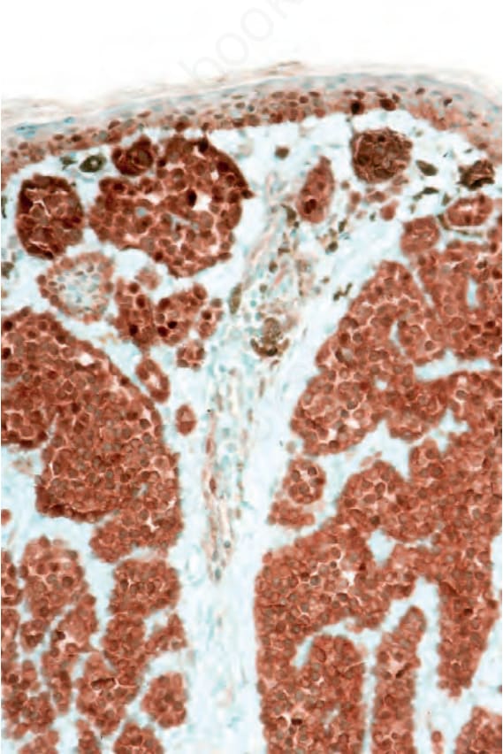
圖 25-60:真皮痣 (dermal nevus):極偶爾的淺表痣細胞表現 cyclin D1。注意僅核染色具意義。
Fig. 25.60 Dermal nevus: very occasional superficial nevus cells express cyclin D1. Note that only the nuclear staining is significant.
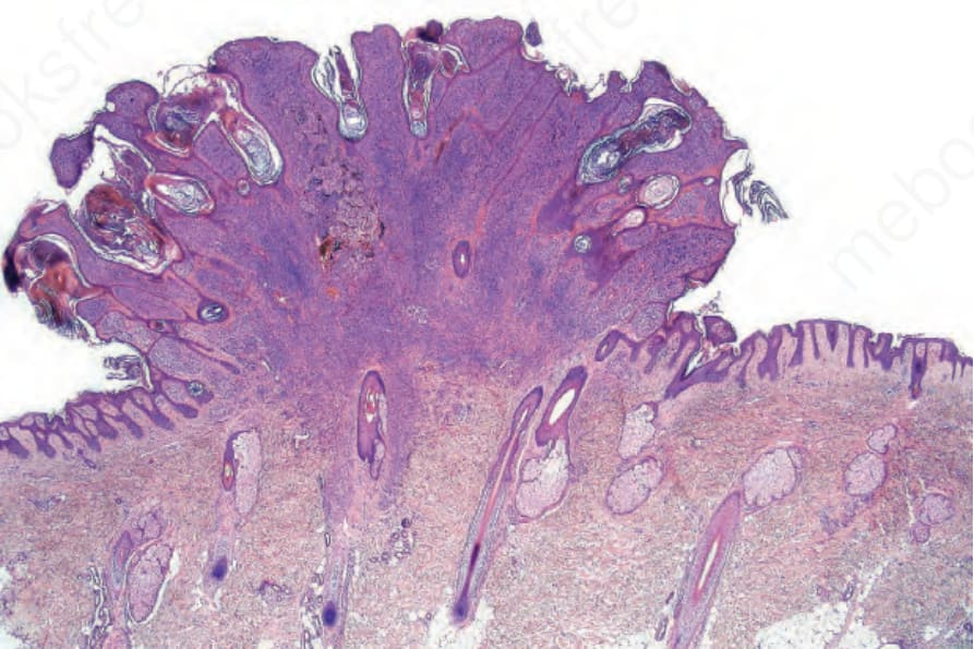
圖 25-61:純系痣 (clonal nevus):此為平庸性複合痣 (banal compound nevus)。注意深部真皮中有明顯的結節,周圍環繞著充滿色素的噬黑色素細胞 (melanophages)。承蒙 W. Grayson, MD,National Health Laboratory Service, Johannesburg, South Africa 提供。
Fig. 25.61 Clonal nevus: this is a banal compound nevus. Note the distinct nodule in the deeper dermis surrounded by pigment-laden melanophages. By courtesy of W. Grayson, MD, National Health Laboratory Service, Johannesburg, South Africa.
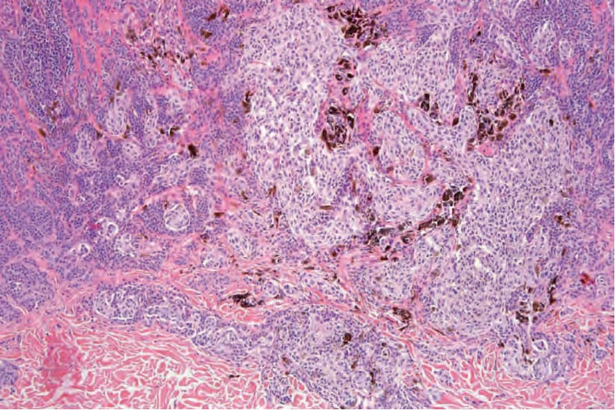
圖 25-62:純系痣 (clonal nevus):中倍視野對比 A型細胞 (type A cells) 之細胞巢與 B型細胞 (type B cells)。巨噬細胞內有大量黑色素 (melanin) 色素。承蒙 W. Grayson, MD,National Health Laboratory Service, Johannesburg, South Africa 提供。
Fig. 25.62 Clonal nevus: medium-power view contrasting the nests of type A cells with the type B cells. There is abundant melanin pigment within macrophages. By courtesy of W. Grayson, MD, National Health Laboratory Service, Johannesburg, South Africa.
-
此變化的原因不明,但其可能屬人工假象 (artifactual),並可能與局部麻醉劑 (local anesthetic) 的效應有關。使用內皮細胞標記 (endothelial cell markers) 的免疫組織化學研究一律為陰性。襯覆腔隙的細胞屬黑色素細胞來源,且以 S100 蛋白標記呈陽性。澱粉樣物質 (amyloid) 罕見可在痣內被辨識出。例外情況下,痣可伴有汗腺上皮 (sweat gland epithelium) 之增生;偶可遇見痣與硬纖維性毛上皮瘤 (desmoplastic trichoepithelioma) 共存。黑色素細胞痣亦曾被報告與毛腺瘤 (trichoadenoma) 相關聯。
-
血管內黑色素細胞 (intravascular melanocytes) 之存在或淋巴管侵犯 (lymphatic involvement) 之證據,是非常令人不安的發現,應促使徹底搜尋 melanoma 的其他特徵。痣巢疝入血管腔 (herniation of a nevus nest into the lumen of a vessel) 不應與血管侵犯 (vascular invasion) 混淆。在良性病灶中,可藉由發現一層內皮 (endothelium) 覆蓋於突出的痣細胞表面來作出此區別 (Fig. 25.55)。
鑑別診斷 (Differential diagnosis)
-
在多數情況下,平庸痣 (banal nevus) 與 melanoma 之間的區別是直截了當的。Melanoma 的低倍檢查可顯示明顯的病灶內轉化 (intralesional transformation),亦即惡性細胞凸顯為一個擴張性、且常界限分明的細胞巢、結節或斑塊,其細胞學上與鄰近的痣細胞不同。其他鑑別特徵包括緻密色素沉著 (dense pigmentation)、缺乏成熟 (lack of maturation)、核多形性 (nuclear pleomorphism)、有絲分裂活性、細胞凋亡 (apoptosis)、淋巴球浸潤 (lymphocytic infiltration),以及深部巢狀生長型態 (deep nested growth pattern),這些在 melanoma 中常明顯可見,但在皮內痣 (intradermal nevi) 中則無。黑色素細胞之向上、表皮內或 pagetoid 擴散是許多 melanoma 中可見的額外特徵。然而,在檢視新生兒痣 (neonatal nevi) 甚至兒童痣 (childhood nevi) 的切片時須謹慎,因為這些痣的表皮上層內可辨識出細胞巢、偶爾單一細胞,有時顯示輕度甚至重度細胞學非典型 (cytological atypia)(見新生兒痣 neonatal nevus)。同樣地,pagetoid 擴散可能是肢端 (acral) 與生殖器 (genital) 痣的特徵(見下文)。網狀纖維 (reticulin fibers) 在 melanoma 細胞中勾勒出細胞巢,而在痣中則傾向環繞個別痣細胞。
-
真皮痣細胞罕見可顯示有絲分裂活性 (Fig. 25.56)。因此其存在應謹慎看待,並搜尋其他提示惡性的特徵。痣樣黑色素瘤 (nevoid melanoma) 在隨意一瞥下可能細胞學上相似。然而仔細檢視可揭示不對稱性、缺乏界限分明、多個真皮有絲分裂、細微的缺乏成熟,以及核仁明顯 (nucleolar prominence)(見痣樣黑色素瘤 nevoid melanoma)。
-
小細胞黑色素瘤 (small cell melanoma) 細胞雖常與 B型痣細胞大小相似,但通常具明顯的嗜伊紅核仁 (eosinophilic nucleoli),且有絲分裂一律存在(見小細胞黑色素瘤 small cell melanoma)。
-
有時在區分侵襲性 melanoma(特別是痣樣與小細胞變異型)與殘餘良性皮內痣細胞時會遭遇困難。後者在評估腫瘤厚度 (tumor thickness) 或侵犯層級 (level of invasion) 時尤其重要。免疫組織化學 (immunohistochemistry) 可能有助於作出此區別。HMB-45 表現在淺表真皮痣中常為陽性,但隨深度而消失 (Figs 25.57 and 25.58)。相對地,在 melanoma 中,腫瘤細胞常於整個病灶中均呈陽性。p53 蛋白在平庸真皮痣中不表現。相對地,其常存在於 melanoma 中。在平庸痣中,少數 Ki-67 與 cyclin D1 陽性細胞可見於較淺表的真皮成分 (Figs 25.59 and 25.60)。在 melanoma 中,它們通常數目多得多,且常存在於病灶的整個厚度中。然而,cyclin D1 染色的型態不應被依賴用於良性與惡性的區別。
-
平庸痣可能與發育不良痣 (dysplastic nevi) 顯示某些組織學重疊。因此它們常為雀斑樣 (lentiginous),且有時顯示肩部形成 (shoulder formation)、延長的表皮突周圍有嗜伊紅性或層板狀纖維化 (eosinophilic or lamellar fibrosis)。相對地,結構紊亂 (architectural disorder)(亦即痣細胞巢不規則地散布於整個表皮)與橋接 (bridging) 則不可見。此外,細胞學非典型 (cytological atypia) 並非平庸痣的特徵。
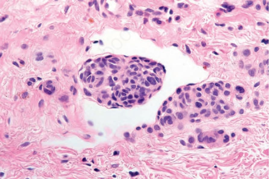
圖 25-55:真皮痣 (dermal nevus):痣細胞陷入血管腔 (invagination of nevus cells into the lumen of a vessel) 不應與真正的血管侵犯 (vascular invasion) 混淆。區別取決於辨識出一層覆蓋於痣樣聚集團塊表面的內皮細胞 (endothelial cells),如此例所示。
Fig. 25.55 Dermal nevus: invagination of nevus cells into the lumen of a vessel should not be confused with true vascular invasion. Distinction depends on identification of a layer of endothelial cells covering the surface of the nevoid aggregate, as shown in this example.
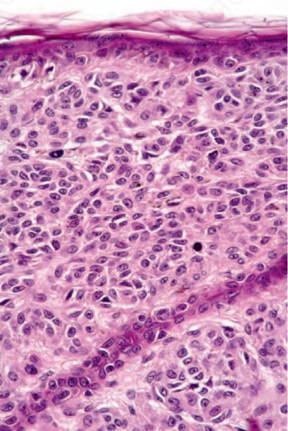
圖 25-56:平庸痣 (banal nevus):辨識出一兩個真皮有絲分裂象 (dermal mitotic figures) 並不總是等同於 melanoma。然而其存在應以相當的關切看待,並搜尋其他有絲分裂或提示 melanoma 的額外特徵。
Fig. 25.56 Banal nevus: identification of one or two dermal mitotic figures is not always synonymous with melanoma. Their presence, however, should be viewed with considerable concern and other mitoses or additional features indicative of melanoma sought.
圖 25-57:真皮痣 (dermal nevus):平庸痣細胞 (banal nevus cells) 的中倍視野。
Fig. 25.57 Dermal nevus: medium-power view of banal nevus cells.
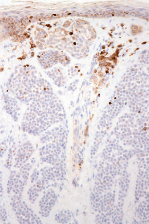
圖 25-59:真皮痣 (dermal nevus):僅有一兩個痣細胞表現 MIB-1。
Fig. 25.59 Dermal nevus: only one or two nevus cells express MIB-1.