Pseudomelanocytic nests
Pseudomelanocytic nests
Clinical features Pseudomelanocytic nests are aggregates of nonmelanocytic cells, or cell fragments within the epidermis mimicking a melanocytic proliferation on histologic examination.1–7 These nests usually develop in the setting of lichenoid
While there is unquestionable malignant transformation on occasions in these lesions, such events are rare; therefore, there is no indication for widespread prophylactic excision of otherwise typical melanocytic nevi (Figs 25.23 and 25.24). It has been estimated that the likelihood of any one nevus evolving into melanoma is roughly 1/100 000.7 Subsequent mortality is of the order of 1/500 000 original nevi. However, the prevalence of nevi is of major epidemiological importance, increased numbers correlating with a greater risk of subsequent development of melanoma, of superficial spreading and nodular subtypes.8–10 Lentigo maligna melanoma does not derive from pre-existent nevi and is not related to their prevalence. In young children and adolescents in whom these lesions are at an early stage of development, increased pigmentation is to be anticipated. However, in adults, evidence of junctional activity is to be viewed with caution; it is those nevi with increasingly marked pigmentation, or appearing de novo in the older age groups, which are often excised for histologic evaluation.
1241 Melanocytic nevus
Clinical features Melanocytic nevi first appear in early childhood and increase in number during the second and third decades.5,6,11–13 In males, the head, neck, and trunk are particularly affected, whereas in females the upper and lower limbs are more often involved.5,12,14,15 Distribution of acquired melanocytic nevi in an agminate (grouped) pattern can occasionally be seen.16 Melanocytic nevi involute during middle age, and most have completely regressed in the elderly. They are more common in individuals with pale skin and light-colored eyes.10,17 Melanocytic nevi are much less frequently seen in Asians and Afro-Caribbeans.10 In these races, the acral sites are particularly affected.1 Dark brown or black hair correlates with increasing numbers of nevi, whereas red hair appears to protect.8,10 Development of melanocytic nevi is related to the extent of sun exposure during the first two decades of life.17–19 Intermittent intense sunlight is of greater importance than chronic exposure.12 In fact, chronic sun exposure correlates with low levels of nevi (i.e., it appears to be protective).14 Increasing nevus counts are found in individuals who tend to sunburn rather than tan following sun exposure and correlate with the degree of freckling.10,20,21 As these factors are also
important in the etiology of melanoma, high levels of freckles and nevi at an early age may be of predictive value.
Melanocytic nevi present a variety of features depending on their stage of evolution. Junctional nevi are usually macular or slightly raised, up to 0.5 cm in diameter and from light to dark brown in color (Figs 25.25–25.27). They are well circumscribed with a regular border and are usually uniformly pigmented, but sometimes the central area is darker. Typically, the skin lines can be clearly discerned on the surface of the lesion.
The compound nevus is raised, sometimes dome-shaped or warty, and often still deeply pigmented (Figs 25.28 and 25.29). Occasionally, there are coarse hairs projecting from its surface; plucking these hairs may traumatize the dermal component of the hair follicle, resulting in granulomatous inflammation, which may cause concern to both the patient and clinician as to the possibility of malignant transformation.8,19
1242 Melanocytic nevi
The intradermal nevus is often devoid of pigment and may present as a dome-shaped nodule, a papillomatous lesion, or a pedunculated skin tag (Figs 25.30 and 25.31).
Nevi may become more highly pigmented under the influence of pregnancy or the oral contraceptive pill.22 There is, however, no evidence that pregnancy in any significant way stimulates their development or alters the biological potential of pre-existent nevi.5
Histologic features There is considerable confusion about the nature of the nevocyte and its distinction from a melanocyte. A nevocyte is merely a melanocyte that has multiplied to form a melanocytic nevus. It has the same electron microscopic appearance as a melanocyte, and identical organelles and enzyme systems; the only significant differences are that the dermal component lacks dendritic processes and with increasing depth melanin synthesis is arrested.
In the earliest stage of development, junctional nests of melanocytes appear in the lower aspect of the epidermis (confined by the basement membrane), usually within the tips or, less often, sides of sometimes broadened and elongated epidermal ridges (lentiginous junctional nevus) (Figs 25.32 and 25.33). The melanocytes may be polygonal and epithelioid, or more
1243 Melanocytic nevus
rarely spindled with clear to pale staining or lightly eosinophilic cytoplasm, and contain uniform round to oval small nuclei with prominent nucleoli (type A nevus cells) (Figs 25.34 and 25.35). The cytoplasm typically contains sparse, evenly distributed, delicate melanin granules. In benign junctional nevi, growth is towards dermal involvement. Any tendency for melanocytes to spread towards the upper reaches of the epidermis (pagetoid spread) is suggestive of malignant change and should be viewed with caution. However, upward migration of melanocytes on its own is not diagnostic of malignancy as this feature may be seen in variants of nevi-like lesions occurring at some special sites (see below). Melanocytes between epidermal ridges may sometimes appear increased in number.2 By definition, junctional nevi are solely intraepidermal; however, in heavily pigmented variants, melanin is typically present in macrophages (melanophages) within the papillary dermis (pigmentary incontinence).
In addition to junctional activity, compound nevi show nests and strands of nevus cells within both the papillary and the superficial reticular dermis (Figs 25.36–25.39). Compound banal nevi are usually fairly well circumscribed and symmetrical. The junctional component does not typically extend beyond the dermal component, i.e., a shoulder is absent in the majority of banal nevi (compare with dysplastic nevus). It should,
however, be noted that a shoulder can sometimes be present in a banal compound nevus, i.e., the presence of a shoulder in itself does not make a nevus dysplastic. Compound nevi may sometimes be associated with marked hyperkeratosis, acanthosis with keratinous pseudocyst formation, and papillomatosis (reminiscent of a seborrheic keratosis), accounting for a warty clinical appearance – the so-called papillomatous (verrucous) melanocytic nevus or keratotic melanocytic nevus (Fig. 25.40).23–25 These nevi are more often found in females, and the trunk is the commonest site affected. The change may be related to estrogens as the nevus cells express pS2.25
The cells of the more superficial component of the dermal lesion may retain cytological characteristics similar to the junctional nevus. The cells in the deeper aspect are much smaller with less cytoplasm and have dense,
1244 Melanocytic nevi
1245 Melanocytic nevus
more darkly staining nuclei resembling lymphocytes (type B nevus cells) (Figs 25.41–25.43). Mitotic activity can occasionally be seen in the dermal component of an acquired melanocytic nevus (see differential diagnosis). Mitotic activity may also be increased in melanocytic nevi in pregnancy. An analysis of dermal mitoses in otherwise banal compound melanocytic nevi has demonstrated the mean number of 0.024 dermal mitoses/mm2.26 Mitoses are normal and never atypical, evenly distributed, usually located in the upper half of the dermis, and do not appear in clusters.26 Although isolated regular mitoses can also be seen in the deep dermal melanocytic component, they are about three times less frequent than mitoses in the upper half of the dermis.26 Importantly, banal nevi with dermal mitoses are significantly more common in younger age groups (e.g., between 0 and 20 years of age).26 Therefore, more than an occasional dermal mitosis in older patients should be viewed with caution.
The dermal nevus, the ‘end stage’ of the melanocytic nevus, is typified by progressively less pigmentation with atrophy (so-called nevus maturation) (Fig. 25.44). This is usually accompanied by the accumulation of loose fibrous tissue. Rarely, dense fibrosis may develop, resulting in separation of individual residual melanocytes (desmoplastic nevus) (see below). In some dermal nevi, there may be worrying nuclear pleomorphism and hyperchromatism. The latter, however, appears smudged, and the nuclei are devoid of nucleoli and mitotic activity (ancient nevus, nevus with senescent atypia).27
In dermal nevi, the melanocytes often develop spindled cell and Schwann cell-like characteristics (neurotization), such as a fibrillar appearance with
pale eosinophilic cytoplasm and wavy nuclei (type C nevus cells) (Fig. 25.45). Cholinesterase positivity may be present, and often Meissner corpuscle-like structures are found (Figs 25.46 and 25.47). The cells are nevertheless still truly melanocytic: ultrastructurally, they contain melanosomes. In addition, they are S100 and dopa positive and do not show Schwann cell morphology, nor do they react with antibodies to myelin basic protein.27 Occasionally, intradermal nevi take on truly neurofibromatous appearances (Fig. 25.48). Other features of ‘maturation’/senescence in dermal nevi include giant cell formation, mucinous degeneration, xanthomatization, and fat accumulation (Figs 25.49–25.51).3 More rarely, changes include calcification and bone formation (usually based on a degenerate hair follicle) (Fig. 25.52). Lesions with bone formation are known as osteonevus of Nanta. A not uncommon observation is the presence of pseudovascular spaces imparting an angiomatous-like appearance (Figs 25.53 and 25.54).
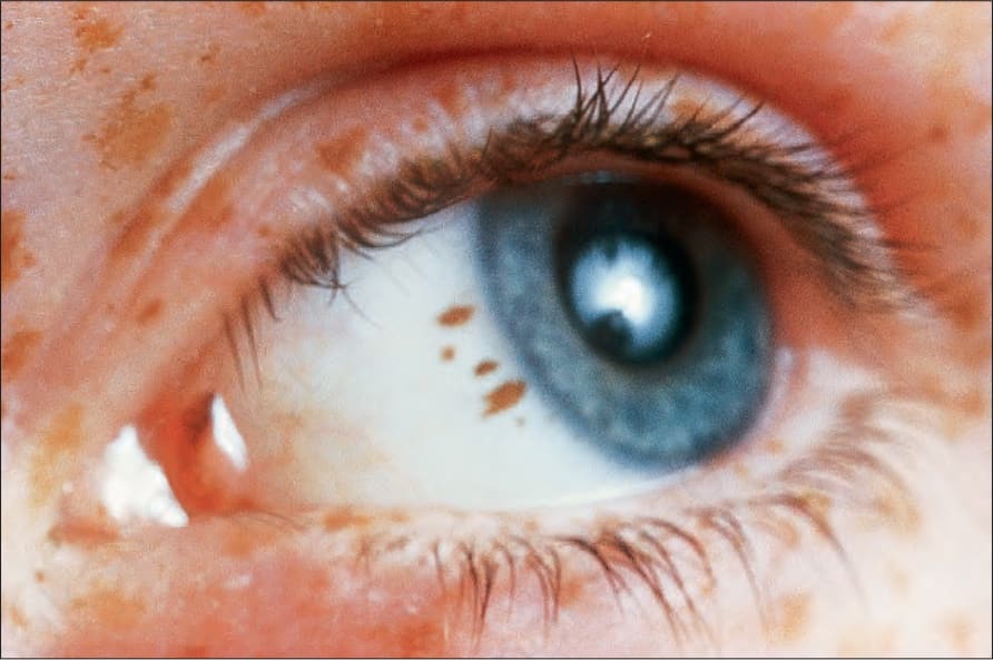
Fig. 25.22 Conjunctival junctional melanocytic nevus: there is uniform pigmentation and a regular border. By courtesy of the late M. Beare, MD, Royal Victoria Hospital, Belfast, N. Ireland.
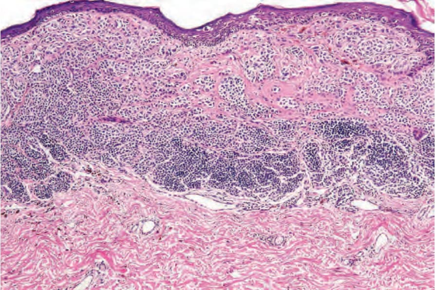
Fig. 25.23 Melanoma: this lesion has arisen within a pre-existent compound melanocytic nevus. The residual benign component is present in the lower field. Compare the pleomorphism of the melanoma with the uniform nevus population.
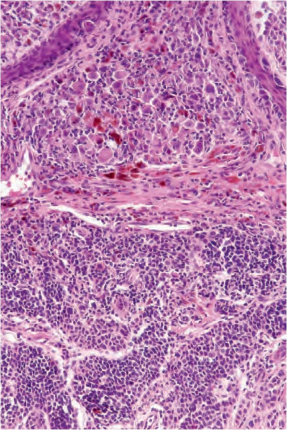
Fig. 25.24 Melanoma: high-power view of Fig. 25.23.
Fig. 25.25 Junctional melanocytic nevus: the lesion is small. Note the uniform coloration. By courtesy of M. Liang, MD, The Children’s Hospital, Boston, USA.
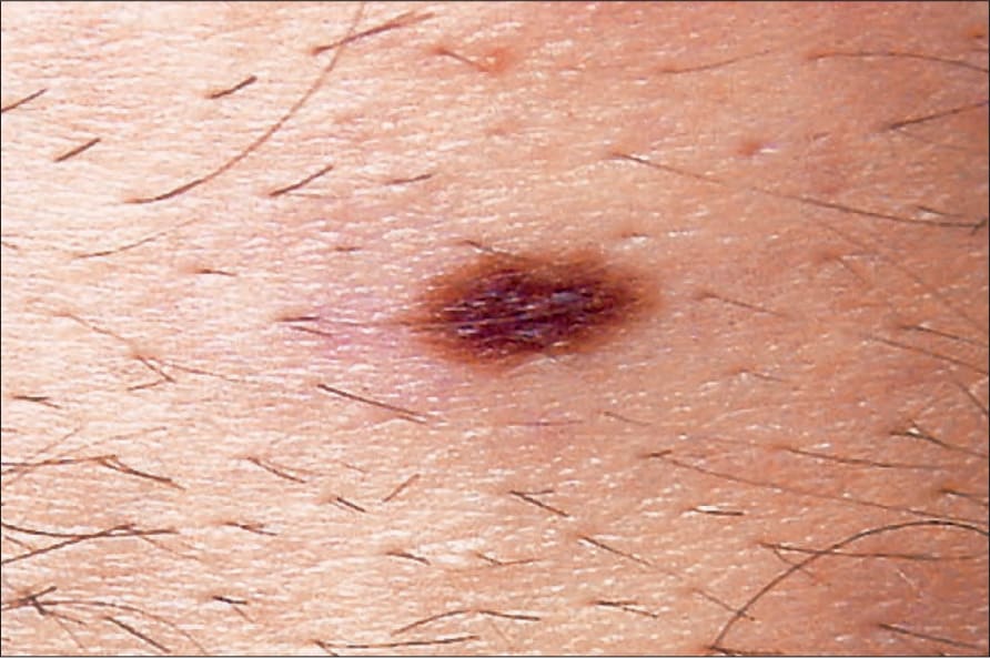
Fig. 25.26 Junctional melanocytic nevus: banal nevi are typically sharply circumscribed. By courtesy of M. Liang, MD, The Children’s Hospital, Boston, USA.
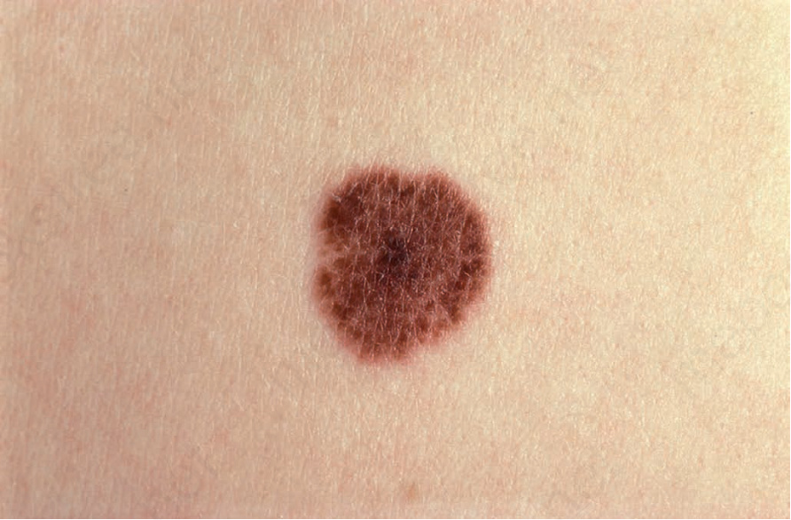
Fig. 25.27 Junctional nevus: the skin markings overlying the nevus are typically present, in contrast to melanoma when they are usually lost. By courtesy of the Institute of Dermatology, London, UK.
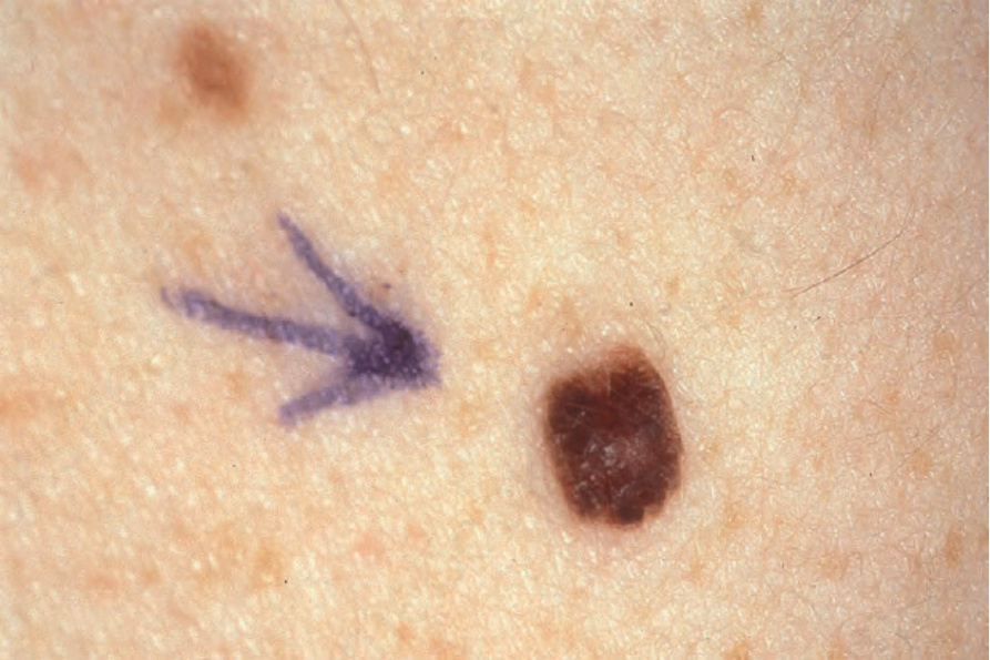
Fig. 25.28 Compound nevus: the nevus has a central raised dome-shaped component. Color is uniform, and the margin is sharply defined. By courtesy of the Institute of Dermatology, London, UK.
Fig. 25.29 Compound nevus: this is a heavily pigmented example. The sharply defined border is a clue to its benign nature. By courtesy of J. Dayrit, MD, Manila, The Philippines.
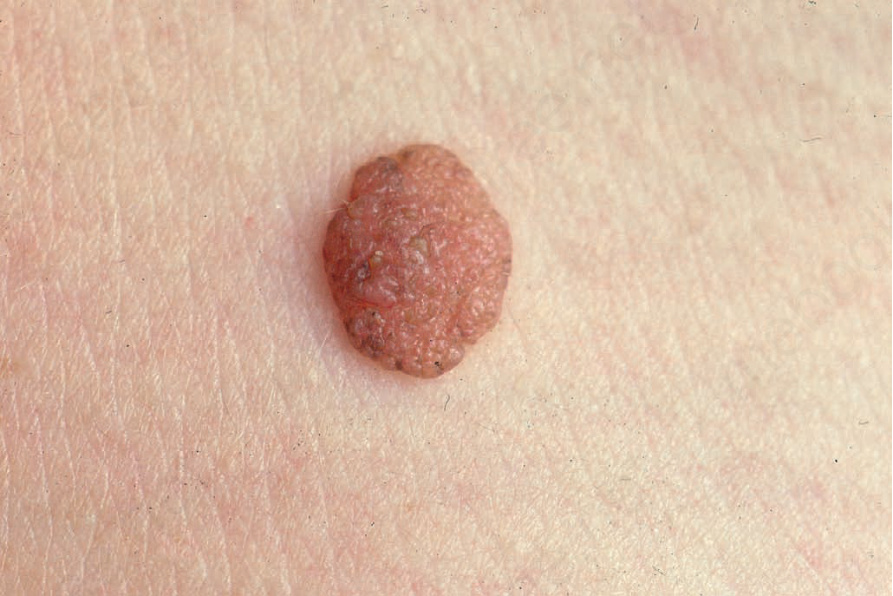
Fig. 25.30 Dermal melanocytic nevus: presentation as a pale, raised, dermal warty, nodule is a common finding. From the collection of the late N.P. Smith, MD, the Institute of Dermatology, London, UK.
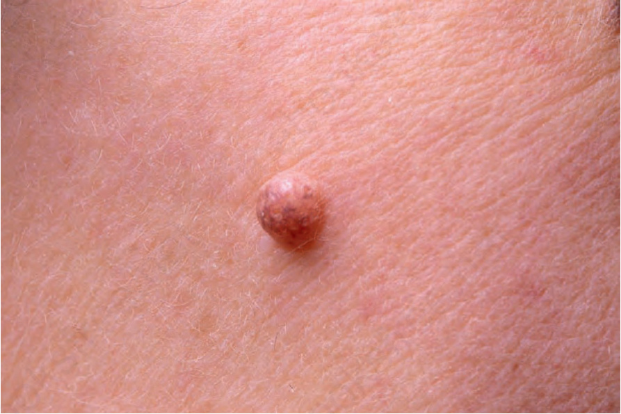
Fig. 25.31 Dermal melanocytic nevus: this example presents a dome-shaped appearance. There is focal residual pigmentation. From the collection of the late N.P. Smith, MD, the Institute of Dermatology, London, UK.
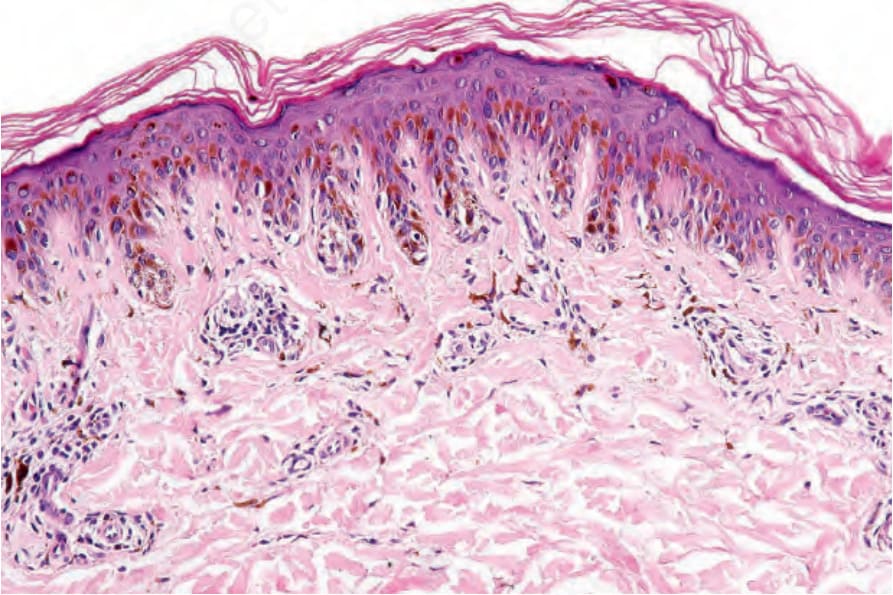
Fig. 25.32 Lentiginous junctional nevus: note the marked elongation of the rete ridges. The junctional nests are present at their tips, a characteristic location in banal nevi.
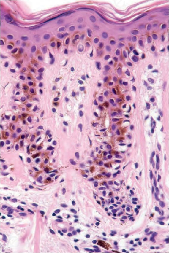
Fig. 25.33 Lentiginous junctional nevus: in this field, nevus cells are distributed as a palisade outlining the margins of the rete. Note the pigment incontinence.
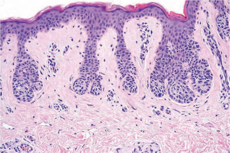
Fig. 25.34 Junctional nevus: in this example, the nests of melanocytes are located at the tips of the rete ridges (compare with dysplastic nevi in which nests are often seen along the sides of the rete and overlying the tips of the dermal papillae).
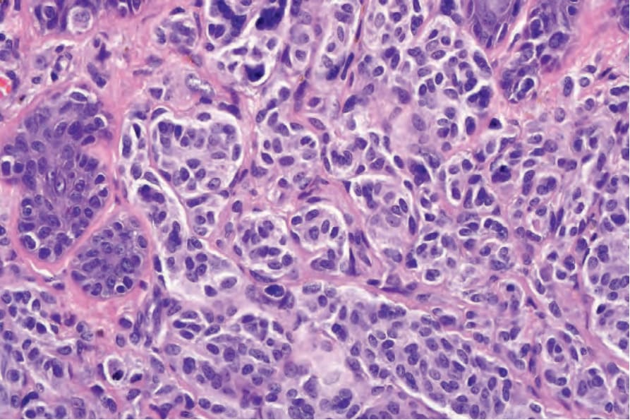
Fig. 25.35 Type A nevus cells: the cytoplasm is pale staining, and nuclei are vesicular and uniform. Nucleoli when visible are typically small.
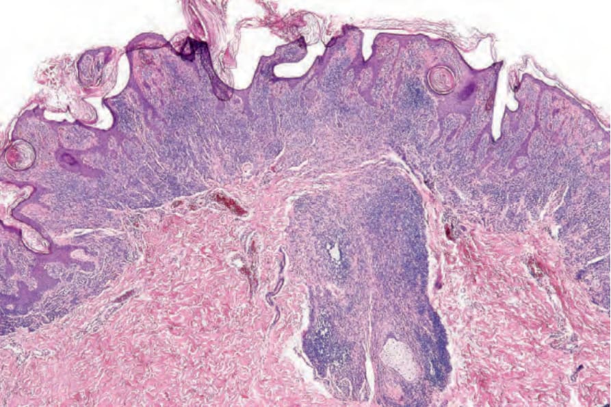
Fig. 25.36 Compound nevus: ensheathing of the hair follicle as shown in this field is more often a feature of congenital lesions, but can sometimes be seen in acquired melanocytic nevi.
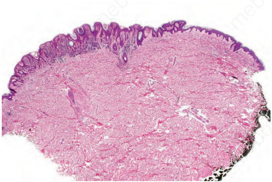
Fig. 25.37 Compound nevus: lesions often have a verrucous or warty appearance.
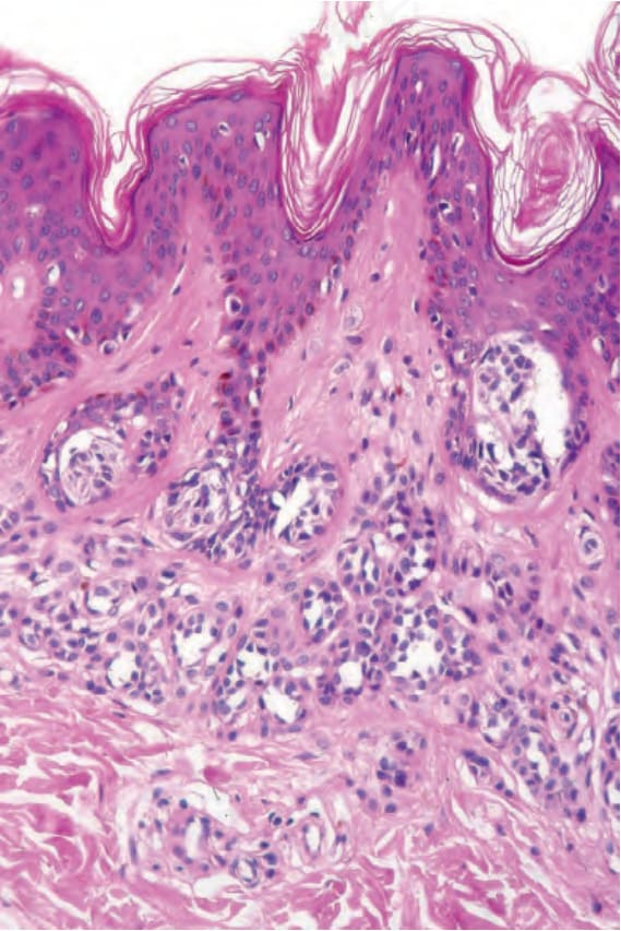
Fig. 25.38 Compound nevus: both junctional and dermal components are present.
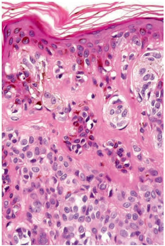
Fig. 25.39 Compound nevus: high-power view of type A nevus cells. The nuclei are uniform and many contain small nucleoli.
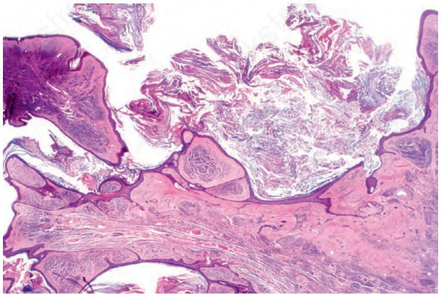
Fig. 25.40 Verrucous compound nevus: this variant shows gross papillomatosis and very marked hyperkeratosis.
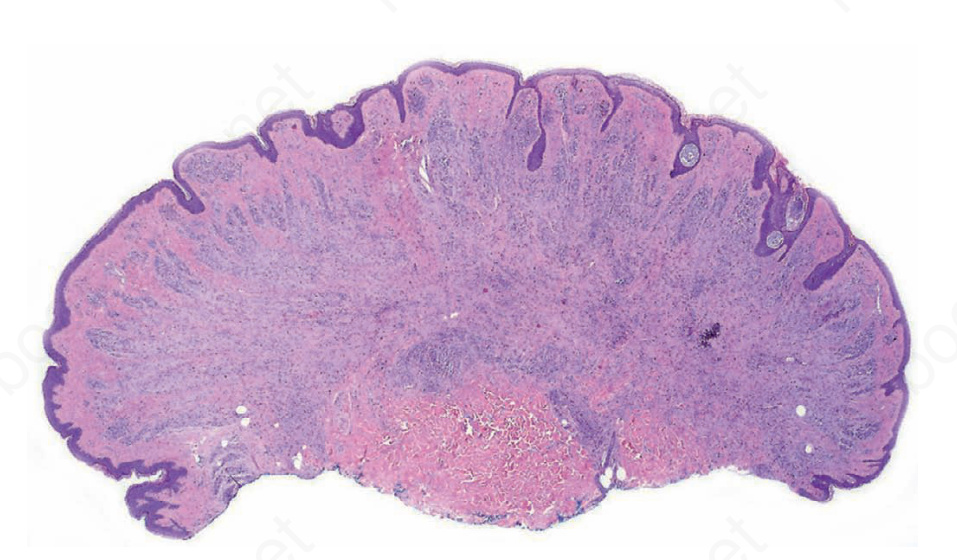
Fig. 25.41 Dermal nevus: at scanning magnification, these lesions typically have a domeshaped or verrucous morphology.
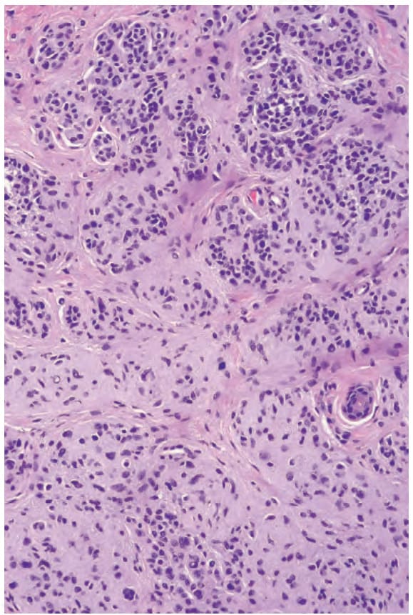
Fig. 25.42 Dermal nevus: the darkly staining superficial nevus cells are type B cells. In the deeper reaches, spindled forms are evident, a feature of maturation.
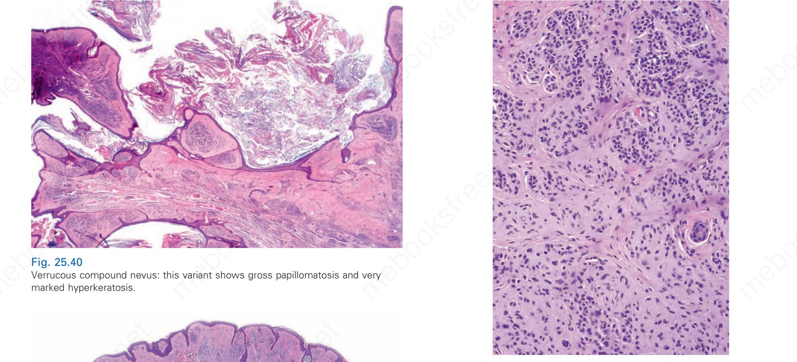
Fig. 25.43 Dermal nevus: type B cells have uniform hyperchromatic nuclei with little cytoplasm.
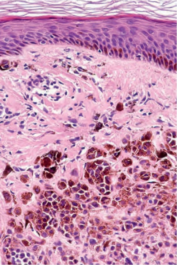
Fig. 25.44 Dermal nevus: melanin pigmentation typically diminishes with depth.
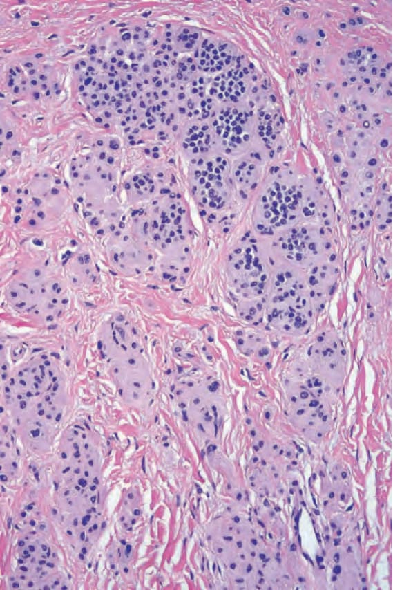
Fig. 25.45 Dermal nevus: this view shows maturation with spindle-shaped type C cells at the base. Nuclei at the top of the lesion are larger than those at the base (maturation). Nests, when present, also diminish in size with depth.
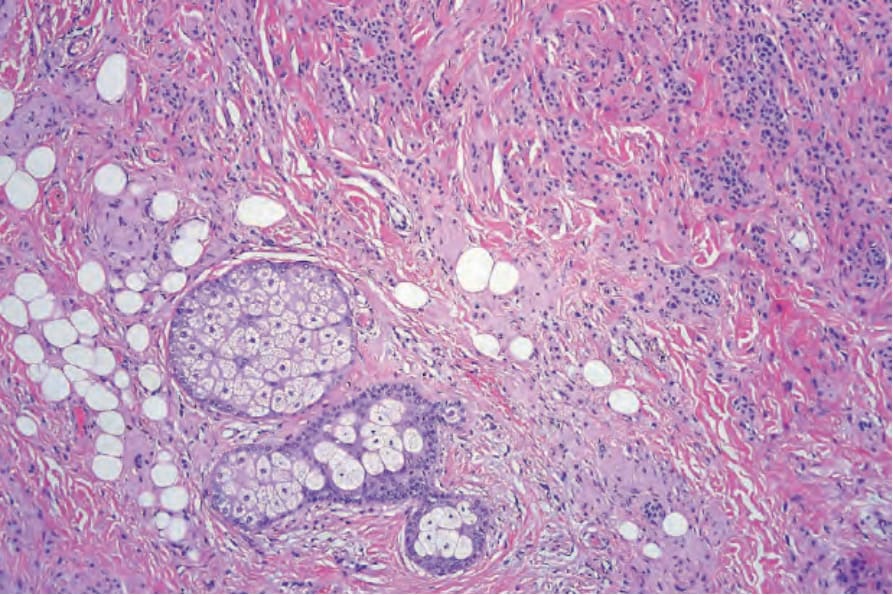
Fig. 25.46 Dermal nevus: neurotization is often accompanied by formation of typically lamellated, Meisner corpuscle-like structures.
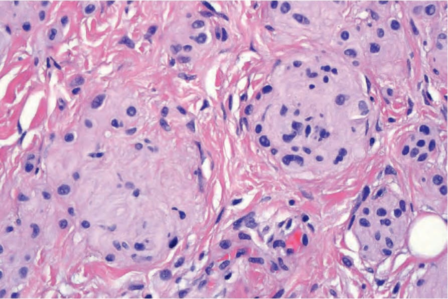
Fig. 25.47 Dermal nevus: high-power view of Meisner corpuscle-like structure.
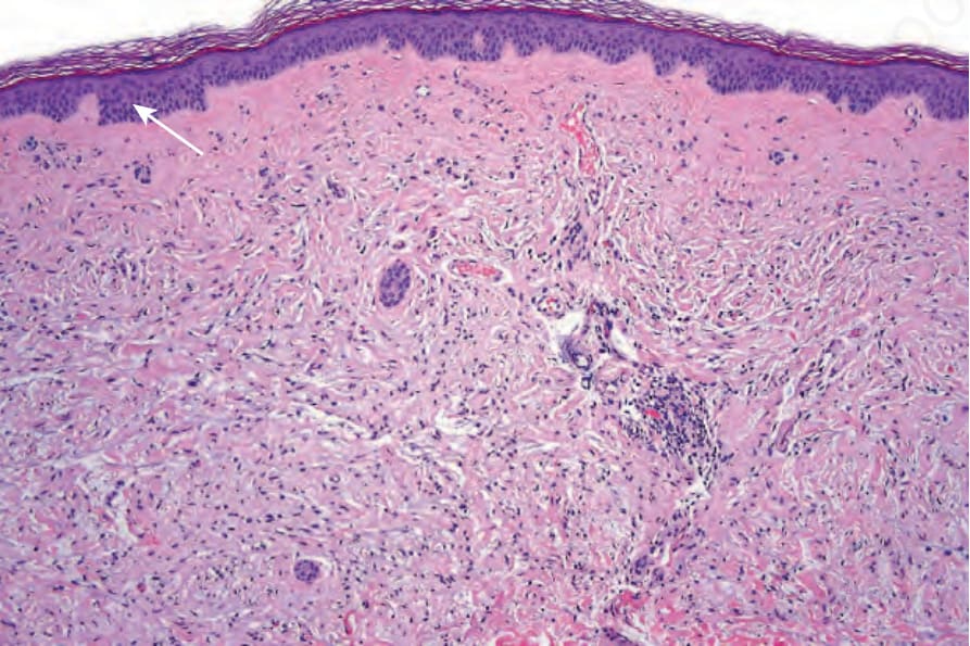
Fig. 25.48 Dermal nevus: this example has undergone extensive neurotization, making it indistinguishable from a neurofibroma. One or two residual nests of nevus cells are evident in the top-left corner of the field (arrowed).
Fig. 25.49 Dermal nevus: multinucleated giant cells often with smudged chromatin are commonly found and are of no significance.
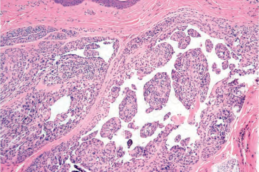
Fig. 25.50 Dermal nevus: very occasionally excessive stromal mucin deposition results in lakes. In extreme cases, this is sometimes known as a myxoid nevus.
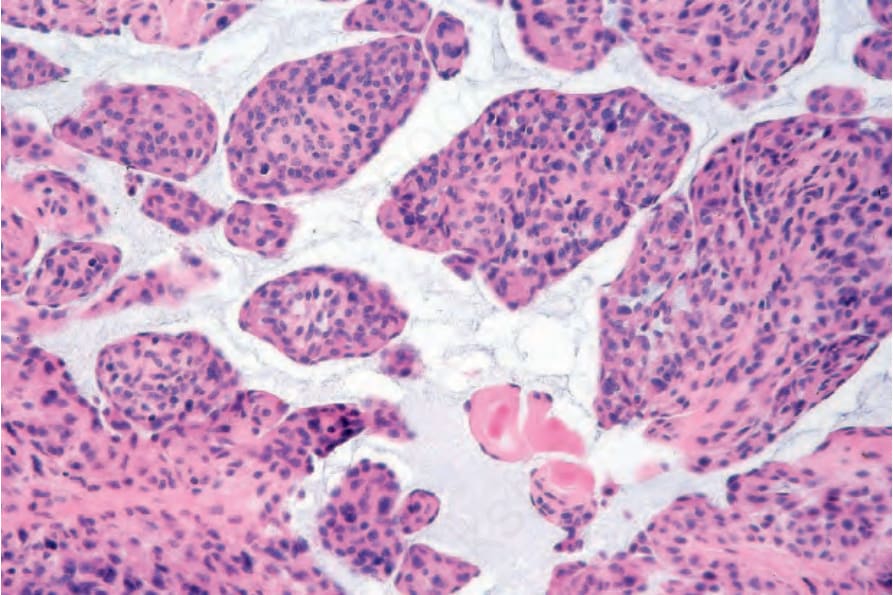
Fig. 25.51 Dermal nevus: high-power view of Fig. 25.57. Note the delicate wisps of mucin.
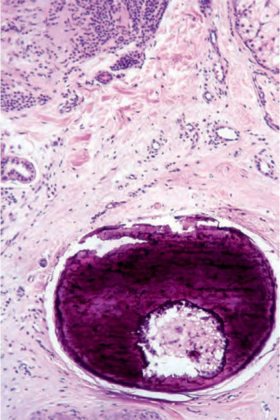
Fig. 25.52 Dermal nevus: focal calcification and even bone formation are not uncommon and usually represent a destroyed hair follicle.
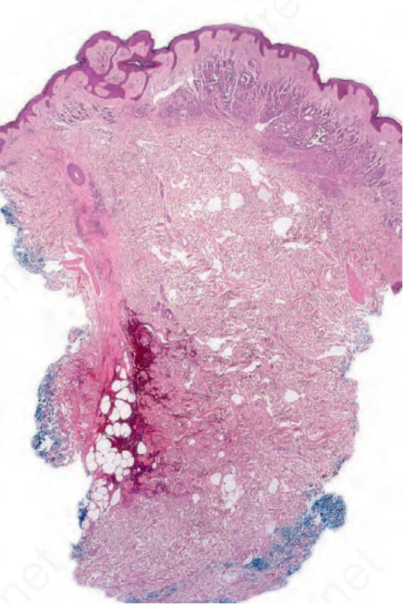
Fig. 25.53 Dermal nevus: the formation of pseudovascular spaces as shown in this example is a common artifact.
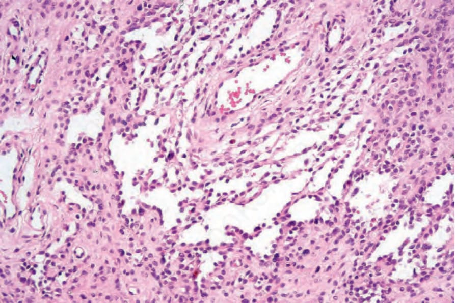
Fig. 25.54 Dermal nevus: the pseudovascular spaces are lined by nevus cells and are negative for vascular markers.
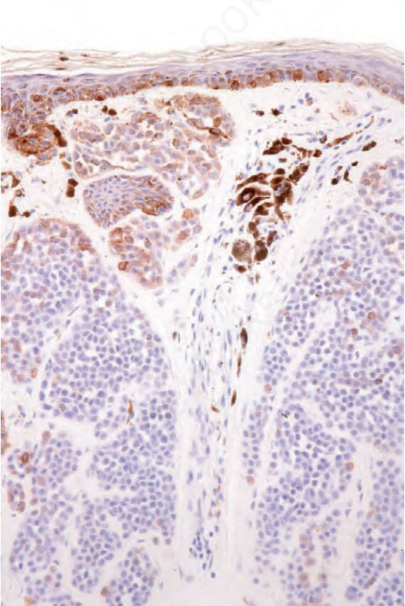
Fig. 25.58 Dermal nevus: the superficial component expresses HMB-45 but this is lost with depth.
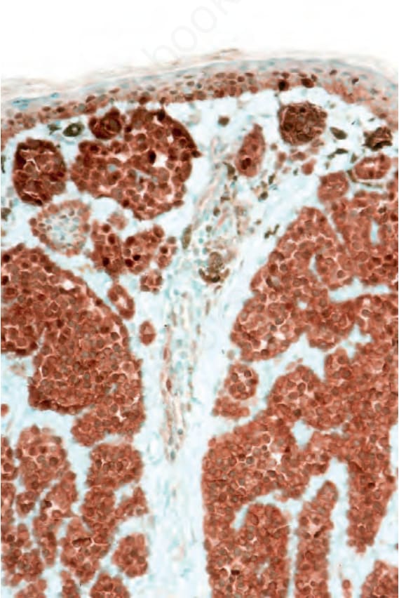
Fig. 25.60 Dermal nevus: very occasional superficial nevus cells express cyclin D1. Note that only the nuclear staining is significant.
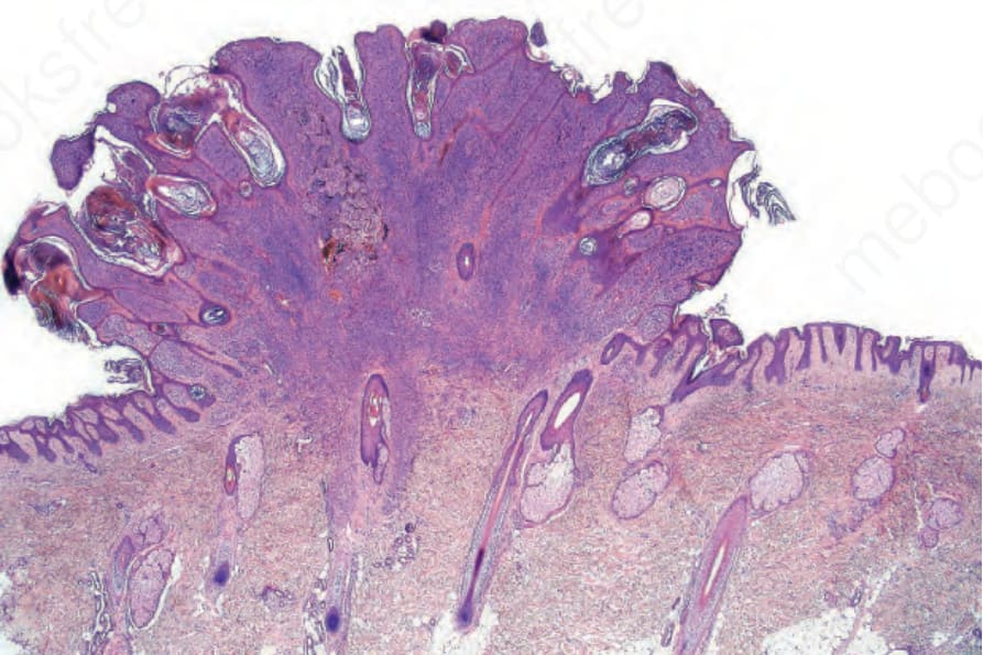
Fig. 25.61 Clonal nevus: this is a banal compound nevus. Note the distinct nodule in the deeper dermis surrounded by pigment-laden melanophages. By courtesy of W. Grayson, MD, National Health Laboratory Service, Johannesburg, South Africa.
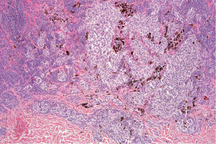
Fig. 25.62 Clonal nevus: medium-power view contrasting the nests of type A cells with the type B cells. There is abundant melanin pigment within macrophages. By courtesy of W. Grayson, MD, National Health Laboratory Service, Johannesburg, South Africa.
1246 Melanocytic nevi
1247 Melanocytic nevus
The cause of this change is unknown, although it is probably artifactual and may relate to the effect of a local anesthetic. Immunohistochemical studies using endothelial cell markers are invariably negative.28,29 The cells lining the spaces are of melanocytic derivation and label positively with S100 protein. Amyloid may rarely be identified within a nevus.30 Exceptionally, a nevus may be accompanied by proliferation of the sweat gland epithelium; coexistence of nevus and desmoplastic trichoepithelioma is occasionally encountered.31–33 Melanocytic nevus has also been reported in association with a trichoadenoma.34
The presence of intravascular melanocytes or evidence of lymphatic involvement is a very disturbing finding and should prompt a thorough search for other features of melanoma. Herniation of a nevus nest into the lumen of a vessel should not be confused with vascular invasion. The
distinction can be made by finding a layer of endothelium covering the protruding nevus cells in the benign lesion (Fig. 25.55).
Differential diagnosis Distinction between a banal nevus and melanoma in the majority of cases is straightforward. Low-power examination of melanoma may reveal obvious intralesional transformation, i.e., the malignant cells stand out as an expansile and often circumscribed nest, nodule, or plaque that is cytologically different from the adjacent nevus cells. Additional distinguishing features include the presence of dense pigmentation, lack of maturation, nuclear pleomorphism, mitotic activity, apoptosis, lymphocytic infiltration, and a deep nested growth pattern, which are commonly evident in melanoma but not in intradermal nevi. Upward, intraepidermal, or pagetoid spread of melanocytes is an additional feature seen in many melanomas. Caution,
1248 Melanocytic nevi
however, is advised when viewing sections from neonatal and even childhood nevi when nests and occasionally single cells, sometimes showing mild or even severe cytological atypia, may be identified within the upper reaches of the epidermis (see neonatal nevus).35 Similarly, pagetoid spread may be a feature of acral and genital nevi (see below). Reticulin fibers outline nests of melanoma cells whereas they tend to surround individual nevus cells.
Dermal nevus cells may rarely show mitotic activity (Fig. 25.56). Their presence should therefore be viewed with caution and other features suggestive of malignancy sought. Nevoid melanoma may be cytologically similar at a casual glance. Careful inspection, however, reveals asymmetry and lack of circumscription, multiple dermal mitoses, subtle lack of maturation, and nucleolar prominence (see nevoid melanoma).
Small cell melanoma cells, although often of a similar size to type B nevus cells, usually have prominent eosinophilic nucleoli, and mitoses are invariably present (see small cell melanoma).
Difficulties are sometimes experienced in differentiating invasive melanoma (particularly the nevoid and small cell variants) from residual benign intradermal nevus cells. The latter is particularly important when assessing tumor thickness or the level of invasion. Immunohistochemistry may be of value in making this distinction. HMB-45 expression is often positive in superficial dermal nevus but is lost with depth (Figs 25.57 and 25.58).36 In
1249 Melanocytic nevus
melanoma, in contrast, the tumor cells are often positive throughout the lesion. p53 protein is not expressed in banal dermal nevi. In contrast, it is frequently present in melanoma.37 In banal nevi, small numbers of Ki-67 and cyclin D1 positive cells may be seen in the more superficial dermal component (Figs 25.59 and 25.60). In melanoma, they are usually much more numerous, and often they are present throughout the thickness of the
lesion.38 The pattern of cyclin D1 staining, however, should not be relied on in the distinction between benign and malignant.
Banal nevi may show some histologic overlap with dysplastic nevi. Thus they are often lentiginous and sometimes show shoulder formation, eosinophilic or lamellar fibrosis around the elongated epidermal ridges. In contrast, architectural disorder (i.e., nests of nevus cells scattered irregularly
1250 Melanocytic nevi
throughout the epidermis) and bridging are not seen. In addition, cytological atypia is not a feature of banal nevi.
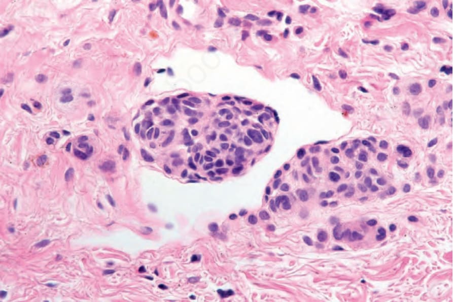
Fig. 25.55 Dermal nevus: invagination of nevus cells into the lumen of a vessel should not be confused with true vascular invasion. Distinction depends on identification of a layer of endothelial cells covering the surface of the nevoid aggregate, as shown in this example.
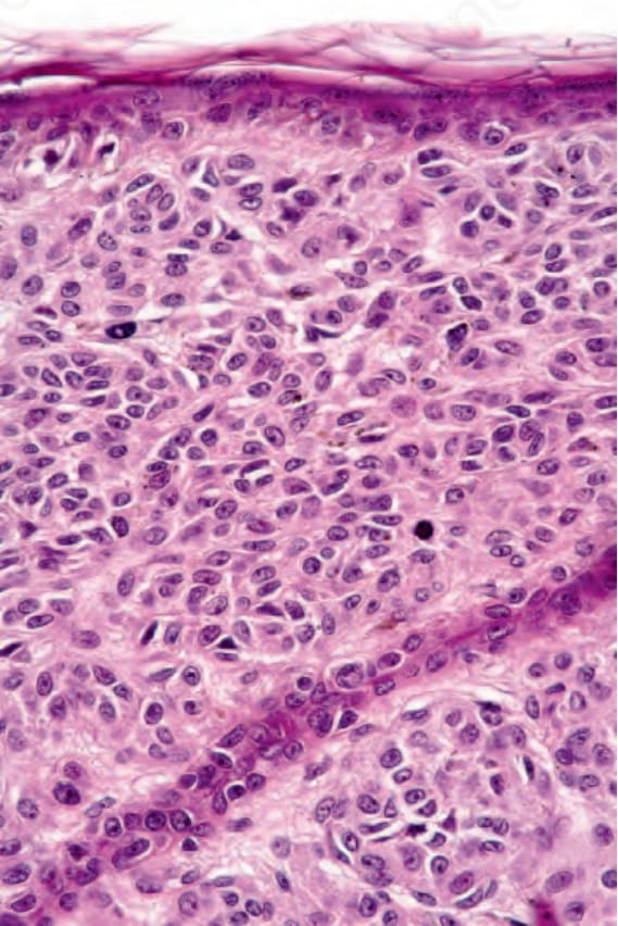
Fig. 25.56 Banal nevus: identification of one or two dermal mitotic figures is not always synonymous with melanoma. Their presence, however, should be viewed with considerable concern and other mitoses or additional features indicative of melanoma sought.
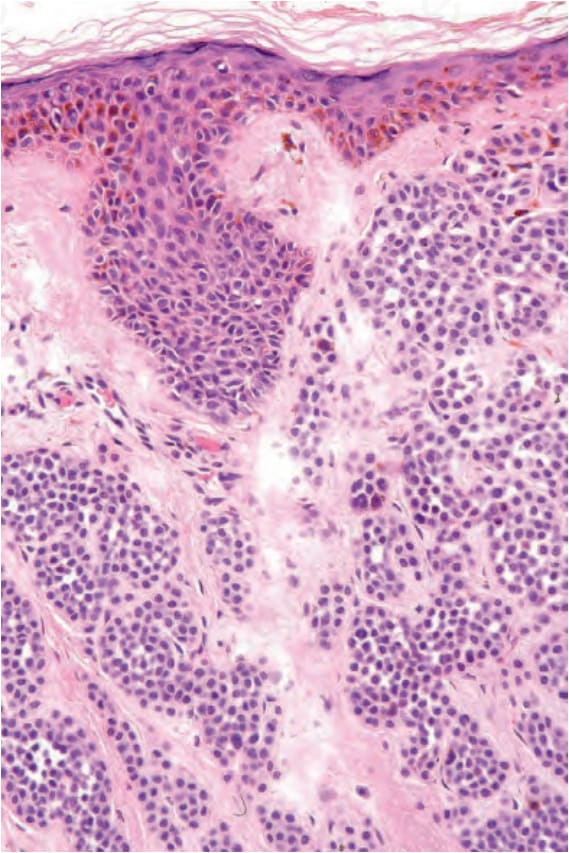
Fig. 25.57 Dermal nevus: medium-power view of banal nevus cells.
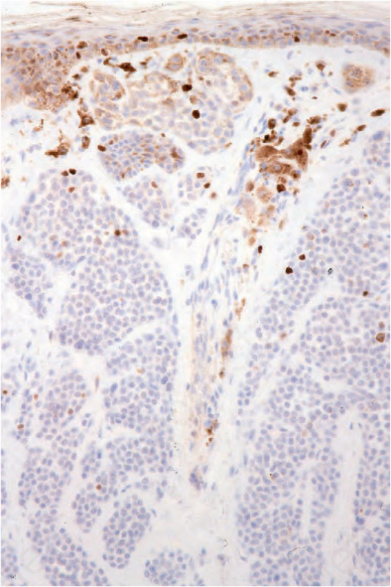
Fig. 25.59 Dermal nevus: only one or two nevus cells express MIB-1.