葉狀微血管瘤/化膿性肉芽腫 (Lobular capillary hemangioma / pyogenic granuloma)
葉狀微血管瘤/化膿性肉芽腫 (lobular capillary hemangioma / pyogenic granuloma)
臨床特徵 (Clinical Features)
葉狀微血管瘤/化膿性肉芽腫 (lobular capillary hemangioma / pyogenic granuloma) 常見於嵌甲 (ingrowing toenails),一般認為是由甲板 (nail plate) 與外側甲褶 (lateral nail fold) 之間的交互作用所誘發。有時可見甲板的改變。其多見於生命的早期數十年,表現為快速生長、疼痛、潰瘍且易出血的外生性 (exophytic) 腫瘤。甲下 (subungual) 位置亦曾被報導。切除後可能有局部復發。多發性甲周 (periungual) pyogenic granuloma 樣病灶可能由全身性藥物所誘發,例如 retinoids、ciclosporine、化療藥物 (chemotherapeutic agents) 及抗反轉錄病毒療法 (antiretroviral therapies)。
此腫瘤體積小(通常小於 1 cm),為膚色或紅藍色的結節 (Fig. 23.71),常伴隨與輕微觸覺刺激或冷暴露相關的陣發性疼痛 (paroxysmal pain)。可出現甲變形 (nail deformity) 與骨質缺損 (osseous defect)。建議的治療為完全外科切除 (complete surgical excision)。復發罕見。
致病機轉與組織學特徵 (Pathogenesis and Histologic Features)
血管球瘤 (glomus tumor) 源自位於血管球體 (glomus bodies) 內的特化平滑肌細胞 (modified smooth muscle cells)。多發性家族性血管球瘤(呈體染色體顯性遺傳模式)已被連結到 glomulin 基因的去活化突變 (inactivating mutations)。在這些「家族性血管球瘤 (familial glomangioma)」中,腫瘤罕見發生於甲下區域。甲下 glomus tumors 與第一型神經纖維瘤病 (neurofibromatosis type I) 之間的關聯曾被報導(伴有 NF1 去活化或染色體拷貝數變化)。MIR143-NOTCH 融合 (fusions) 曾在一系列良性與惡性 glomus tumors(包含部分肢端病灶)中被描述。轉位 t(1;5) 僅在一例散發性腫瘤中被報導。
組織學特徵 (Histologic Features)
在許多病例中,組織學顯示一團茂盛的小血管腔道 (exuberant small vascular channels)。表皮增生 (hyperplasic) 且常潰瘍化。真皮內有微血管 (capillaries) 的增生,並伴隨顯著的混合性發炎細胞浸潤 (mixed inflammatory cell infiltrate)。中性球 (neutrophils) 與漿細胞 (plasma cells) 通常為主。間質 (stroma) 可為疏鬆且水腫,或在後期呈纖維化。罕見情況下,甲部病灶呈息肉狀 (polypoid) 並顯示發育良好的葉狀結構 (lobular architecture)。這些小葉 (lobules) 由微血管與小靜脈 (venules) 的聚集組成,伴或不伴可辨識的管腔,內襯飽滿的內皮細胞 (plump endothelial cells),並被一層 smooth-muscle actin 陽性的周細胞 (pericytes) 所環繞。
Glomus tumors 由血管球細胞 (glomus cells)、血管與平滑肌細胞所組成。依這些成分的相對比例,可分為三組:「實質性血管球瘤 (solid glomus tumor)」、「血管球血管瘤 (glomangioma)」與「血管球血管肌瘤 (glomangiomyoma)」。
Glomus tumor 通常界線清楚 (well circumscribed),但有些細胞團簇可見於主腫瘤團塊以外的血管周圍。Solid glomus tumor 由環繞微血管的血管球細胞片狀排列所組成。Glomus cells 一致且圓形,具有淡嗜伊紅性 (pale eosinophilic) 細胞質與位於中央的圓形細胞核 (Fig. 23.72)。一層由 PAS 凸顯的基底膜 (basal lamina) 環繞每個細胞。在 glomangioma 中,血管成分顯著,由眾多擴張的血管腔隙所組成。Glomangiomyoma 的特徵為從血管球細胞逐漸過渡到類似成熟平滑肌細胞的拉長細胞。Glomus tumors 對 smooth muscle actin 與第四型膠原蛋白 (type IV collagen) 呈陽性。h-Caldesmon、desmin 與 CD34 亦可能呈陽性。
鑑別診斷 (Differential Diagnosis)
在老年病人或後天免疫缺乏症候群 (acquired immunodeficiency syndrome) 病人中,鑑別診斷包括結節型卡波西氏肉瘤 (nodular Kaposi sarcoma),其特徵為顯著的紡錘狀細胞 (spindle cell) 成分以及對人類疱疹病毒第八型 (human herpesvirus 8) 呈陽性。
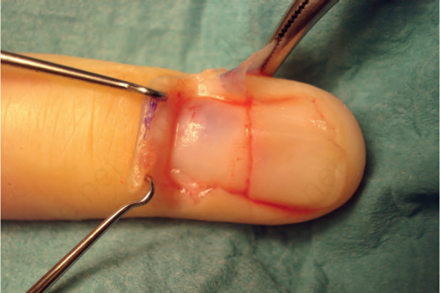
圖 23-71:血管球瘤 (glomus tumor):在近端甲板 (proximal nail plate) 深部可見一界線不清的藍色腫瘤。Courtesy of B. Richert, MD, Université de Liège, Belgium。
Fig. 23.71 Glomus tumor: there is an ill-defined bluish tumor deep to the proximal nail plate. Courtesy of B. Richert, MD, Université de Liège, Belgium.
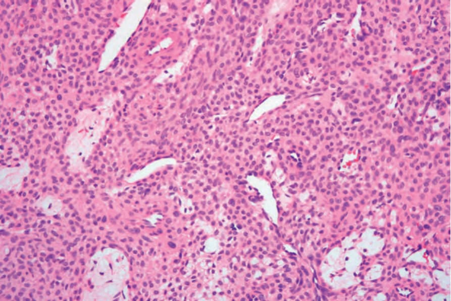
圖 23-72:血管球瘤 (glomus tumor):中倍視野顯示薄壁擴張的血管,周圍環繞典型的血管球細胞 (glomus cells)。
Fig. 23.72 Glomus tumor: medium-power view showing thin-walled dilated vessels surrounded by typical glomus cells.