臨床特徵 (Clinical Features)
- 甲母質瘤 (onychomatricoma) 是一種罕見的良性甲母質腫瘤,具有獨特的臨床與組織學特徵,最早於 1992 年由 R. Baran 描述。類似的甲母質腫瘤也曾以 ‘onychoblastoma’、‘unguioblastoma’ 及 ‘unguioblastic fibroma’ 之名被報導。
- 主要發生於手指,較少見於腳趾,無性別好發傾向。通常發生於高加索 (Caucasian) 中年與老年病人。
- 病灶為單發,罕為多發。其特徵為甲板增厚並伴有明顯的縱向脊狀隆起 (longitudinal ridging)、沿整個甲板長度的黃色變色 (xanthonychia)、多發性線狀出血 (splinter hemorrhages),以及甲板橫向過度彎曲的傾向 (Fig. 23.61)。
- 亦曾描述不尋常的臨床表現,如縱向黑甲 (longitudinal melanonychia) 或翼狀胬肉 (pterygium)。
- 建議的治療為手術切除。可能觀察到復發。onychomatricoma 與甲癬 (onychomycosis) 之合併並不少見。
組織學特徵 (Histologic Features)
- 拔甲後可見一個帶蒂的絨毛狀甲母質腫瘤,具有特徵性的遠端指狀突起 (distal digitations),延伸進入近端甲板的多個孔洞中 (Fig. 23.62)。這導致甲板增厚並呈漏斗狀。遠端甲板修剪 (distal nail plate clipping) 時若顯示多個腔隙 (lacunar spaces),可能是診斷的線索。
(以下段落於原文中順序錯置,依原文逐字保留、不重排。)
-
陽性的纖維母細胞 (fibroblastic cells) 排列方向隨機,並伴肥大細胞 (mast cells) 數量增加。部分病例曾報導具有奇異深染、多形性核 (bizarre hyperchromatic and pleomorphic nuclei) 的細胞與多核巨細胞 (multinucleated giant cells)(Fig. 23.64)。母質呈膠原性 (collagenous)、黏液膠原性 (myxocollagenous) 或黏液性 (myxoid)。在病灶較深部,間質通常細胞較少,膠原束較粗,並沿同一水平軸向排列。間質細胞表現 CD34。在極少數病例中,曾檢測 CD10(onychodermis 的標記),並報導其在間質中瀰漫性表現。
-
此纖維上皮性腫瘤 (fibroepithelial tumor) 具有「海葵」(anemone) 的構造,且腫瘤遠端與近端區域的組織學特徵相當不同。
-
遠端區域 (distal zone) 的特徵為多個「手套指」(glove finger) 乳頭狀突起,覆以母質型上皮 (matrix-type epithelium),此上皮缺乏顆粒層 (stratum granulosum),並透過嗜伊紅性的角化生成帶 (eosinophilic keratogenous zone) 進行角化 (Fig. 23.63)。近端區域 (proximal zone) 相當於蒂部 (peduncle),在橫切面上呈圓頂狀。其襯以乳頭瘤狀的母質型上皮,並有垂直走向、深入間質的內陷 (invaginations)。這些內陷以特徵性的 V 形構造圍繞著光學上空無一物的腔隙 (optically empty cavities)。在缺乏「手套指」乳頭狀突起的破碎或不完整標本上,辨識出具有 V 形凹陷的母質型上皮對於診斷 onychomatricoma 至關重要。間質中度至高度富含細胞,由 CD34 陽性、排列方向隨機的纖維母細胞(接續上文錯置段落)所組成。
鑑別診斷 (Differential Diagnosis)
- 鑑別診斷包括甲母質的纖維角化瘤 (fibrokeratoma) 與纖維瘤 (fibroma)。兩者的特徵皆為缺乏多發性乳頭狀突起,並具有顆粒層 (granular layer) 與角化過度 (hyperkeratosis)。
- 根據 Perrin 的意見,乳頭瘤型的 onychocytic matricoma 可能與 onychomatricoma 相似,但其甲板上的孔洞較小、基底樣細胞 (basaloid cells) 較多,且缺乏 onychomatricoma 典型的纖維性間質。
- onychomatricoma(尤其是其近端部分)也可能與淺表肢端纖維黏液瘤 (superficial acral fibromyxoma;AFM; acral fibromyxoma) 混淆;然而後者缺乏具特徵性 V 形內陷的母質型上皮。
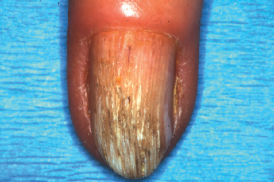
圖 23-61:甲母質瘤 (onychomatricoma):注意甲板增厚並伴有縱向脊狀隆起 (longitudinal ridging)、黃色變色與過度彎曲。
Fig. 23.61 Onychomatricoma: note the thickening of the nail plate with longitudinal ridging, yellow discoloration, and excessive curvature.
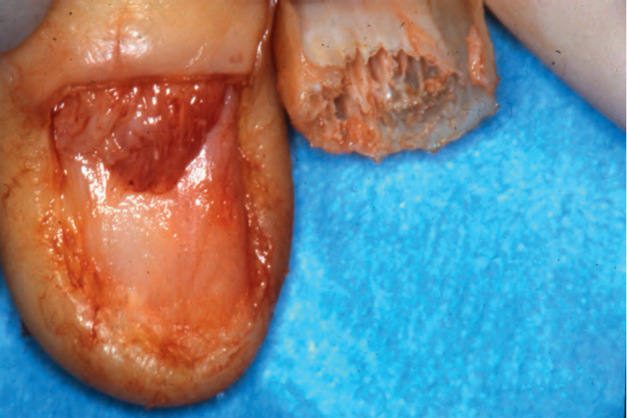
圖 23-62:甲母質瘤 (onychomatricoma):拔除甲板後可見典型的大體外觀。
Fig. 23.62 Onychomatricoma: the typical gross appearances are seen after avulsion of the nail plate.
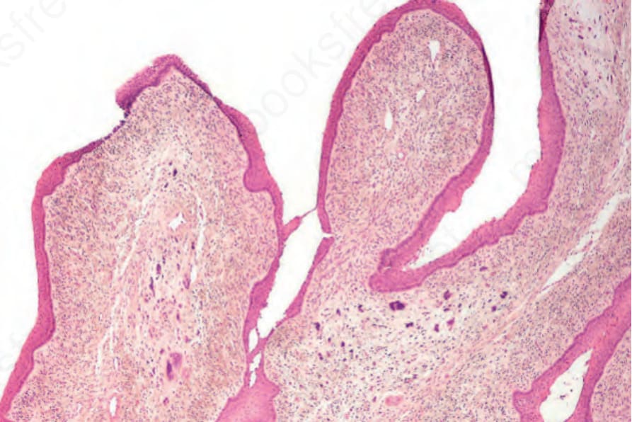
圖 23-63:甲母質瘤 (onychomatricoma):掃描視野顯示覆以母質型上皮 (matrix type epithelium) 的「手套指」(glove finger) 乳頭狀突起。
Fig. 23.63 Onychomatricoma: scanning view showing the ‘glove finger’ papillary projections covered by matrix type epithelium.
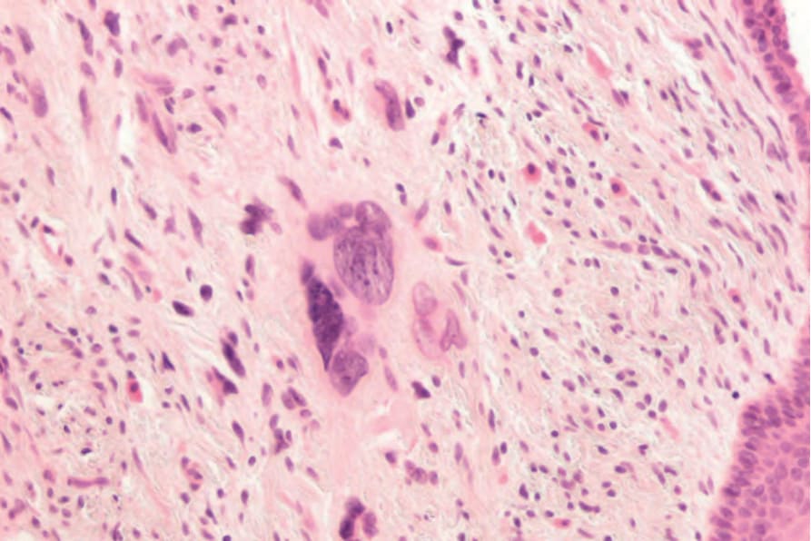
圖 23-64:甲母質瘤 (onychomatricoma):間質高度富含細胞,並有明顯的核多形性 (nuclear pleomorphism)。注意顯著的肥大細胞 (mast cells)。
Fig. 23.64 Onychomatricoma: the stroma is highly cellular with marked nuclear pleomorphism. Note the conspicuous mast cells.
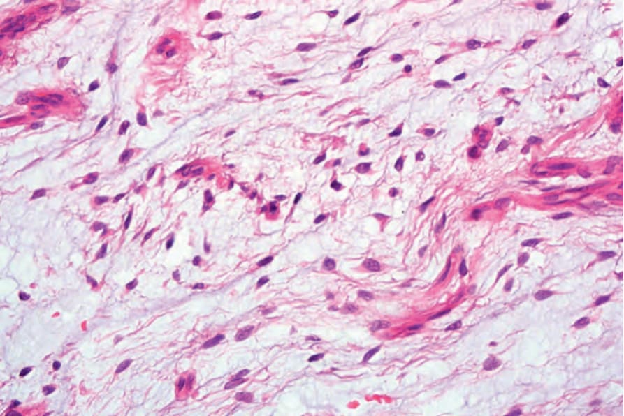
圖 23-65:Koenen 腫瘤 (tuberous sclerosis):典型病灶從第四指的近端甲褶 (proximal nail fold) 冒出,並見指甲上多條縱向凹陷。Courtesy of B. Richert, MD, PhD, Université Libre de Bruxelles, Belgium.
Fig. 23.65 Koenen tumors (tuberous sclerosis): typical lesion emerging from the proximal nail fold of the fourth finger and multiple longitudinal depressions in the fingernails. Courtesy of B. Richert, MD, PhD, Université Libre de Bruxelles, Belgium.