藍痣 (Blue Nevus)
藍痣 (blue nevus)
臨床特徵 (Clinical Features)
- 文獻中已發表十例有完整紀錄之甲器 (nail apparatus) 藍痣 (blue nevus)。其中三例為先天性,其餘則出現於年齡 20 至 64 歲之成人。其臨床特性整理於 Table 23.2。
- 在痣 (nevi) 中,巢 (nests) 位於甲基質 (matrix),有時亦見於近端甲褶 (PNF) 之腹側部分及/或甲下皮 (hyponychium)。後者僅在縱向切片 (longitudinal biopsy) 上可見。巢通常不見於甲床 (nail bed)。絕大多數的甲痣 (ungual nevi) 屬交界型 (junctional)。在複合痣 (compound nevi) 中,真皮內巢最常見於甲下皮 (hyponychium)。
- 對稱性 (symmetry) 在甲痣中並非有用之特徵,因切片往往為部分性且尺寸甚小。此外,由於甲的結構,痣在縱向切片中本身即呈不對稱。再者,邊緣 (margins) 通常無法評估。巢往往稀疏,但罕見病例可顯示強烈的黑色素細胞增生 (melanocytic hyperplasia),伴有融合性巢,常由大型上皮樣黑色素細胞 (epithelioid melanocytes) 構成(Figs 23.37 與 23.38)。15% 的病灶可見一些具核異型性 (nuclear atypia) 之黑色素細胞,並可見輕度的黑色素細胞向上遷移 (melanocytic upward migration)。
組織病理特徵 (Histopathology)
- 常見型藍痣 (common blue nevus) 見於七例、細胞型藍痣 (cellular blue nevus) 見於兩例、非典型藍痣 (atypical blue nevus) 見於一例、聯合性藍痣 (combined blue nevus) 見於兩例(Table 23.2)。
鑑別診斷 (Differential Diagnosis)
- 罕見情況下,部分消退的甲下黑色素瘤 (subungual melanoma) 在部分切片中可能類似細胞型藍痣 (cellular blue nevus)。
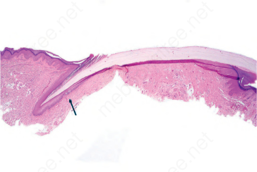
圖 23-37:交界型黑色素細胞痣 (junctional melanocytic nevus):掃描視野顯示甲基質內兩個交界型巢(箭頭所示)。
Fig. 23.37 Junctional melanocytic nevus: scanning view showing two junctional nests in the nail matrix (arrowed).
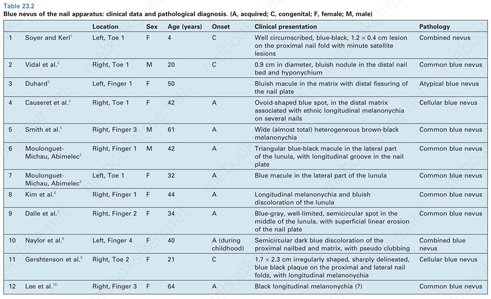
表 23-2:甲器藍痣 (blue nevus of the nail apparatus):臨床資料與病理診斷。(A,後天性 acquired;C,先天性 congenital;F,女性 female;M,男性 male)
Table 23.2 Blue nevus of the nail apparatus: clinical data and pathological diagnosis. (A, acquired; C, congenital; F, female; M, male)