Blue nevus
Blue nevus
Clinical features Ten cases of well-documented blue nevus in the nail apparatus have been published. Three cases were congenital, the others presented in adults aged 20 to 64 years. Their clinical characteristic are summarized in Table 23.2.1–10
In nevi, nests are located in the matrix, and sometimes also in the ventral part of the PNF and/or the hyponychium. The latter are only visible on longitudinal biopsy. Nests are not usually observed in the nail bed. The vast majority of ungual nevi are junctional.2,3 In compound nevi, dermal nests are most often observed in the hyponychium. Symmetry is not a useful feature in ungual nevi because biopsies are often partial and small in size. Moreover, nevi are asymmetrical in longitudinal biopsies because of the nail architecture.3 Also, margins cannot usually be evaluated. Nests tend to be scarce, but rare cases display intense melanocytic hyperplasia with confluent nests often consisting of large epithelioid melanocytes (Figs 23.37 and 23.38). The presence of some melanocytes with nuclear atypia can be seen in 15% of lesions and a mild degree of melanocytic upward migration in
Histologic features Common blue nevus was observed in seven cases, cellular blue nevus in two cases, atypical blue nevus in one case, and a combined blue nevus in two cases (Table 23.2).
Differential diagnosis Rarely, partially regressed subungual melanoma may mimic a cellular blue nevus in partial biopsies.11
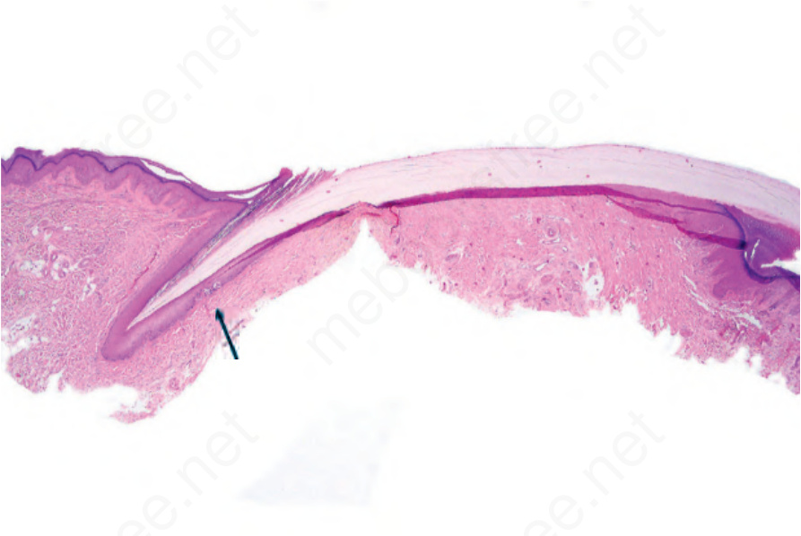
Fig. 23.37 Junctional melanocytic nevus: scanning view showing two junctional nests in the nail matrix (arrowed).
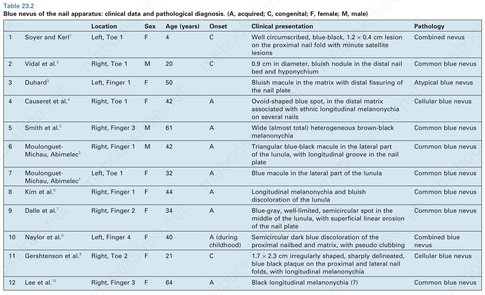
Table 23.2 Blue nevus of the nail apparatus: clinical data and pathological diagnosis. (A, acquired; C, congenital; F, female; M, male)