項部瘢痕疙瘩性痤瘡 (Acne keloidalis nuchae)
臨床特徵 (Clinical Features)
- 項部瘢痕疙瘩性痤瘡 (acne keloidalis nuchae,又稱 folliculitis keloidalis nuchae) 是非洲裔男性中最常見的瘢痕性禿髮 (scarring alopecia) 形式。病灶侵犯後頸部 (nape of the neck),男女比例為二十比一。
- acne keloidalis 此一命名其實並不正確,因為本病既不伴隨痤瘡 (acne) 病灶,組織學研究中也未見 keloidal collagen。
- 本病以局限性的毛囊性丘疹 (follicular papules) 起始,病灶逐漸增大並融合,形成瘢痕疙瘩樣斑塊 (keloidal plaques),隨後出現毛髮脫落 (Fig. 22.164)。本病也曾見於非洲裔及拉丁美洲裔女性,並與 keratosis follicularis spinulosa decalvans、tufted hair folliculitis、acanthosis nigricans、使用 tacrolimus 與 sirolimus 的腎臟移植病人、ciclosporine A(白人及非洲裔病人皆有)、lithium carbonate、抗癲癇藥物 (antiepileptic medications),以及使用美式足球頭盔 (football helmets) 有關聯。
致病機轉與組織學特徵 (Pathogenesis and Histologic Features)
- 病因未明。最可能反映的是對局部刺激現象的反應,並併發續發性細菌感染 (secondary bacterial infection)。捲曲的毛髮被剪得過短後重新長回,又向內長入皮膚,誘發發炎反應,被認為是可能的成因。雖然同樣的現象也曾被推測為 pseudofolliculitis barbae 的致病機轉,但這兩種疾病之間並無關聯。
- 本病也曾被視為一種經表皮排除 (transepidermal elimination) 障礙。相關因素包括高濃度的 testosterone、seborrheic dermatitis,以及後頸部 mast cells 密度增加。
- 評估 acne keloidalis 最佳的方式是採用垂直切片 (vertical sections)(Fig. 22.165)。組織學上,初期病灶與 folliculitis decalvans 相似,於峽部 (isthmus) 及皮脂腺 (sebaceous glands) 周圍可見嗜中性球浸潤 (neutrophilic infiltrate),這些皮脂腺最終消失。漏斗部 (infundibulum) 擴張。其後可見厚帶狀的緻密膠原蛋白 (compact collagen),混雜程度不一的淋巴漿細胞性發炎細胞浸潤 (lymphoplasmacytic inflammatory cell infiltrate),主要集中於毛囊漏斗部與峽部 (Fig. 22.166)。如前所述,並不會觀察到真正的 keloidal collagen。伴隨外毛根鞘 (external root sheath) 變薄與層狀纖維增生 (lamellar fibroplasia)(見 Fig. 22.120),最終毛囊破壞導致游離的毛幹 (free hair shafts) 進入真皮,並在該處引發強烈的發炎細胞反應 (Fig. 22.167)。有時也可遇到類似 dissecting cellulitis 所見的膿瘍 (abscesses) 與瘻管 (fistulous tracts)。毛囊失去其皮脂腺,多個毛囊可能融合在一起,形成含有多根毛幹的單一毛囊開口 (polytrichia)。
鑑別診斷 (Differential Diagnosis)
- dissecting cellulitis 的鑑別診斷包括 folliculitis decalvans、頭皮無菌性結節 (aseptic nodules of the scalp,即 pseudocyst of the scalp),以及感染性病程。
- dissecting cellulitis 典型可見的波動性化膿性結節 (fluctuating suppurative nodules) 伴瘻管,並非 folliculitis decalvans 的特徵。組織學上,後者的特徵為嗜中性球性發炎浸潤,主要侵犯毛囊上半部,並伴有淋巴球性浸潤及對毛幹的局灶性異物肉芽腫 (foreign body granulomata)。頭皮無菌性結節為單一結節性病灶,排出膿性物質後遺留囊性腔洞。與 dissecting cellulitis 相反,其禿髮為可逆性,預後極佳。
- central centrifugal scarring alopecia 是主要的鑑別診斷。雖然臨床表現與侵犯部位差異甚大,但組織學上有相當程度的重疊。亦曾有一例 tinea capitis 在女性病人身上類似 acne keloidalis 的報告。
- 對外觀正常但鄰近異常病灶的皮膚進行切片,可能顯示出毛囊破壞與纖維化的證據。

圖 22-120:複合毛囊 (compound follicles)。上圖,正常複合毛囊於漏斗部 (infundibulum) 層次的橫切面。注意顆粒細胞層 (granular cell layer)、不明顯的毛囊周圍纖維化層 (perifollicular fibrotic layer) 與極少的淋巴球浸潤。下圖,瘢痕性禿髮 (scarring alopecia) 中的複合毛囊,於峽部 (isthmus) 層次的水平切片。可見廣泛的毛囊周圍纖維化 (perifollicular fibrosis) 與發炎浸潤。左下圖的毛囊取自一例 acne keloidalis nuchae,右圖則取自一例 folliculitis decalvans。
Fig. 22.120 Compound follicles. Upper panel, compound normal follicles transversally cut at the level of the infundibulum. Note the granular cell layer and discrete perifollicular fibrotic layer and minimal lymphocytic infiltrate. Lower panel, compound follicles in scarring alopecia. Horizontal sections at the level of the isthmus. There is extensive perifollicular fibrosis and an inflammatory infiltrate. The follicles on the left lower panel are from a case of acne keloidalis nuchae and those on the right panel from a case of folliculitis decalvans.
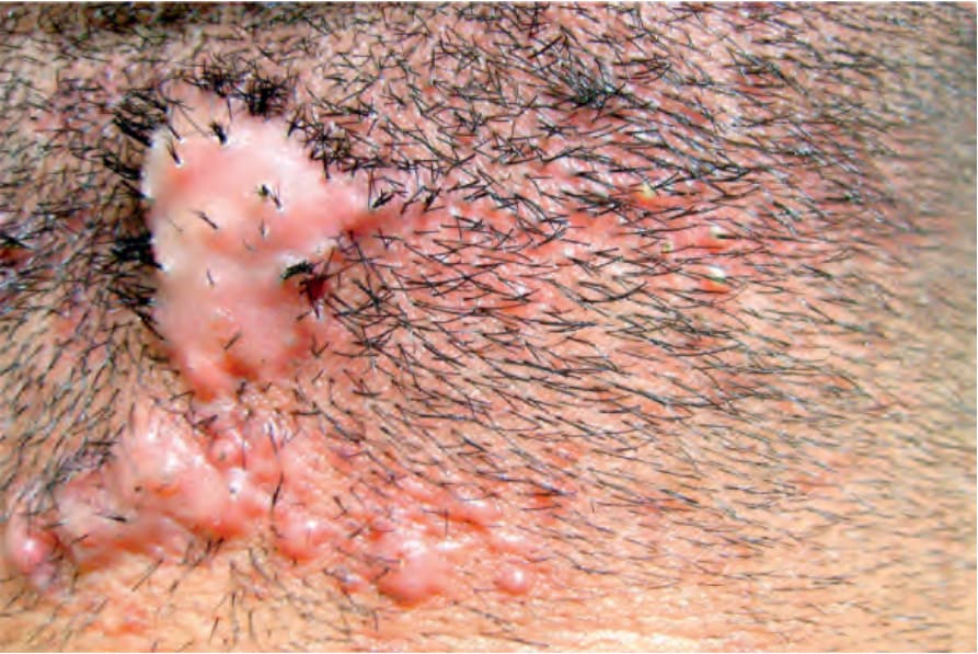
圖 22-164:項部瘢痕疙瘩性痤瘡 (acne keloidales nuchae):丘疹已融合形成瘢痕疙瘩樣斑塊 (keloidal plaque),亦可見膿疱 (pustules)。圖片由 C. Velázquez, MD, CES, Medellín, Colombia 提供。
Fig. 22.164 Acne keloidales nuchae: papules have coalesced forming a keloidal plaque. Pustules are also seen. Courtesy of C. Velázquez, MD, CES, Medellín, Colombia.
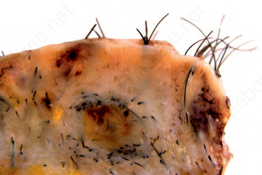
圖 22-165:項部瘢痕疙瘩性痤瘡 (acne keloidales nuchae):手術檢體顯示真皮強烈纖維化 (intense fibrosis) 伴毛囊方向扭曲。表皮表面可見一些 tufted follicles。
Fig. 22.165 Acne keloidales nuchae: surgical specimen showing intense fibrosis of the dermis with distortion of the orientation of the hair follicles. The epidermal surface shows some tufted follicles.
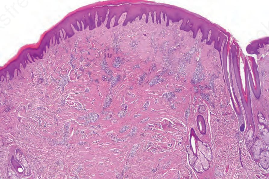
圖 22-166:項部瘢痕疙瘩性痤瘡 (acne keloidales nuchae):掃描倍率下顯示真皮瘢痕化 (dermal scarring) 與多個游離毛幹碎片 (free hair shaft fragments)。
Fig. 22.166 Acne keloidales nuchae: scanning magnification showing dermal scarring and multiple free hair shaft fragments.
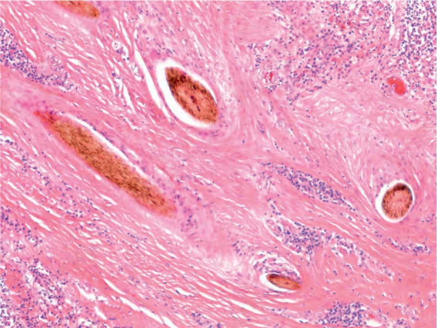
圖 22-167:項部瘢痕疙瘩性痤瘡 (acne keloidales nuchae):高倍視野顯示毛幹碎片 (hair shaft fragments) 被緻密纖維化 (dense fibrosis) 包圍。
Fig. 22.167 Acne keloidales nuchae: high-power view of hair shaft fragments surrounded by dense fibrosis.