解剖學 (Anatomy)
引言
為正確評估頭皮切片,病理醫師不僅要熟稔各類禿髮 (alopecias) 的組織學,也必須通曉健康毛囊 (hair follicle) 複雜且不斷變化的解剖構造,以及其在垂直切面 (vertical sections) 與水平切面 (horizontal sections) 中的特徵。與正常毛囊週期 (follicular cycle),以及易感個體中終毛毛囊 (terminal follicles) 微小化 (miniaturization) 相關的型態變異十分重要。以下章節將逐一回顧這些面向。
不確定毛 (Indeterminate hairs)
這些不確定毛 (indeterminate hairs) 究竟應歸屬於哪一類毛囊,目前尚無明確共識。我們認同部分作者的建議,將其中一半歸入 terminal hair follicles,另一半歸入 vellus hairs。
毛幹與內根鞘的辨識
毛幹 (hair shaft) 與內根鞘 (inner root sheath) 在莖部 (stem) 層次,亦即恰位於膨大部 (bulge) 下方處,於垂直與水平切面中均易辨識,可用常規染色程序,包括蘇木精伊紅染色 (hematoxylin and eosin)、甲苯胺藍 (toluidine blue) 與彈性纖維染色 (elastic stains)(圖 22.15,並參見圖 22.13)。terminal hairs 與 vellus hairs 的比例約為 7 : 1。區分 terminal hair follicles 與 vellus hair follicles 對於雄性禿 (androgenetic alopecia) 與顳部三角形禿髮 (temporal triangular alopecia) 的診斷至關重要。比例為 4 : 1 或更低時,提示 androgenetic alopecia。依定義,毛囊與 vellus hairs 並不延伸至皮下組織;因此,為確定毛囊的最終數量,特別是 vellus hair 的數量,必須在峽部 (isthmus) 層次製作淺層切面 (superficial sections)。基於此原因,深層切面與淺層切面中毛囊的定量結果並不相同。淺層切面所見的毛囊數,可能比深層切面所計數者多出五到六個。如前所述,terminal follicles 與 vellus hairs 的比例可能因不同族群而異(表 22.2)。須謹記,毛幹在切片過程中經常脫失。在這些情形中,可由內根鞘所界定之空腔的直徑推估毛幹的大小。
毛囊單位 (Follicular units)
毛囊上段的水平切面顯示,頭皮的毛囊呈群聚分布,形成稱為毛囊單位 (follicular units) 的解剖結構,其由 terminal hairs 與 vellus hairs、皮脂腺 (sebaceous glands) 及豎毛肌 (arrector pili muscles) 所組成。毛囊在毛囊單位中的分布,在漏斗部 (infundibulum) 與峽部 (isthmus) 層次最能清楚觀察。圍繞外根鞘 (outer root sheath) 的結締組織,傾向於圍繞由三到六個毛囊所組成的離散單位濃縮聚集,其中一或兩個為 vellus hairs(圖 22.11,並參見圖 22.6)。
終毛毛囊 (Terminal hair follicles)
Terminal hair follicles 在水平切面中容易辨認,因為它們遠大於 vellus hairs(圖 22.12)。這些毛囊較粗、有色素,且通常下降至皮下脂肪(參見圖 22.8)。它們含有一根毛幹,其直徑大於內根鞘的厚度,一般超過 0.06 mm(圖 22.13,並參見圖 22.6)。
毛囊的分段
毛囊在解剖與功能上可分為兩個截然不同的節段。這兩個部分之間的差異,可由以下事實解釋:毛囊上段非常穩定,不受毛髮成熟與脫落的影響;而毛囊下段則主動參與毛髮生長,並依其在毛囊週期中所處的階段而發生顯著的型態變化。
毛囊上段由毛囊口 (follicular ostium)、漏斗部 (infundibulum) 與峽部 (isthmus) 構成(圖 22.16)。毛囊口是毛莖通常以二或三根成群露出的實際開口。其消失在臨床上可見於瘢痕性禿髮 (scarring alopecias)。漏斗部由毛囊口向下延伸至皮脂腺導管的開口。它呈圓錐形,管壁由表皮構成,正常情況下其周圍有輕微的同心性纖維化 (concentric fibrosis) 與淋巴球發炎浸潤 (inflammatory lymphocyte infiltrate),尤其在非洲裔人士中可見。此現象在評估發炎性瘢痕性禿髮時可能造成混淆(圖 22.17)。
毳毛 (Vellus hairs) 與微小化毛
真正的毳毛 (true vellus hairs) 細、短、無色素,其特徵為毛球 (hair bulb) 僅延伸至網狀真皮的上層或中層(圖 22.14)。豎毛肌通常無法偵測到。毛幹直徑等於或小於內根鞘的厚度(圖 22.15)。其典型上小於 0.03 mm。
類毳毛 (vellus-like hairs,即微小化毛 miniaturized hairs) 與 vellus hairs 非常相似。它們對應於因雄性素 (androgens) 作用而微小化的 terminal hair follicles。它們可能很難與 true vellus hairs 區分,唯一的例外是微小化的毛囊會遺留下一條星狀塌陷的纖維組織痕跡,即毛囊星 (follicular stellae)。在頭皮切片中,當提及 vellus hairs 而未限定其來源時,依慣例係指微小化毛與 true vellus hairs 兩者。
不確定毛 (indeterminate hairs) 具有介於 terminal hairs 與 vellus hairs 之間的中間型態,被視為微小化過程中的一個中間步驟。不確定毛毛幹的直徑介於 0.03 與 0.06 mm 之間。
峽部與下段
峽部從皮脂腺導管的開口延續至豎毛肌附著於毛囊膨大部 (hair bulge) 之處(圖 22.18)。豎毛肌透過彈性肌腱 (elastic tendons) 附著於膨大部,並向上延伸至其在鄰近乳頭真皮 (papillary dermis) 中的上方附著點。
下段從豎毛肌的附著點延伸至毛球的下部,並有兩個組成部分:莖部(球上區 suprabulbar zone),其從豎毛肌附著點延伸至 Adamson 緣 (Adamson fringe);以及稱為毛球 (bulb) 的下部(圖 22.19 與 22.20)。在生長期 (anagen) 的毛囊中,莖部是毛囊中最長的結構,具有清楚分化的內、外根鞘 (internal and external radicular sheath)(圖 22.21)。毛球由基質細胞 (matrix cells) 與圍繞真皮乳頭 (dermal papilla) 的基底黑色素細胞 (basal melanocytes) 所組成(圖 22.22)。
毛幹的層次組成
毛幹由三層構成:髓質 (medulla)、皮質 (cortex) 與表皮層/毛小皮 (cuticle)。髓質代表毛幹的中央區域,在人類中並非恆定存在,但在其他動物中常為毛髮的重要組成(圖 22.23)。皮質是最厚的一層,負責毛幹的強度。它由硬角蛋白 (hard keratin) 的中間絲 (intermediate filaments) 構成,這些中間絲排列成微原纖維 (microfibrils),並相互交織形成稱為巨原纖維 (macrofibrils) 的纜索狀結構。毛小皮 (cuticle) 構成毛幹的外層,負責抵抗環境所造成的磨損。它由與表皮成直角排列的鱗片 (scales) 組成。這些鱗片與內根鞘的毛小皮相互嵌合以維持完整性,在毛球區則呈現為單層(圖 22.24,並參見圖 22.21)。毛髮呈直線或彎曲的形狀,取決於內根鞘 (internal radicular sheath) 呈彎曲或直線的形狀。在水平切面中,非洲裔個體的毛髮呈彎曲、橢圓形,且偏心地位於毛囊管道 (follicular channel) 之內。在高加索 (Caucasian) 個體中,毛髮呈圓形,且位於毛囊管道的中央。在 terminal anagen hair follicle 的水平切面中,於球上區可辨識出不同的層次。由中心向周邊,這些層次包括:
- 毛幹(包含毛髮的髓質、皮質與毛小皮),
- 內根鞘(包含內根鞘的毛小皮層、Huxley 層、Henle 層與伴隨層 companion layer),
- 外根鞘 (outer root sheath),
- 玻璃膜層與外側纖維層 (vitreous and external fibrous layer,即毛囊周圍結締組織鞘 perifollicular connective tissue sheath)(圖 22.24)。這些層次會隨顯微切面的層次而顯著變化。在毛球層次,毛幹的毛小皮與內根鞘的所有層次均進行毛透明蛋白角化 (trichohyaline keratinization)。在膨大部,內根鞘開始消失,並在峽部由源自外根鞘的毛根鞘角蛋白 (trichilemmal keratin) 所取代(參見圖 22.18)。
外根鞘在漏斗部與表皮相連續,並在該處形成具有表皮角蛋白籃編狀排列 (basket weave arrangement) 的顆粒層 (granular layer)(參見圖 22.17)。朝向毛球方向,外根鞘的細胞含有豐富的肝醣 (glycogen)(圖 22.21,並參見圖 22.24)。
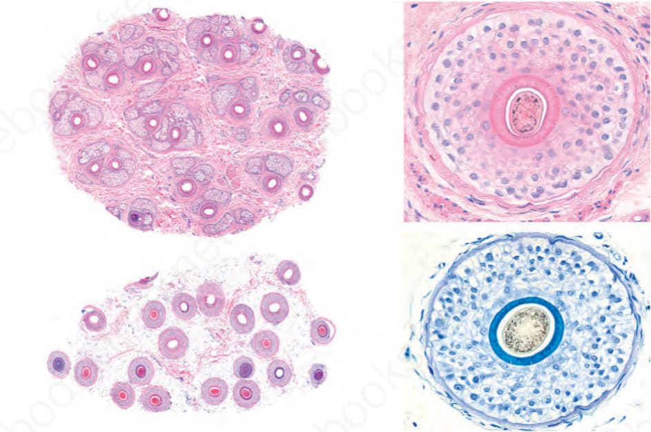
圖 22.6:正常毛髮切片:水平切面。左側於掃描放大倍率下可見所有毛囊。右側於以 H&E 與 toluidine blue 染色的水平切面中可見毛囊的所有組成成分。(Normal hair biopsy: horizontal section. On the left, all the hair follicles are visible at scanning magnification. On the right, all components of the hair follicle are seen in horizontal sections stained with H&E and toluidine blue.)
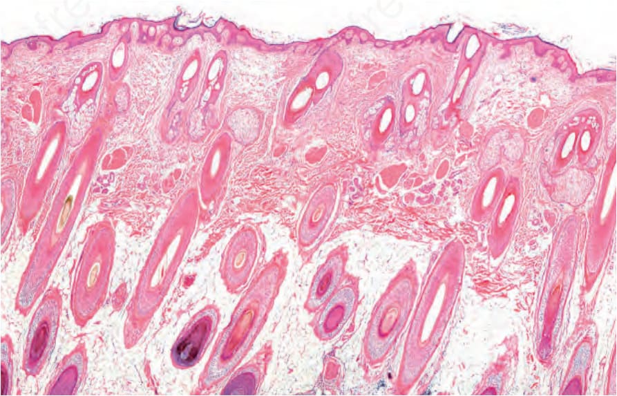
圖 22.8:頭皮切片:在此垂直切面中,未見任何完整的毛囊。由於它們全都不完整,需要追加切面以取得可接受的三維重建。(Scalp biopsy: in this vertical section, no whole hair follicles are seen. Since they are all incomplete, additional sections are required to obtain an acceptable three-dimensional reconstruction.)
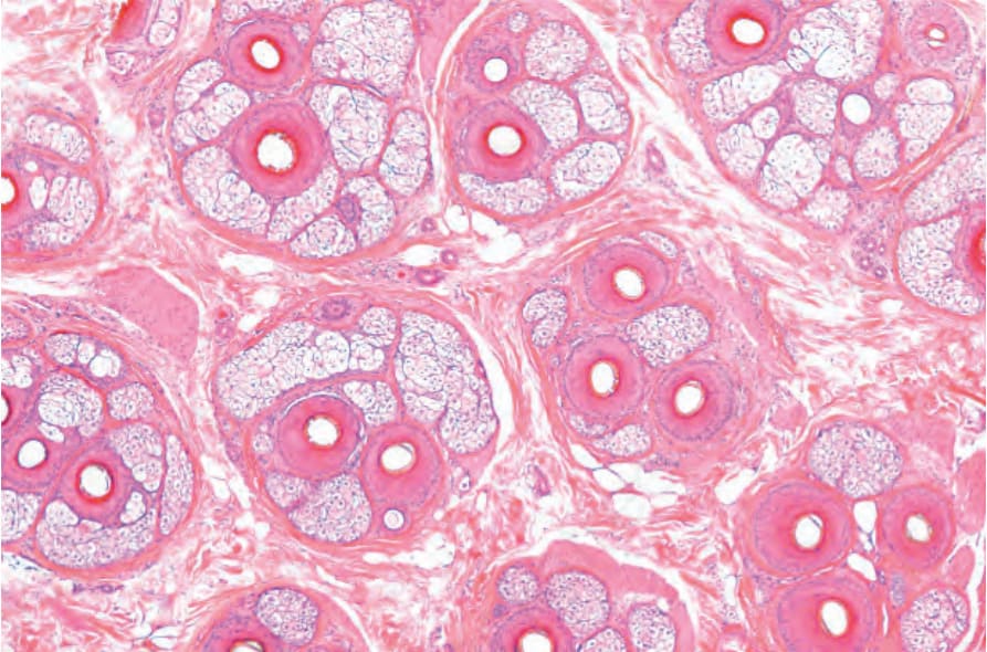
圖 22.11:毛髮切片,水平切面:毛囊單位由外膜膠原 (adventitial collagen) 的濃縮所清楚界定(Masson trichrome 染色)。(Hair biopsy, horizontal section: the follicular units are clearly delineated by condensation of the adventitial collagen [Masson trichrome stain].)
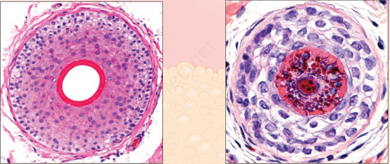
圖 22.12:終毛生長期與毳毛,垂直與水平切面:terminal hair follicles 抵達脂肪並深埋其中。Vellus hairs 與微小化毛囊位於真皮內。注意 terminal hairs 與 vellus hairs 中毛幹與內根鞘相對大小的差異。(Terminal anagen and vellus hair, vertical and horizontal sections: the terminal hair follicles reach the fat and are deeply embedded within it. The vellus hairs and miniaturized follicles are located within the dermis. Note the differences between the relative size of the hair shaft and the inner root sheath in terminal and vellus hairs.) Courtesy of M. Mejia, MD, Universidad Pontificia Bolivariana, Medellín, Colombia.
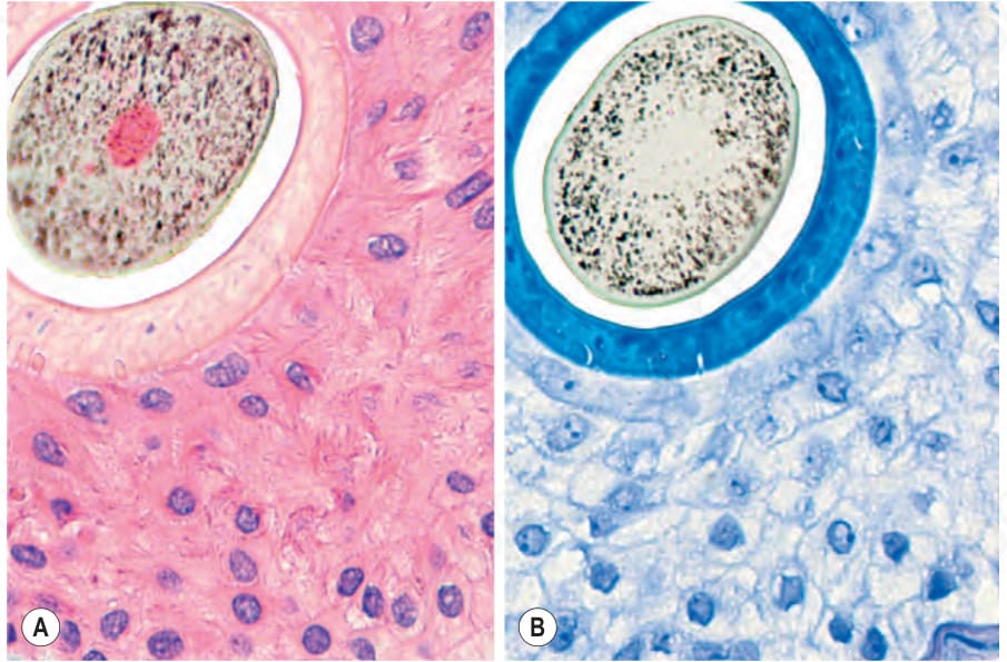
圖 22.13:終毛生長期毛髮,水平切面:比較毛幹的直徑與薄內根鞘的厚度,(A) 以 hematoxylin and eosin 染色呈粉紅色,(B) 以 toluidine blue 染色呈深藍色。(Terminal anagen hair, horizontal section: compare the diameter of the hair shaft with the thickness of the thin internal root sheath [A] in pink with hematoxylin and eosin and in [B] dark blue with the toluidine blue stain.)
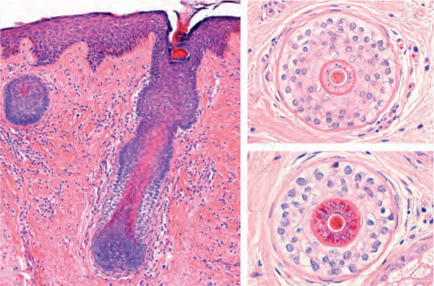
圖 22.14:毳毛,垂直與水平切面:短的 vellus hairs 位於淺層真皮。在水平切面中,毛幹的直徑與內根鞘相同或更細。(Vellus hair, vertical and horizontal sections: short vellus hairs are located in the superficial dermis. In the horizontal sections, the diameter of the hair shaft is the same as or thinner than the inner root sheath.)
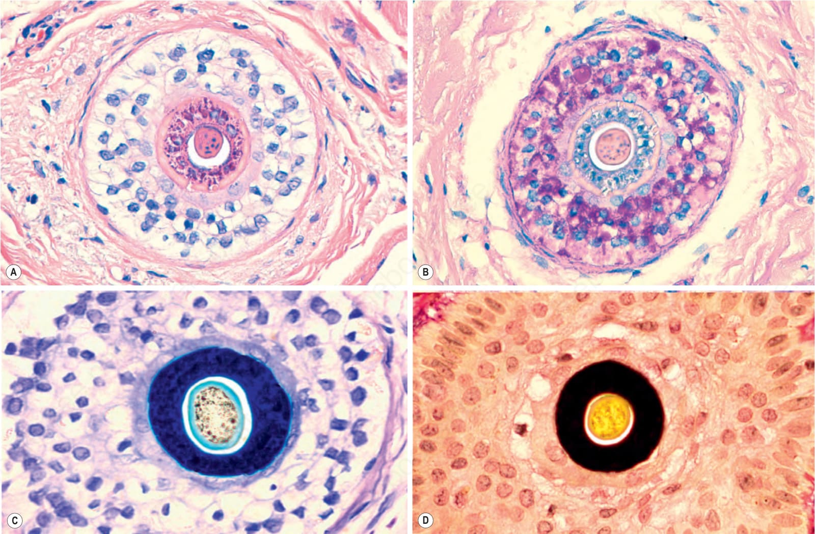
圖 22.15:毳毛,水平切面:注意毛幹相對於內根鞘的口徑:(A) 粉紅色,hematoxylin and eosin;(B) 灰色,PAS;(C) 藍色,toluidine blue;(D) 黑色,elastic tissue stain。(Vellus hair, horizontal section: note the caliber of the hair shaft compared to the inner root sheath in: [A] pink, hematoxylin and eosin; [B] gray, PAS; [C] blue, toluidine blue; and [D] black, elastic tissue stain.)
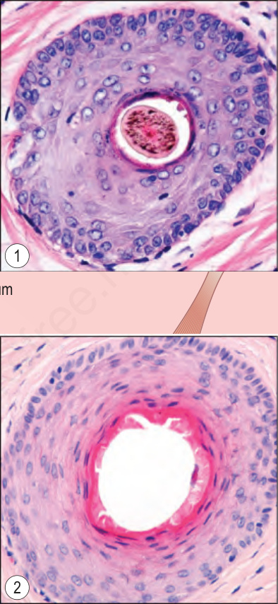
圖 22.16:終毛生長期毛髮,上段的垂直與水平切面:注意毛囊口 (ostium)、漏斗部 (infundibulum) 與峽部 (isthmus)。插圖顯示通過 (1) 漏斗部與 (2) 峽部的水平切面。漏斗部的外根鞘由類似表皮的鱗狀上皮 (squamous epithelium) 所構成。(Terminal anagen hair, vertical and horizontal sections of the upper segment: note the ostium, infundibulum and isthmus. The insets show horizontal sections through [1] the infundibulum and [2] the isthmus. The outer root sheath of the infundibulum is composed of squamous epithelium similar to the epidermis.) Courtesy of M. Mejia, MD, Universidad Pontificia Bolivariana, Medellín, Colombia.
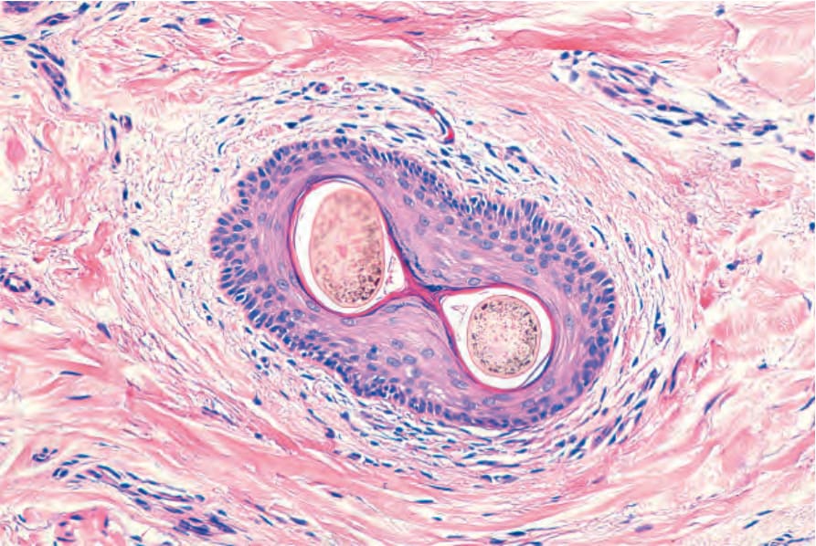
圖 22.17:漏斗部。水平切面。管壁由鱗狀複層角化上皮 (squamous stratified keratinized epithelium) 所構成。周圍真皮被一些淋巴球浸潤。有兩根毛幹自單一毛囊口露出。(Infundibulum. Horizontal section. The wall is made up of squamous stratified keratinized epithelium. The surrounding dermis is infiltrated by some lymphocytes. There are two hair shafts emerging from a single ostium.)
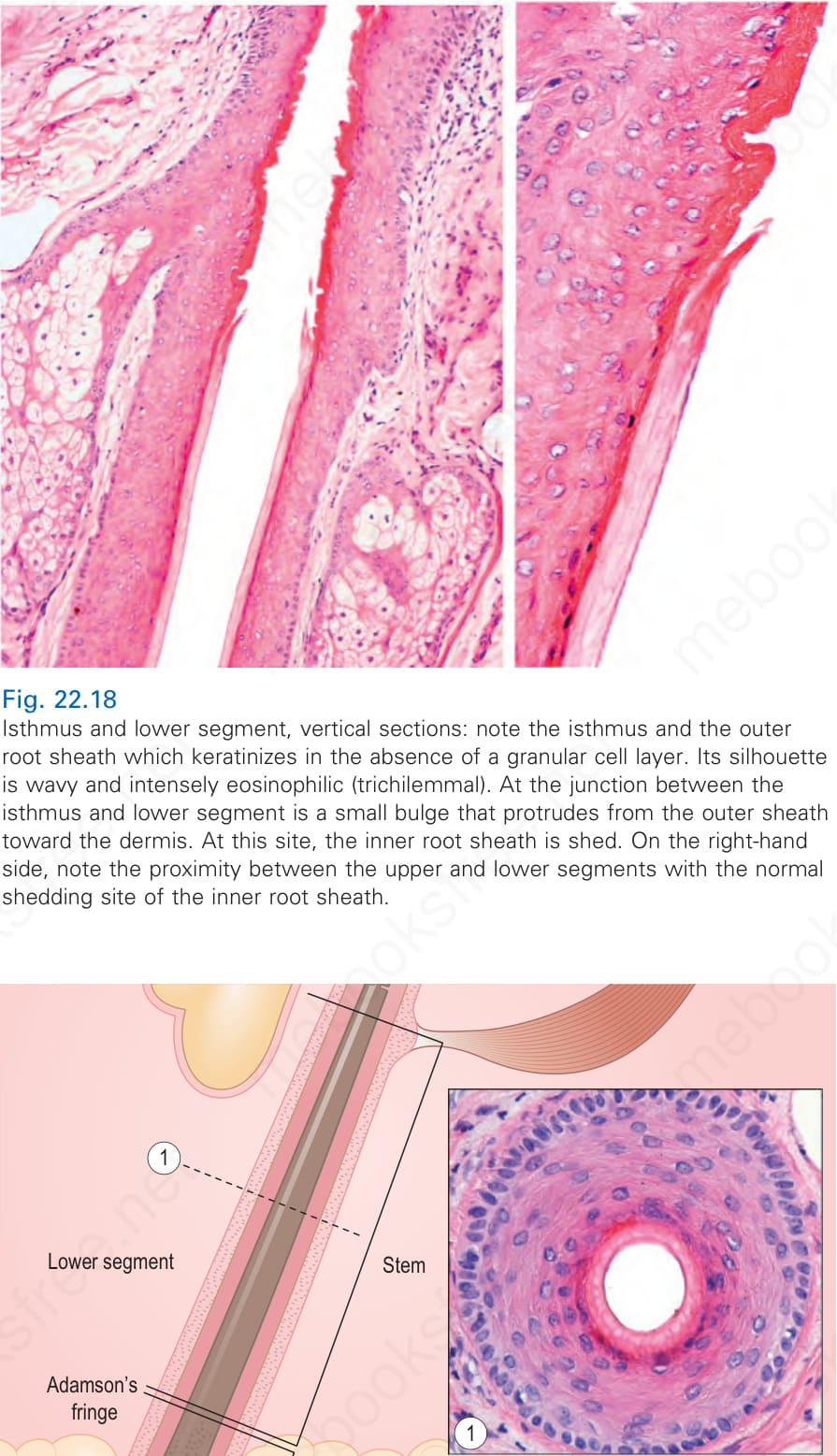
圖 22.18:峽部與下段,垂直切面:注意峽部與在缺乏顆粒細胞層情況下角化的外根鞘。其輪廓呈波浪狀且強烈嗜伊紅 (trichilemmal)。在峽部與下段之交界處有一個小膨大部,由外鞘向真皮突出。在此部位,內根鞘脫落。在右側,注意上段與下段的接近程度,以及內根鞘正常的脫落部位。(Isthmus and lower segment, vertical sections: note the isthmus and the outer root sheath which keratinizes in the absence of a granular cell layer. Its silhouette is wavy and intensely eosinophilic [trichilemmal]. At the junction between the isthmus and lower segment is a small bulge that protrudes from the outer sheath toward the dermis. At this site, the inner root sheath is shed. On the right-hand side, note the proximity between the upper and lower segments with the normal shedding site of the inner root sheath.)
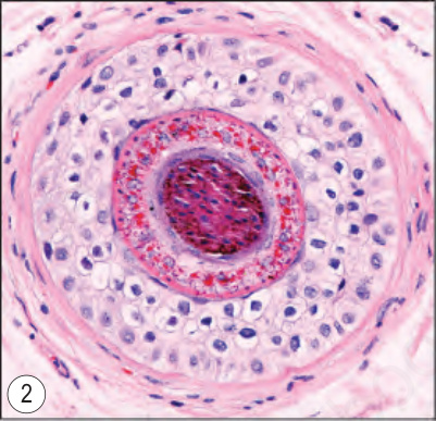
圖 22.19:終毛生長期毛髮,下段的垂直與水平切面:注意毛幹與位於 Adamson 緣深部的毛球。水平切面顯示 (1) 莖部與 (2) 毛球上部。注意毛幹與內根鞘中細胞核的消失。(Terminal anagen hair, vertical and horizontal sections of the lower segment: note the shaft and the hair bulb deep to Adamson fringe. The horizontal sections show [1] the stem and [2] the upper part of the bulb. Note the loss of nuclei in the hair shaft and the inner root sheath.) Courtesy of M. Mejia, MD, Universidad Pontificia Bolivariana, Medellín, Colombia.
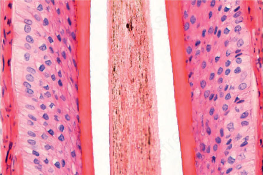
圖 22.21:終毛生長期毛髮,下段:在此垂直切面中,粉紅色的內根鞘(無核 anucleated)與外根鞘(有核 nucleated)形成清楚對比。毛小皮具有鋸齒狀邊緣,其方向與內根鞘相反。外根鞘的細胞因此層次顯著的細胞質內肝醣而具有透明的細胞質。(Terminal anagen hair, lower segment: the pink inner root sheath [anucleated] clearly contrasts with the outer root sheath [nucleated] in this vertical section. The cuticle has a serrated border orientated in the opposite direction to that of the inner root sheath. The cells of the outer root sheath have clear cytoplasm due to prominent intracytoplasmic glycogen at this level.)
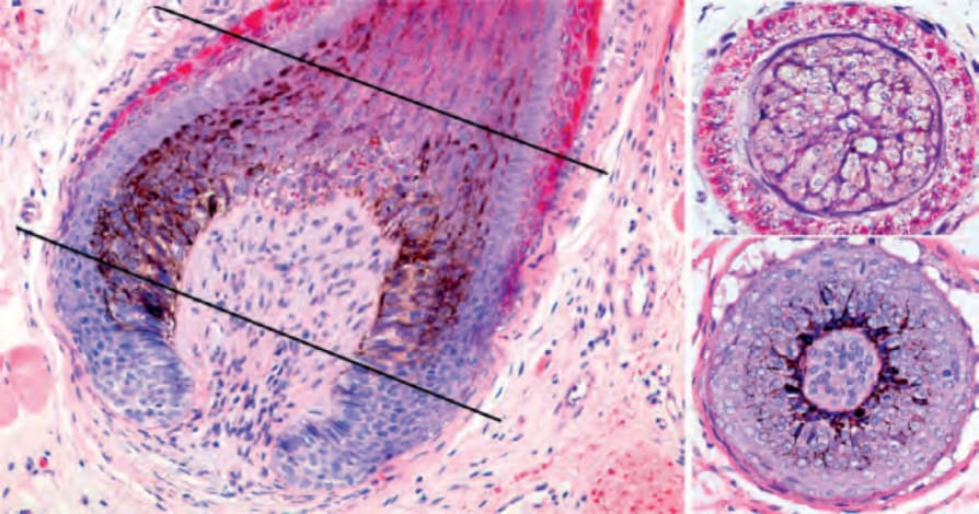
圖 22.22:終毛生長期毛髮:毛球與毛囊色素單位 (hair follicle pigmentary unit),垂直與水平切面。在垂直與水平切面中,注意基質上細胞 (supramatricial cells) 含有由圍繞真皮乳頭的樹突狀黑色素細胞 (dendritic melanocytes) 所轉移的細胞質內色素。後者由結締組織與血管所組成。(Terminal anagen hair: hair bulb and hair follicle pigmentary unit, vertical and horizontal sections. In the vertical and horizontal sections, note the supramatricial cells with intracytoplasmic pigmentation transferred from the dendritic melanocytes that surround the dermal papilla. The latter is composed of connective tissue and blood vessels.)
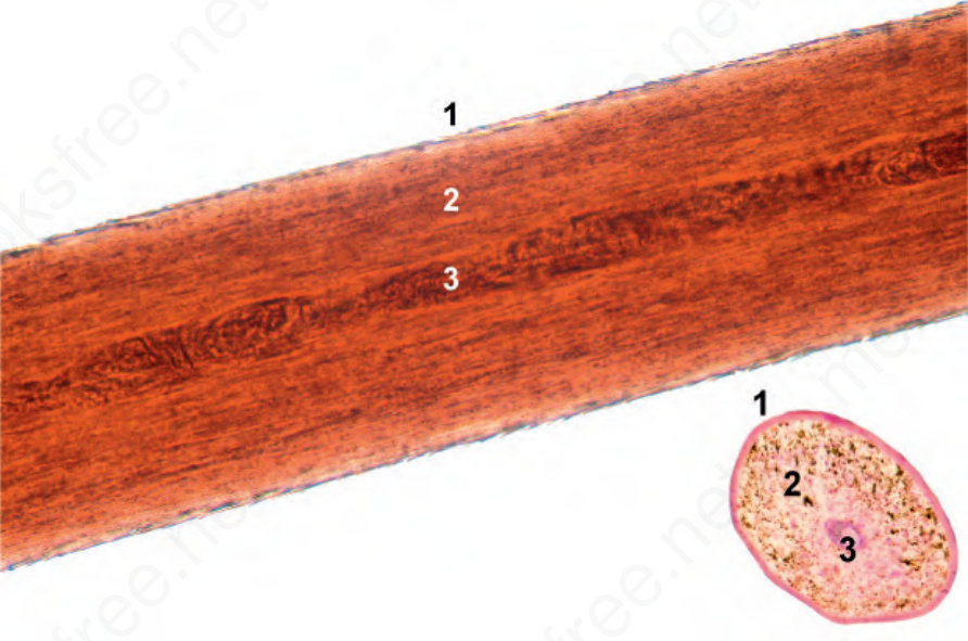
圖 22.23:毛幹:注意皮質 (2) 與毛小皮 (1)。有時,髓質容易被觀察到 (3)。(Hair shaft: Note the cortex [2] and the cuticle [1]. Sometimes, the medulla is easily visualized [3].)
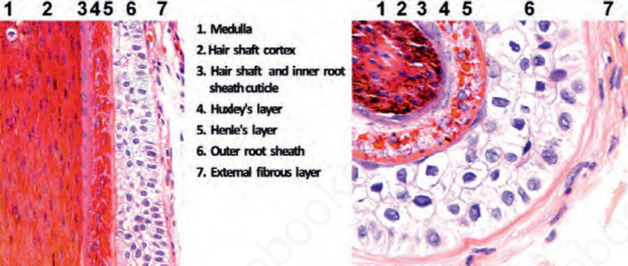
圖 22.24:終毛生長期毛囊:恰位於 Adamson 緣下方的水平與垂直切面。注意毛囊的不同層次。(1) 髓質 medulla,(2) 皮質 cortex,(3) 毛髮的毛小皮與內根鞘的毛小皮 cuticle of the hair and cuticle of the inner root sheath,(4) Huxley 層,(5) Henle 層,(6) 外根鞘 outer root sheath,(7) 玻璃膜層與外側纖維層(毛囊周圍結締組織鞘 perifollicular connective tissue sheath)。(Terminal anagen hair follicles: horizontal and vertical sections immediately below the Adamson fringe. Note the different layers of the hair follicle. [1] Medulla, [2] cortex, [3] cuticle of the hair and cuticle of the inner root sheath, [4] Huxley layer, [5] Henle layer, [6] outer root sheath, and [7] vitreous and external fibrous layer [perifollicular connective tissue sheath].)
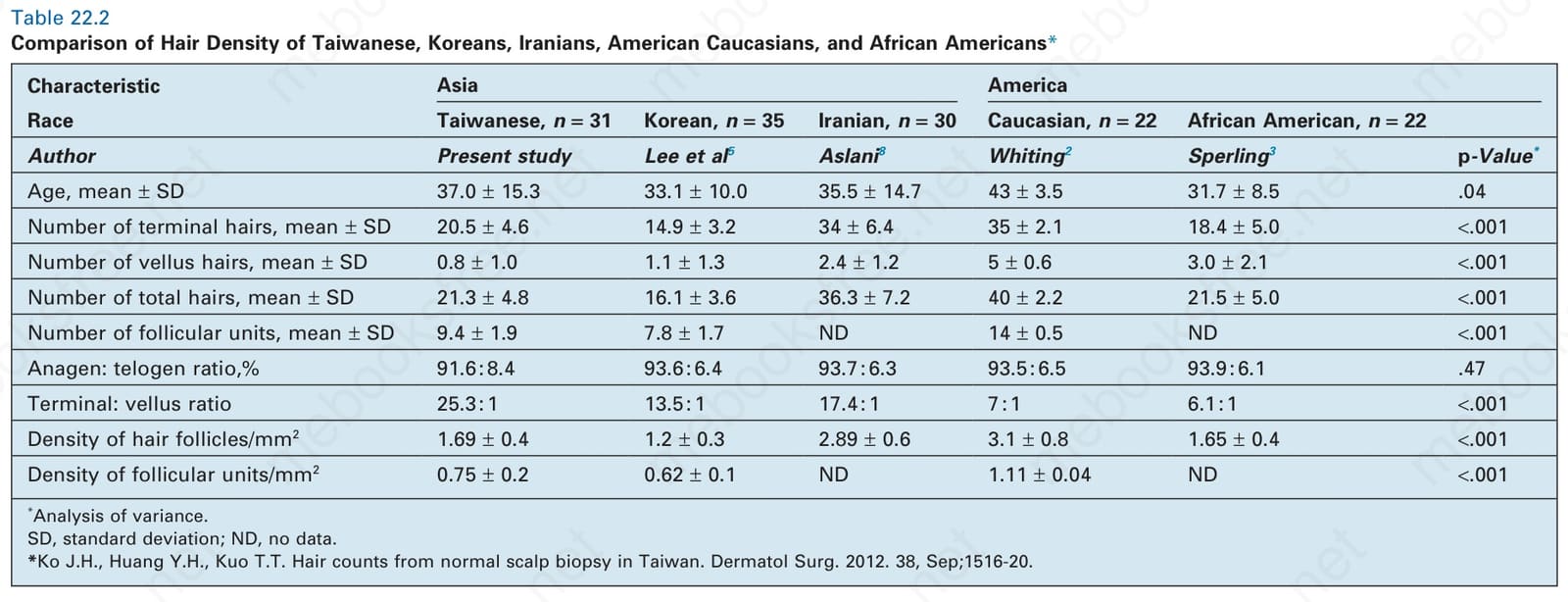
表 22.2:台灣人、韓國人、伊朗人、美國高加索人與非裔美國人毛髮密度之比較。(Comparison of Hair Density of Taiwanese, Koreans, Iranians, American Caucasians, and African Americans.)