Anatomy
Anatomy
To correctly evaluate a scalp biopsy, it is important for the pathologist to be well versed not only in the histology of the different types of alopecias but also in the complex and changing anatomy of the healthy hair follicle and its features in both vertical and horizontal sections. There are important morphological variations related to the normal follicular cycle and to the miniaturization of the terminal follicles in susceptible individuals. In the following section, these aspects will be individually reviewed.
of hair follicles do indeterminate follicles belong to. We concur with the suggestion of some authors who recommend assigning half of these hairs to terminal hair follicles and the other half to vellus hairs.4
The hair shaft and the inner root sheath are easily identified in vertical and horizontal sections at the level of the stem, immediately below the bulge, with conventional staining procedures including hematoxylin and eosin, toluidine blue, and elastic stains (Fig. 22.15 and see Fig. 22.13). The ratio between terminal and vellus hairs is approximately 7 : 1. Distinguishing between terminal and vellus hair follicles is critical for the diagnosis of androgenetic and temporal triangular alopecia. A ratio of 4 : 1 or less is suggestive of androgenetic alopecia. By definition, hair follicles and vellus hairs do not reach the subcutaneous tissue; therefore, to determine the final number of hair follicles and in particular vellus hair, superficial sections must be done at the level of the isthmus. For this reason, the quantification of follicles in deep and superficial sections is different. Superficial sections may reveal five to six more follicles than those counted in deep sections. As previously mentioned, the ratio of terminal follicles to vellus hairs may vary between different ethnic groups (Table 22.2).5 It is important to bear in mind that the hair shaft is frequently lost during the biopsy process. In these cases, its size may be extrapolated from the diameter of the empty space delimited by the inner root sheath.
Horizontal sections of the upper segment of the hair follicle show that hair follicles in the scalp are grouped, forming anatomic structures known as follicular units, which are composed of terminal and vellus hairs, sebaceous glands, and arrector pili muscles. The distribution of hair follicles in follicular units is better appreciated at the level of the infundibulum and isthmus. The connective tissue surrounding the outer root sheath tends to condense around discrete units comprising three to six hair follicles, one or two of which represent vellus hairs (Fig. 22.11 and also see Fig. 22.6).
Terminal hair follicles are easy to recognize in horizontal sections because they are much larger than vellus hairs (Fig. 22.12). These follicles are thick, pigmented, and usually descend to the subcutaneous fat (see Fig. 22.8). They contain a hair shaft, the diameter of which is greater than the thickness of the inner root sheath, and generally measures in excess of 0.06 mm (Fig. 22.13 and also see Fig. 22.6).
The hair follicle can be anatomically and functionally divided into two distinct segments. Differences between these two elements are explained by the fact that the upper portion of the hair follicle is very stable and not affected by maturation and shedding of the hair. The lower portion of the hair follicle is actively involved in hair growth and undergoes considerable morphological change according to its stage in the hair cycle.
The upper segment of the hair follicle consists of the follicular ostium, the infundibulum, and the isthmus (Fig. 22.16). The ostium is the physical orifice through which hair stems usually emerge in groups of two or three. The loss of it is clinically observed in scarring alopecias. The infundibulum extends from the ostium downwards to the opening of the sebaceous gland duct. It is cone shaped, its walls formed by the epidermis, and normally, around it there is mild concentric fibrosis and an inflammatory lymphocyte infiltrate, particularly seen in people of African descent. This phenomenon may cause confusion when evaluating inflammatory scarring alopecias (Fig. 22.17). 6
True vellus hairs are thin, short, and nonpigmented, and are characterized by a hair bulb that only extends to the upper or mid-reticular dermis (Fig. 22.14). The arrector pili muscle is usually undetected. The hair shaft diameter is equal to or less than the thickness of the inner root sheath (Fig. 22.15). It is typically less than 0.03 mm.
Vellus-like hairs (miniaturized hairs) are very similar to vellus hairs. They correspond to terminal hair follicles that have miniaturized owing to the effect of androgens. They may be very difficult to differentiate from true vellus hairs, except for the fact that the miniaturized follicles leave a trail of stellate collapsed fibrous tissue, the follicular stellae. In a scalp biopsy, when reference is made to vellus hairs without qualifying its origin, it refers by convention to both miniaturized and to true vellus hairs.
Indeterminate hairs have an intermediate morphology between that of terminal and vellus hairs and are considered an intermediate step in the process of miniaturization. The diameter of an indeterminate hair shaft is between 0.03 and 0.06 mm. There is no clear consensus as to which group
The isthmus continues from the opening of the sebaceous gland duct to the site of attachment of the arrector pili muscle at the hair bulge (Fig. 22.18). The arrector pili muscle attaches to the bulge area through elastic tendons and extends to its upper attachment in the adjacent papillary dermis.
The lower segment extends from the arrector pili insertion to the lower part of the hair bulb and has two components: the stem (suprabulbar zone)
1058 Diseases of the hair
Terminal anagen hair Vellus hair
A B
1059 Hair cycle
A B
C D
that extends from the arrector pili insertion to the Adamson fringe and the lower portion called the bulb (Figs 22.19 and 22.20). In a hair follicle in anagen, the stem is the longest structure in the hair follicle with a clearly differentiated internal and external radicular sheath clearly (Fig. 22.21). The bulb comprises the matrix cells and the basal melanocytes that surround the dermal papilla (Fig. 22.22).7–9
A hair shaft is composed of three layers: the medulla, the cortex, and the cuticle. The medulla, which represents the central region of the hair shaft, is not consistently present in humans, but it is often an important component of hair in other animals (Fig. 22.23). The cortex is the thickest layer and is responsible for the strength of the shaft. It is composed of intermediate filaments of hard keratin that are arranged in microfibrils, which intertwine to form cable-like structures called macrofibrils. The cuticle, which constitutes the outer part of the shaft, is responsible for the resistance to the wear and tear produced by the environment. It consists of scales orientated at right angles to the epidermis. These interlock with the cuticle of the internal root sheath maintaining integrity and in the bulbar zone appears as a single layer (Fig. 22.24 and see Fig. 22.21). The linear or curved shape of the hair is determined by the curved or linear shape of the internal radicular sheath. In horizontal sections, the hair in individuals of African descent is curved, elliptical, and eccentrically located within the follicular channel. In Caucasian individuals the hair is circular and centrally located within the follicular channel. In horizontal sections of a terminal anagen hair follicle, different layers can be identified in the suprabulbar area. From the center to the periphery these comprise:
• Hair shaft (including the medulla, the cortex, and the cuticle of the hair),
• Inner root sheath (including the cuticular layer of the inner root sheath, Huxley layer, Henle layer, and companion layer),
• Outer root sheath,
• Vitreous and external fibrous layer (perifollicular connective tissue sheath) (Fig. 22.24). These layers change noticeably depending on the level of the microscopic section. The cuticle of the hair shaft and all layers of the internal root sheath at the bulb level undergo trichohyaline keratinization. At the bulge, the inner root sheath starts to disappear and in the isthmus is replaced with trichilemmal keratin derived from the external root sheath (see Fig. 22.18).10
The external root sheath is continuous with the epidermis at the infundibulum and where it forms a granular layer with a basket weave arrangement of the epidermal keratin (see Fig. 22.17). Toward the bulb, the cells of the outer root sheath cells contain abundant glycogen (Fig. 22.21 and see Fig. 22.24).
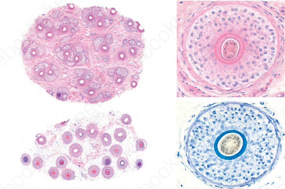
Fig. 22.6 Normal hair biopsy: horizontal section. On the left, all the hair follicles are visible at scanning magnification. On the right, all components of the hair follicle are seen in horizontal sections stained with H&E and toluidine blue.
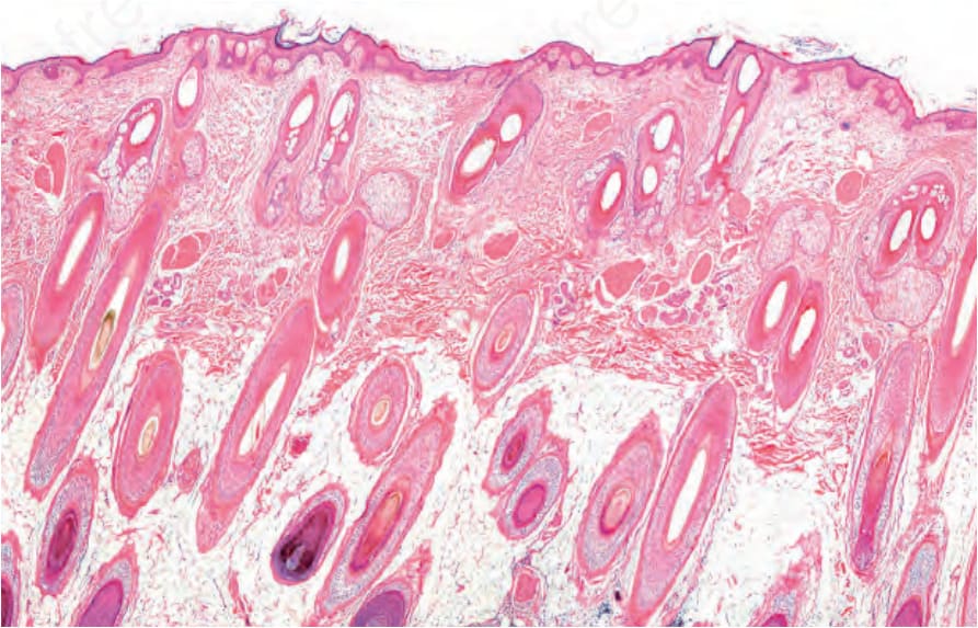
Fig. 22.8 Scalp biopsy: in this vertical section, no whole hair follicles are seen. Since they are all incomplete, additional sections are required to obtain an acceptable threedimensional reconstruction.
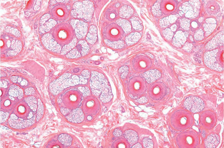
Fig. 22.11 Hair biopsy, horizontal section: the follicular units are clearly delineated by condensation of the adventitial collagen (Masson trichrome stain).
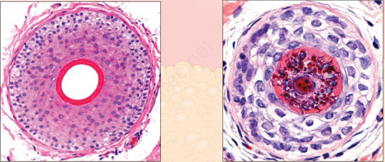
Fig. 22.12 Terminal anagen and vellus hair, vertical and horizontal sections: the terminal hair follicles reach the fat and are deeply embedded within it. The vellus hairs and miniaturized follicles are located within the dermis. Note the differences between the relative size of the hair shaft and the inner root sheath in terminal and vellus hairs. Courtesy of M. Mejia, MD, Universidad Pontificia Bolivariana, Medellín, Colombia.
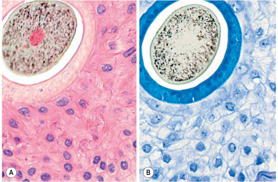
Fig. 22.13 Terminal anagen hair, horizontal section: compare the diameter of the hair shaft with the thickness of the thin internal root sheath (A) in pink with hematoxylin and eosin and in (B) dark blue with the toluidine blue stain.
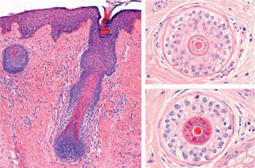
Fig. 22.14 Vellus hair, vertical and horizontal sections: short vellus hairs are located in the superficial dermis. In the horizontal sections, the diameter of the hair shaft is the same as or thinner than the inner root sheath.
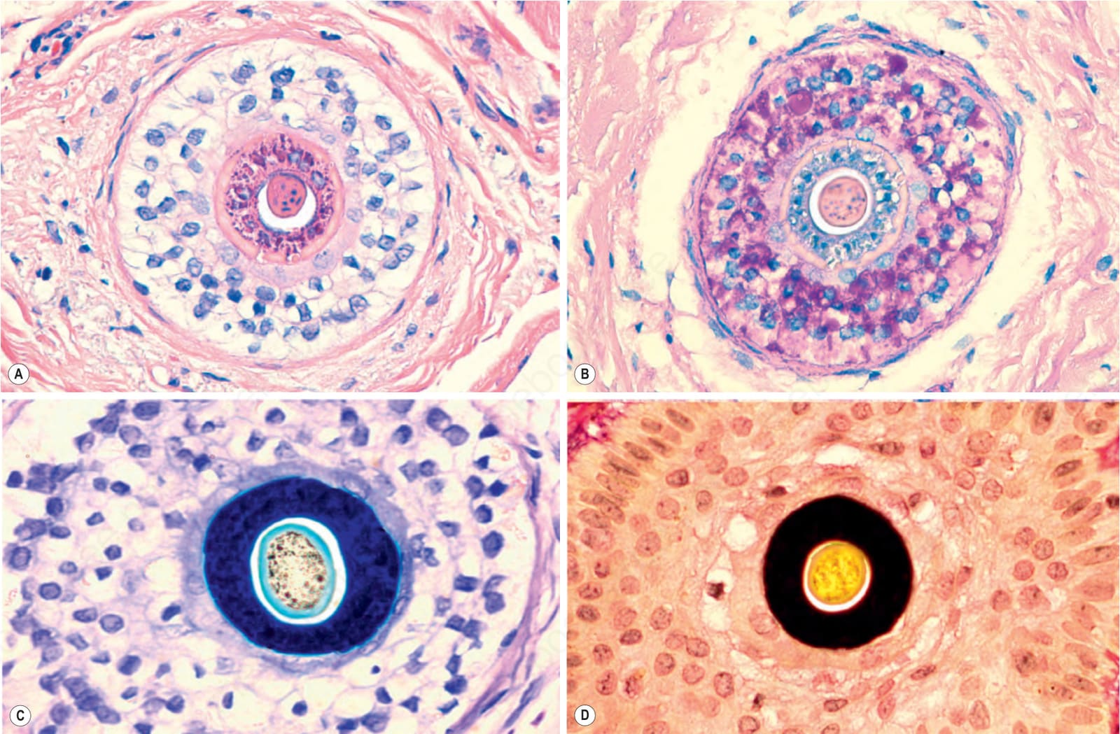
Fig. 22.15 Vellus hair, horizontal section: note the caliber of the hair shaft compared to the inner root sheath in: (A) pink, hematoxylin and eosin; (B) gray, PAS; (C) blue, toluidine blue; and (D) black, elastic tissue stain.
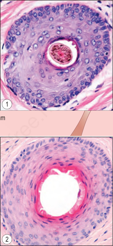
Fig. 22.16 Terminal anagen hair, vertical and horizontal sections of the upper segment: note the ostium, infundibulum and isthmus. The insets show horizontal sections through (1) the infundibulum and (2) the isthmus. The outer root sheath of the infundibulum is composed of squamous epithelium similar to the epidermis. Courtesy of M. Mejia, MD, Universidad Pontificia Bolivariana, Medellín, Colombia.
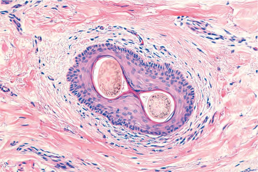
Fig. 22.17 Infundibulum. Horizontal section. The wall is made up of squamous stratified keratinized epithelium. The surrounding dermis is infiltrated by some lymphocytes. There are two hair shafts emerging from a single ostium.
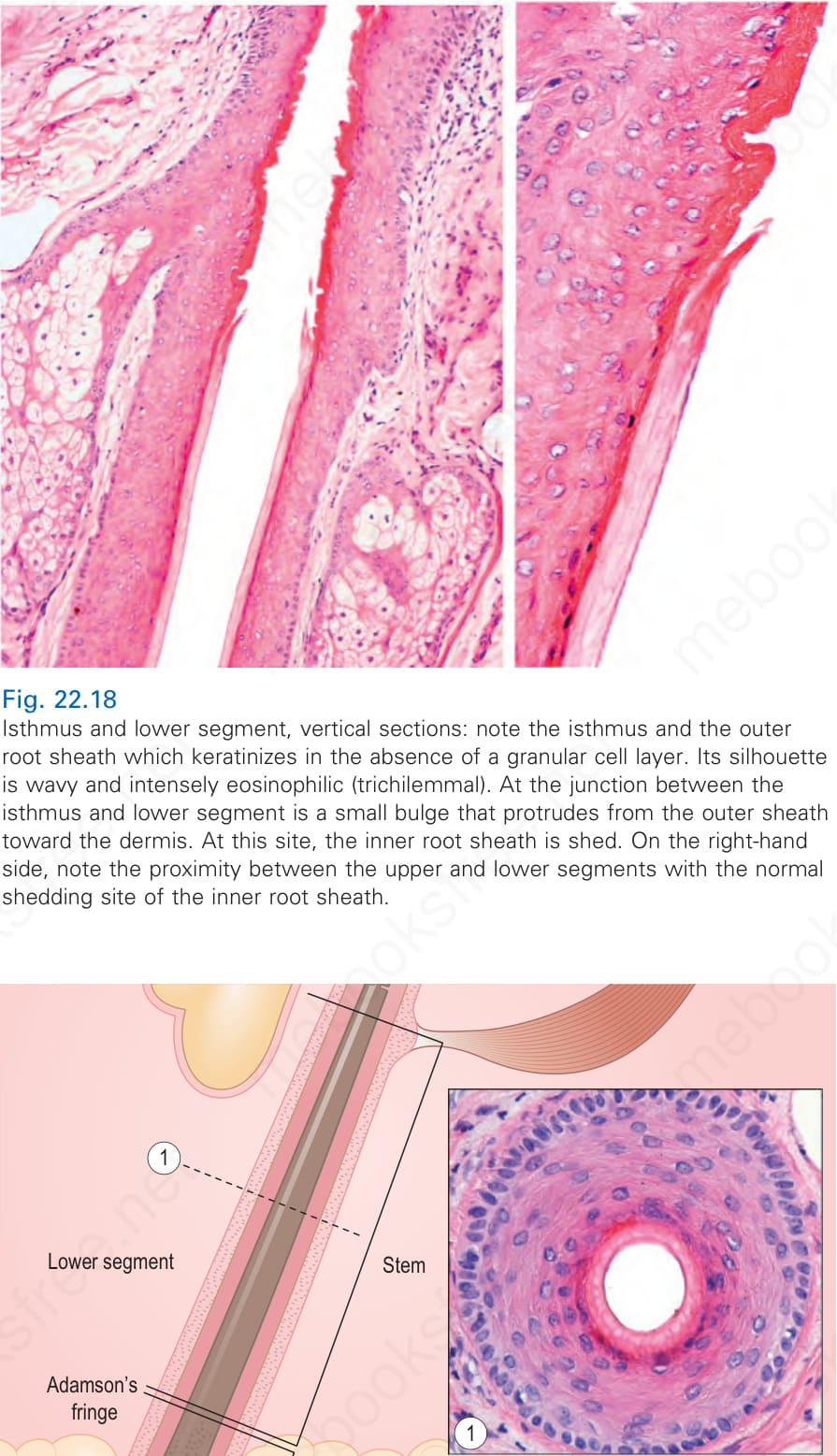
Fig. 22.18 Isthmus and lower segment, vertical sections: note the isthmus and the outer root sheath which keratinizes in the absence of a granular cell layer. Its silhouette is wavy and intensely eosinophilic (trichilemmal). At the junction between the isthmus and lower segment is a small bulge that protrudes from the outer sheath toward the dermis. At this site, the inner root sheath is shed. On the right-hand side, note the proximity between the upper and lower segments with the normal shedding site of the inner root sheath.
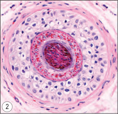
Fig. 22.19 Terminal anagen hair, vertical and horizontal sections of the lower segment: note the shaft and the hair bulb deep to Adamson fringe. The horizontal sections show (1) the stem and (2) the upper part of the bulb. Note the loss of nuclei in the hair shaft and the inner root sheath. Courtesy of M. Mejia, MD, Universidad Pontificia Bolivariana, Medellín, Colombia.
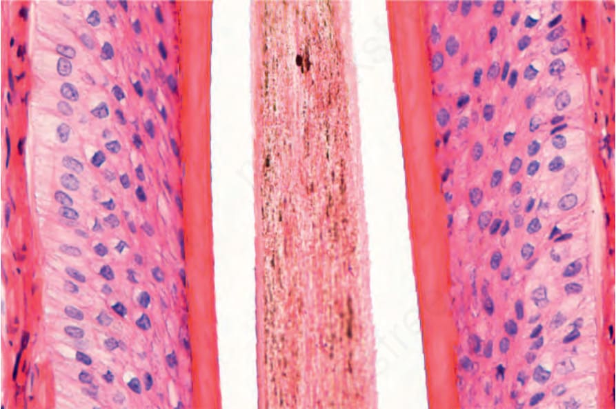
Fig. 22.21 Terminal anagen hair, lower segment: the pink inner root sheath (anucleated) clearly contrasts with the outer root sheath (nucleated) in this vertical section. The cuticle has a serrated border orientated in the opposite direction to that of the inner root sheath. The cells of the outer root sheath have clear cytoplasm due to prominent intracytoplasmic glycogen at this level.
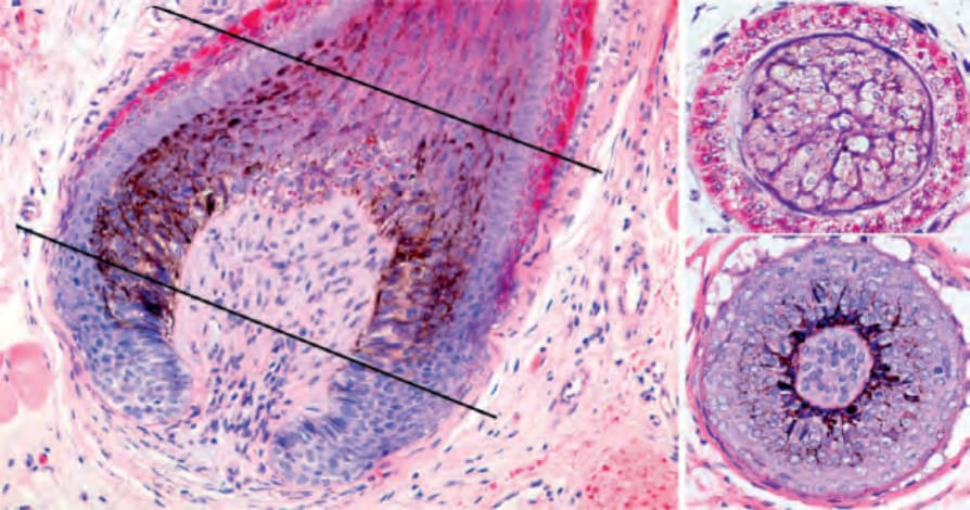
Fig. 22.22 Terminal anagen hair: hair bulb and hair follicle pigmentary unit, vertical and horizontal sections. In the vertical and horizontal sections, note the supramatricial cells with intracytoplasmic pigmentation transferred from the dendritic melanocytes that surround the dermal papilla. The latter is composed of connective tissue and blood vessels.
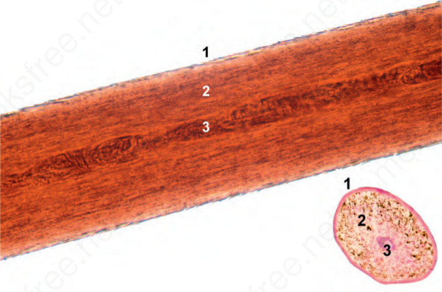
Fig. 22.23 Hair shaft: Note the cortex (2) and the cuticle (1). Sometimes, the medulla is easily visualized (3).
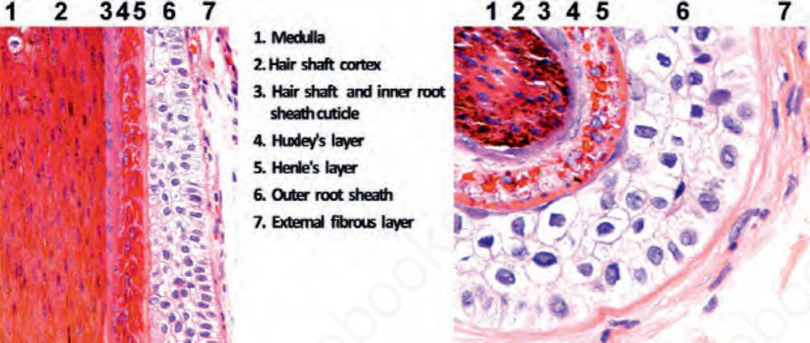
Fig. 22.24 Terminal anagen hair follicles: horizontal and vertical sections immediately below the Adamson fringe. Note the different layers of the hair follicle. (1) Medulla, (2) cortex, (3) cuticle of the hair and cuticle of the inner root sheath, (4) Huxley layer, (5) Henle layer, (6) outer root sheath, and (7) vitreous and external fibrous layer (perifollicular connective tissue sheath).
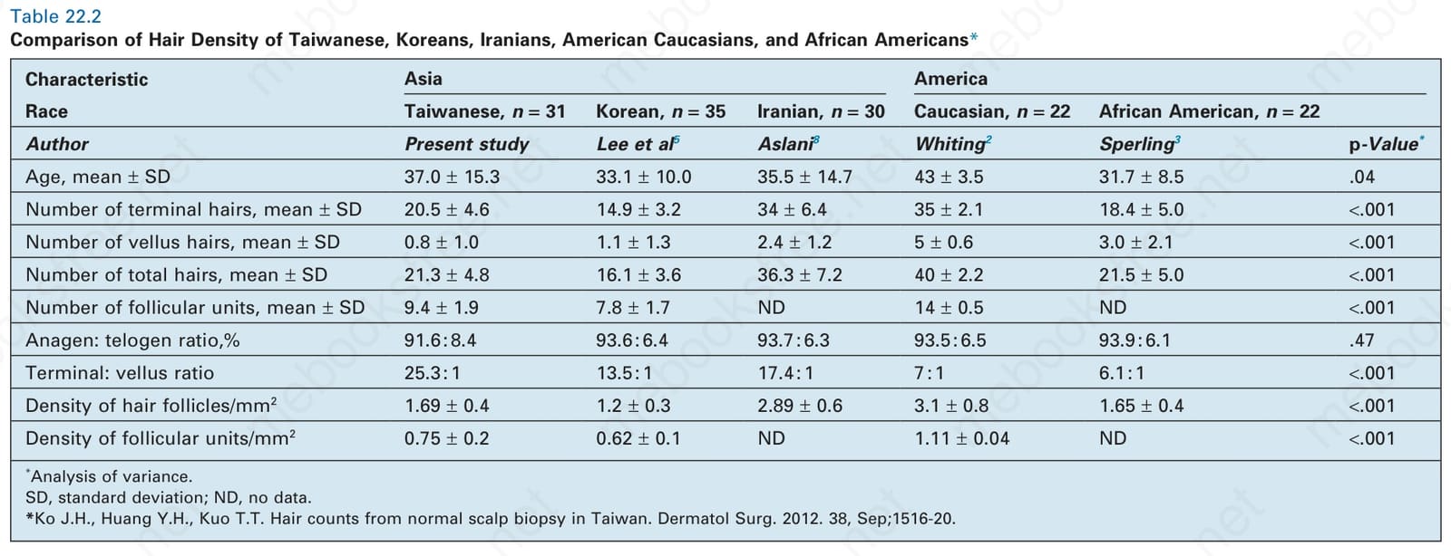
Table 22.2 Comparison of Hair Density of Taiwanese, Koreans, Iranians, American Caucasians, and African Americans*