臨床特徵 (Clinical Features)
- 足菌腫 (mycetoma)(希臘文 mykes,意為真菌;oma,意為腫瘤)是一種皮膚與皮下組織的慢性流膿性感染,特徵為多發性竇道 (sinus tracks) 與滲出液中出現顆粒 (granules)。
- 其可由細菌引起(放線菌性足菌腫 actinomycetoma)(例如 Nocardia,見前述),或(較少見地)由真菌引起(真菌性足菌腫 eumycetoma)。
- mycetoma 大致侷限於熱帶地區,主要分布在南緯 15° 與北緯 30° 之間(即所謂的「足菌腫帶 mycetoma belt」)。
- 此疾病最常發生於足部,但其他部位亦可受侵犯 (Fig. 18.345)。
- 病灶的形成必須經由輕微外傷反覆接種 (inoculation),最初表現為丘疹 (papule),逐漸增大而成為流膿性結節 (discharging nodule)。此過程會擴展至鄰近皮膚,且流膿性瘻管 (fistulae) 無法癒合 (Fig. 18.346)。
- 受侵犯區域因發炎與纖維化而變形,下方骨骼亦可能受累。
- mycetoma 最常見於 20 至 50 歲者,並呈現明顯的男性優勢。其與反覆性職業性外傷有關。
- 由真菌引起的 mycetoma 一般較細菌性病灶發炎程度較輕、侵犯較不深層。
致病機轉與組織學特徵 (Pathogenesis and Histologic Features)
- 最常見的真菌性致病原因包括:
- Madurella mycetomatis,
- Madurella grisea,
- Pseudallescheria boydii,
- Pyrenochaeta romeroi,
- Leptosphaeria senegalensis,
- Neotestudina rosatti。
- 細菌性致病原因包括 Nocardia、Actinomyces 與 Streptomyces 等屬。真菌性原因僅佔 mycetoma 病例中的少數。其中,M. mycetomatis 是全世界最重要者。
- 然而,一項來自印度、共 73 例的分析顯示,在 4 年期間內,馬杜拉菌性足菌腫 (maduromycotic mycetoma) 相對於放線菌性足菌腫 (actinomycotic mycetoma) 的比例呈下降趨勢。
- 在醫源性免疫抑制 (iatrogenically immunosuppressed) 的個體中,已有由 Scedosporium apiospermum(其有性世代 teleomorph Pseudallescheria boydii 的無性對應型)、Cladophialophora bantiana 與 Phaeoacremonium fuscum 所引起感染的記載。S. apiospermum 真菌性足菌腫 (eumycetoma) 亦可發生於免疫健全的宿主。另有一例關於 Diaporthe phaseolorum (Phomopsis phaseoli) 真菌性足菌腫的報告,發生於一名感染人類 T 細胞淋巴趨向性病毒 1 型 (human T-cell lymphotropic virus 1, HTLV-1) 的病人。
- 所有致病微生物均會產生顆粒 (granules),其構型與顏色可能有助於辨識致病原 (Figs 18.347–18.350; Table 18.3)。雖然透過培養進行確切的菌種鑑定 (speciation) 常具挑戰性,但較新的 PCR 技術頗具前景。
- M. mycetomatis 感染中所見顆粒的黑色,是由於黑色素 (melanin) 的產生所致,此可能對該微生物提供對抗抗真菌劑作用的保護。在編碼幾丁質分解酵素 chitotriosidase 的基因中之一個多型性 (polymorphism),已被連結至發生 M. mycetomatis 足菌腫風險增加。
- 顆粒見於化膿 (suppuration) 區域內,並被柵欄狀組織球 (palisaded histiocytes) 包圍。顆粒由排列有序、緊密成團的菌絲 (hyphae) 組成,可能伴隨黏附的嗜中性球 (neutrophils) 或結晶基質 (crystalline matrix)。在此之外可見多核巨細胞 (multinucleate giant cells),周邊則為一圈水腫性肉芽組織 (edematous granulation tissue)。
- 特殊染色的運用,對於區分放線菌性 (actinomycetic) 與真菌性 (eumycetic) 的足菌腫致病原具有價值。後者以 PAS 與銀染色 (silver stains) 呈陽性,但 Gram 染色呈陰性 (Fig. 18.351)。有時可見 Splendore-Hoeppli 現象 (Fig. 18.352)。
治療與預後 (Treatment & Prognosis)
- 對治療的反應不一,但當深層組織受侵犯時,病情更為棘手。
A
馬杜拉菌性(真菌性)Maduromycotic (eumycetic) 放線菌性 Actinomycetic
黑色 Black Madurella mycetomatis Madurella grisea Pyrenochaeta romeroi Phialophora jeanselmei Leptosphaeria senegalensis Leptosphaeria tompkinsii
黃色或黃白色 Yellow or yellowish-white
Allescheria boydii Acremonium sp. Fusarium sp. Neotestudina rosatti Actinomadura madurae Streptomyces somaliensis
B
紅色 Red Actinomadura pelletieri
白色或不可見 White or not visible
Nocardia brasiliensis Nocardia caviae Nocardia asteroides
經許可轉載自 Magaña, M. and Magaña-Garcia, M. (1989) Dermatologic Clinics, 7, 203–217。
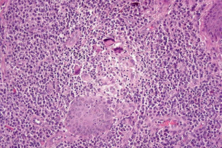
圖 18-343:著色芽生菌病 (chromoblastomycosis):常可見肉芽腫 (granulomata)。
Fig. 18.343 Chromoblastomycosis: granulomata are commonly present.
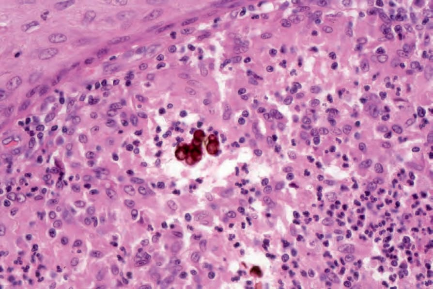
圖 18-344:著色芽生菌病 (chromoblastomycosis):呈棕色染色(有時有分隔)的細胞具有特異性診斷意義 (pathognomonic)。
Fig. 18.344 Chromoblastomycosis: the brown-staining (sometimes septate) cells are pathognomonic.
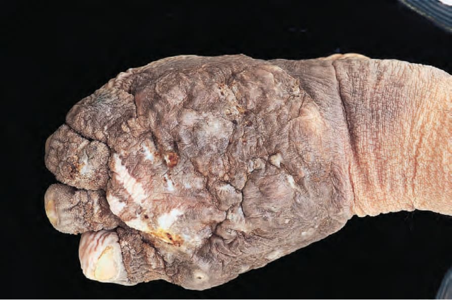
圖 18-345:足菌腫 (mycetoma):足部明顯腫脹且變形,存在多處引流性竇道 (draining sinuses)。
Fig. 18.345 Mycetoma: the foot is grossly swollen and misshapen. Numerous draining sinuses are present.
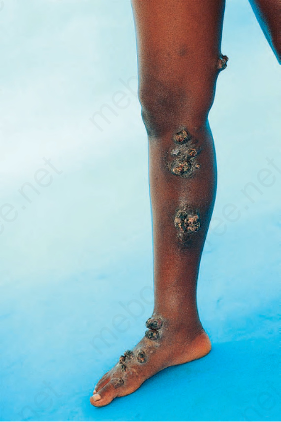
圖 18-346:足菌腫 (mycetoma):可見多發性潰瘍性疣狀結節 (ulcerated verrucous nodules)。承蒙 N.C. Dlova, MD, Nelson R. Mandela School of Medicine, University of KwaZulu-Natal, South Africa 提供。
Fig. 18.346 Mycetoma: multiple ulcerated verrucous nodules are present. By courtesy of N.C. Dlova, MD, Nelson R. Mandela School of Medicine, University of KwaZulu-Natal, South Africa.
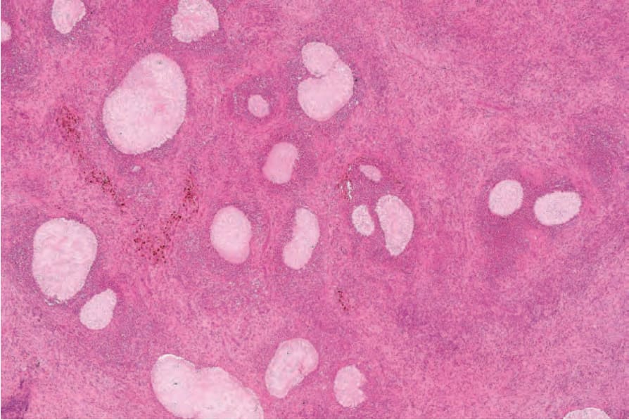
圖 18-347:足菌腫 (mycetoma):特徵性顆粒 (granules) 出現於嗜中性球膿瘍 (neutrophil abscesses) 內,周邊有組織球/巨細胞柵欄狀排列 (histiocytic/giant cell palisade)。
Fig. 18.347 Mycetoma: characteristic granules are present in neutrophil abscesses. There is a peripheral histiocytic/giant cell palisade.
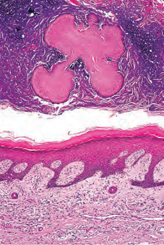
圖 18-348:足菌腫 (mycetoma):本例中,在上覆痂皮 (crust) 內可見一菌落 (colony)。
Fig. 18.348 Mycetoma: in this example, a colony is present in the overlying crust.
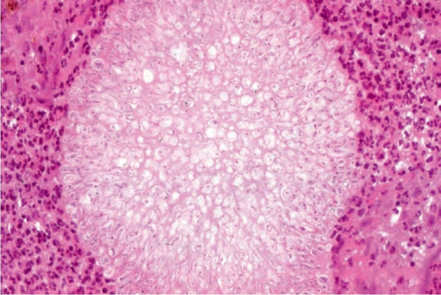
圖 18-349:足菌腫 (mycetoma):本高倍視野中可清楚見到顆粒 (granule) 的內部結構。
Fig. 18.349 Mycetoma: the internal structure of the granule is clearly visible in this high-power view.
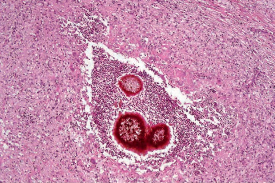
圖 18-350:足菌腫 (mycetoma):色素性顆粒 (pigmented granules) 為 Madurella mycetomatis 感染的特徵。
Fig. 18.350 Mycetoma: pigmented granules are characteristic of Madurella mycetomatis infection.
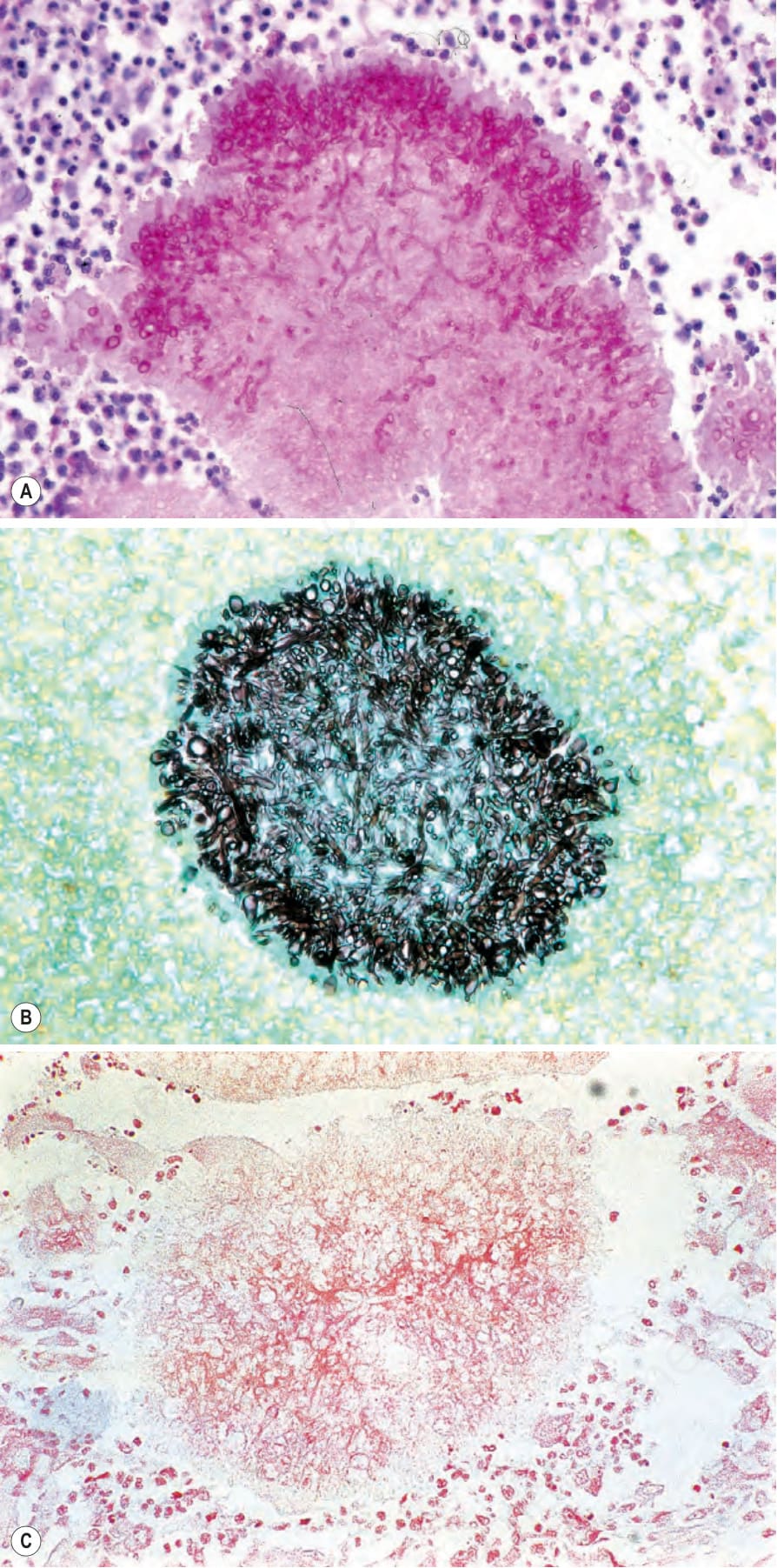
圖 18-351:(A–C) 足菌腫 (mycetoma):特殊染色的運用可輕易確認此變異型的真菌性本質。(A) periodic acid-Schiff;(B) methenamine silver;(C) Gram。
Fig. 18.351 (A–C) Mycetoma: the use of special stains readily confirms the fungal nature of this variant. (A) Periodic acid-Schiff; (B) methenamine silver; (C) Gram.
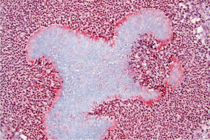
圖 18-352:足菌腫 (mycetoma):此 fibrin 染色凸顯出 Splendore-Hoeppli 現象。
Fig. 18.352 Mycetoma: this fibrin stain highlights the Splendore-Hoeppli phenomenon.
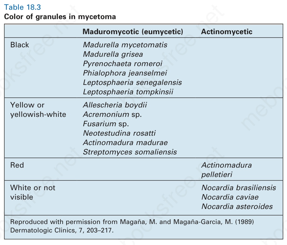
表 18-3:足菌腫顆粒的顏色 (color of granules in mycetoma)。
Table 18.3 Color of granules in mycetoma