臨床特徵 (Clinical Features)
桿菌性血管瘤病 (bacillary angiomatosis, BA) 是一種血管增生性 (vasoproliferative) 病灶,容易與化膿性肉芽腫 (pyogenic granuloma) 或卡波西氏肉瘤 (Kaposi sarcoma) 混淆,主要 (但非僅限) 見於皮膚。病灶亦曾於骨骼、軟組織、肝臟、淋巴結與脾臟被描述。病人可能出現全身性表現,包括發燒、倦怠 (malaise)、肝脾腫大 (hepatosplenomegaly) 與淋巴結病變 (lymphadenopathy)。雖然此病最初被認為是 AIDS 特有的疾病,但也曾在其他免疫低下狀態 (例如腎臟移植受贈者) 甚至外觀正常的個體中被描述。病人表現為廣泛、數量眾多 (有時達數百個) 的血紅色、表面平滑的淺表丘疹,以及膚色或暗紫色的皮下結節 (Figs 18.130 and 18.131)。此病可由 Bartonella henselae (引起貓抓病 (cat scratch disease) 的病原體) 所致,或較少見地由 B. quintana (引起戰壕熱 (trench fever) 的病原體) 所致。
致病機轉與組織病理特徵 (Pathogenesis and Histologic Features)
B. henselae 感染是經由貓咬或貓抓、或貓蚤叮咬而獲得,而 B. quintana 感染則是經由體蝨 (body lice) 傳播。然而,BA 病人似乎很少能夠佐證 (corroborate) 此類病史。一項來自約翰尼斯堡 (Johannesburg) 的 PCR 為基礎之研究顯示,在某 HIV 門診的 188 名就診者中,Bartonella 菌血症 (bacteremia) 的比率為 10%,其中僅一人表現出 BA 的臨床特徵。在病原體初步於紅血球內細胞內定殖 (intracellular colonization) 後,血管內皮細胞 (vascular endothelial cells) 是其主要的標靶。Bartonella 的 VirB 第四型分泌系統 (VirB type IV secretion system) 不僅在建立紅血球內感染 (intraerythrocytic infection) 方面、也在介導該病原體與內皮細胞之交互作用方面扮演關鍵角色。血管新生 (angiogenesis) 是經由多種機轉的組合所增強,包括抑制細胞凋亡 (apoptosis)、釋放趨化激素 (chemokines) 如 IL-8、以及活化缺氧誘導因子-1 (hypoxia-inducible factor-1, HIF-1)。
組織學顯示由微血管 (capillaries) 組成的小葉 (lobules),伴隨明顯、常呈立方形 (cuboidal) 的血管內皮細胞,有時圍繞著擴張 (ectatic) 的血管,其間散布著呈現白血球碎裂 (leukocytoclasis) 的嗜中性多形核白血球 (neutrophil polymorphs) 以及紫色的桿菌顆粒 (purplish granules of bacilli),後者以 Warthin-Starry 反應最易辨識 (Figs 18.132–18.135)。Giemsa 染色亦可用於辨識這些病原體,但由於上述兩種染色都難以判讀且常難以操作,已發展出一種 PCR 方法 (見下文)。有時可見實質性的內皮細胞增生 (solid endothelial cell proliferation)。可能出現異型性 (atypia) 與有絲分裂象 (mitoses)。淺表病灶呈息肉樣 (polypoid) 構造,且可能伴隨類似化膿性肉芽腫的表皮領圈 (epidermal collarette)。偶爾可見潰瘍。亦曾描述相關的假上皮瘤樣表皮增生 (pseudoepitheliomatous epidermal hyperplasia)。
雖然在某些病灶的周邊可見到梭形內皮細胞 (spindled endothelial cells) 切割膠原蛋白 (collagen dissection) 的現象,但在卡波西氏肉瘤 (Kaposi sarcoma) 中所見到的含鐵血黃素沉積 (hemosiderin deposition) 與透明小球 (hyaline globules) 在此並不明顯。晚期、退化中 (involuting) 的病灶顯示血管化真皮 (vascularized dermis) 的廣泛纖維化,以及極少的伴隨核碎裂 (karyorrhexis) 的多形核白血球浸潤。此類病例需要高度的懷疑指數 (high index of suspicion),因為細菌可能難以呈現。由於此病大多數病人為免疫低下的 HIV 感染個體,謹慎的做法是仔細檢查切片以尋找其他伺機性病原體 (opportunistic pathogens),例如 CMV 或分枝桿菌 (mycobacteria)。
內皮細胞可以用對抗第八凝血因子相關抗原 (factor VIII-related antigen)、CD31 與 CD34 的抗體進行標記。組織學上,肝臟與脾臟的侵犯表現為紫斑病 (peliosis)。然而,典型的細菌亦同時存在。辨識此感染並將其與卡波西氏肉瘤及其他血管增生性病灶區別開來極為重要,特別是因為它很容易對抗生素治療有反應。
在超微結構 (ultrastructurally) 上,這些病原體呈現為真皮內的桿菌聚集體 (aggregates of bacilli)。這些細菌具有三層 (trilaminar) 細胞壁。
鑑別診斷 (Differential Diagnosis)
BA 必須與祕魯疣 (verruga peruana)、化膿性肉芽腫 (pyogenic granuloma)、上皮樣血管瘤 (epithelioid hemangioma) 與卡波西氏肉瘤 (Kaposi sarcoma) 區別。化膿性肉芽腫與 Bartonella 感染無關。尤其是卡波西氏肉瘤的小葉狀微血管瘤 (lobular capillary hemangioma;即化膿性肉芽腫) 樣變異型可能與 BA 混淆。雖然罕見,但曾有 BA 與卡波西氏肉瘤併存的描述。以 PCR 或免疫組織化學檢測 HHV-8 是區別卡波西氏肉瘤與 BA 的有用方法;前者一律與 HHV-8 相關,而後者已被發現為 HHV-8 陰性。此外,在疑似 BA 的病例中,由於這些病原體難以培養,可使用 PCR 來確認 Bartonella spp. 的存在。
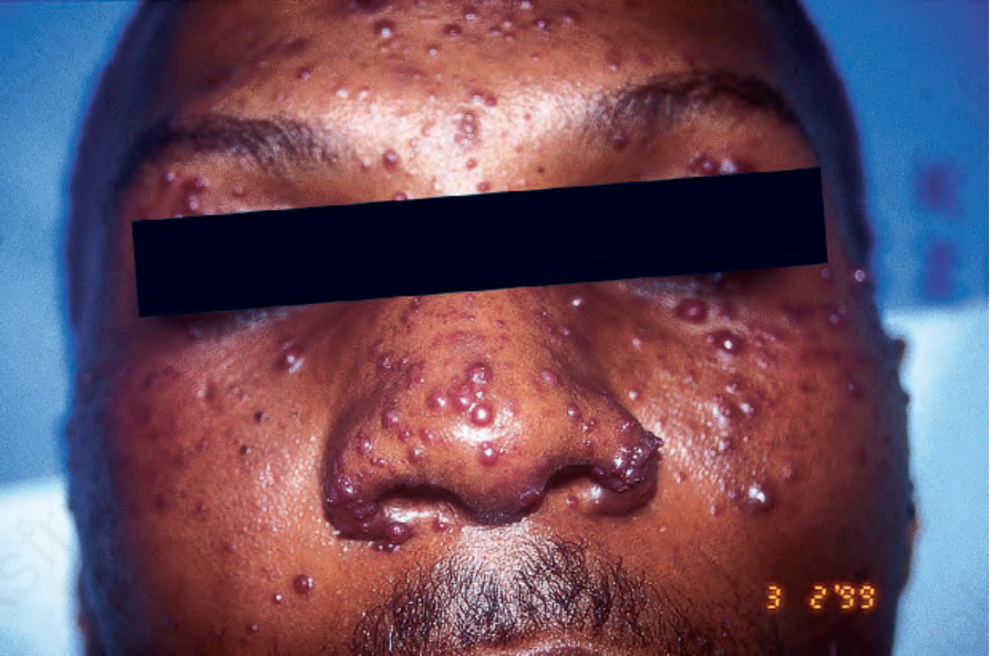
圖 18-130:桿菌性血管瘤病 (bacillary angiomatosis):可見眾多丘疹與結節。By courtesy of N.C. Dlova, MD, Nelson R. Mandela School of Medicine, University of KwaZulu-Natal, South Africa.
Fig. 18.130 Bacillary angiomatosis: numerous papules and nodules are present. By courtesy of N.C. Dlova, MD, Nelson R. Mandela School of Medicine, University of KwaZulu-Natal, South Africa.
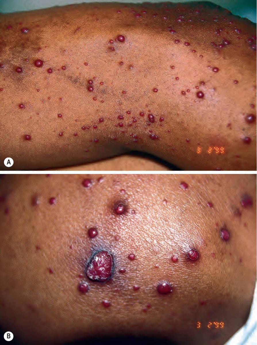
圖 18-131:(A, B) 桿菌性血管瘤病 (bacillary angiomatosis):鮮紅色的著色是其特徵。By courtesy of N.C. Dlova, MD, Nelson R. Mandela School of Medicine, University of KwaZulu-Natal, South Africa.
Fig. 18.131 (A, B) Bacillary angiomatosis: the bright red coloration is characteristic. By courtesy of N.C. Dlova, MD, Nelson R. Mandela School of Medicine, University of KwaZulu-Natal, South Africa.
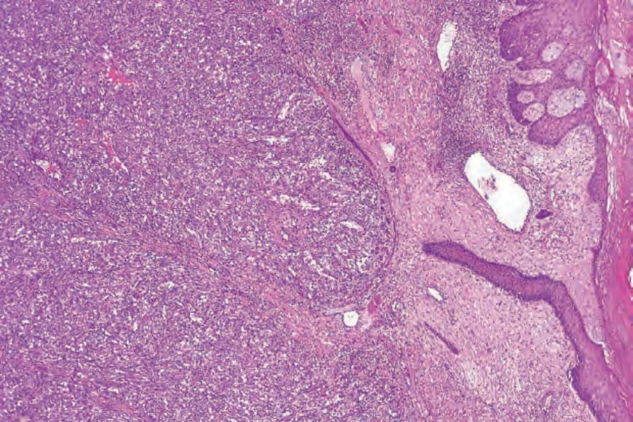
圖 18-132:桿菌性血管瘤病 (bacillary angiomatosis):可見緻密的結節狀微血管增生病灶 (nodular capillary proliferative lesion);注意擴張的血管 (ectatic vessels)。
Fig. 18.132 Bacillary angiomatosis: there is a dense nodular capillary proliferative lesion; note the ectatic vessels.
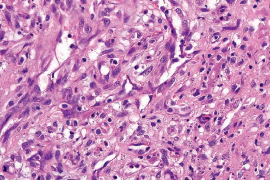
圖 18-133:桿菌性血管瘤病 (bacillary angiomatosis):內皮細胞 (endothelial cells) 腫脹。可見明顯的嗜中性球 (neutrophils)。
Fig. 18.133 Bacillary angiomatosis: the endothelial cells are swollen. Conspicuous neutrophils are evident.
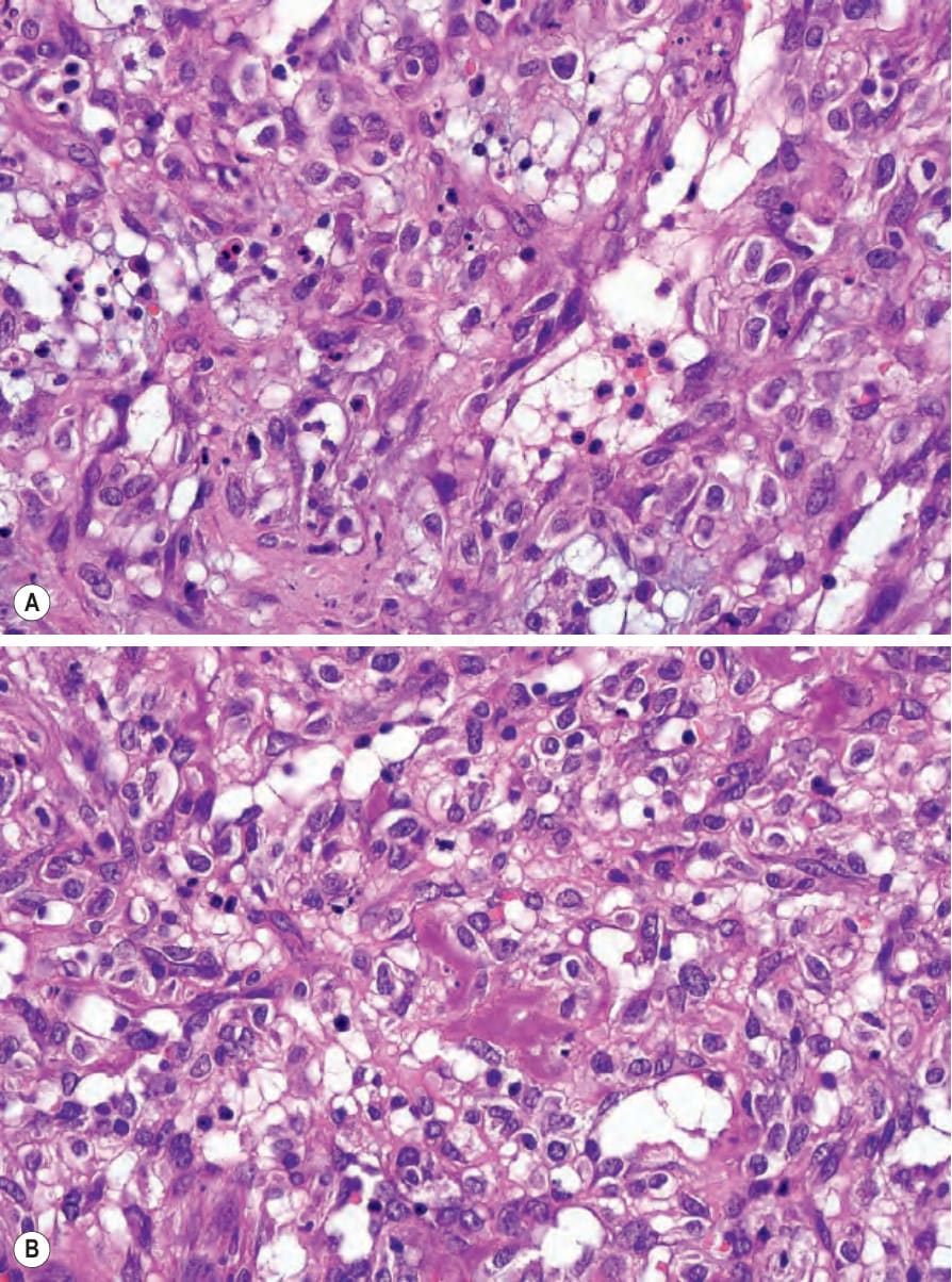
圖 18-134:(A, B) 桿菌性血管瘤病 (bacillary angiomatosis):亦可見淋巴球 (lymphocytes) 與組織球 (histiocytes)。注意視野中央紫色的細菌菌落 (purple colony of bacteria)。
Fig. 18.134 (A, B) Bacillary angiomatosis: lymphocytes and histiocytes are also present. Note the purple colony of bacteria in the center of the field.
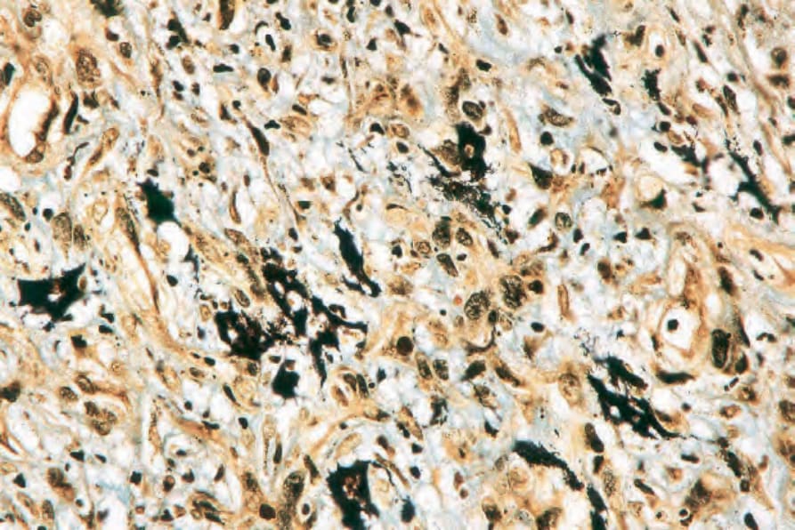
圖 18-135:桿菌性血管瘤病 (bacillary angiomatosis):以 Warthin-Starry 染色可輕易辨識這些病原體。
Fig. 18.135 Bacillary angiomatosis: the organisms are easily identified with the Warthin-Starry stain.
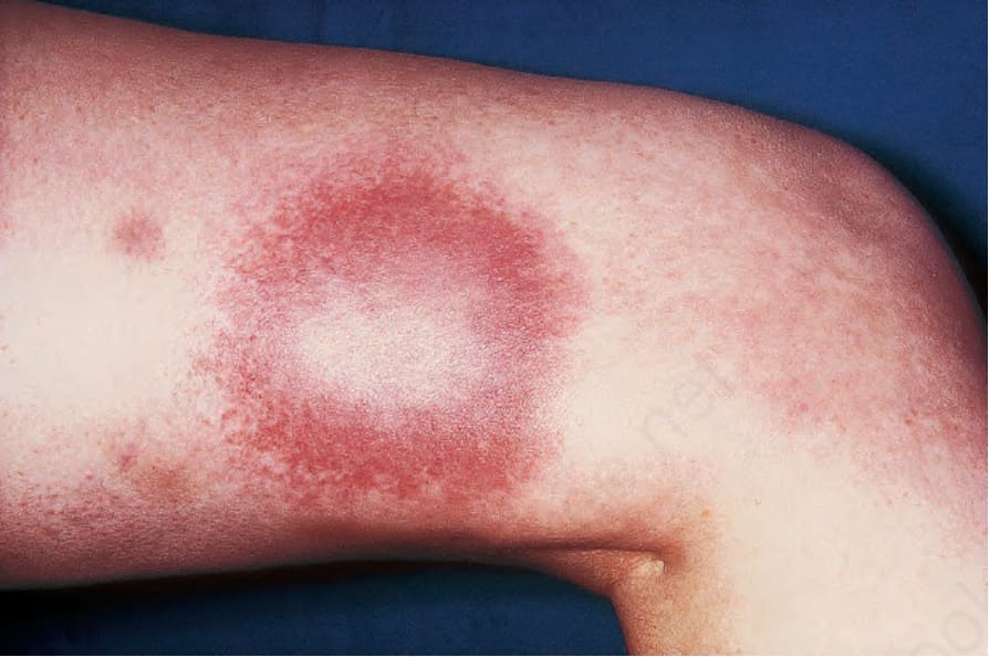
圖 18-136:萊姆病 (Lyme disease):此環狀、紅斑性病灶於蜱叮咬部位周圍 (數週後) 出現。By courtesy of R.A. Marsden, MD, St George’s Hospital, London, UK.
Fig. 18.136 Lyme disease: this annular, erythematous lesion developed (several weeks later) around the site of a tick bite. By courtesy of R.A. Marsden, MD, St George’s Hospital, London, UK.