尖型濕疣 (Condyloma acuminatum)
臨床特徵 (Clinical Features)
-
大多數有性行為的人,一生中至少會有一次可檢出的 HPV 感染。估計每年約有 1400 萬人感染生殖器 HPV。近期一項針對肛門生殖器疣 (anogenital warts) 的系統性文獻回顧顯示,男性與女性的發生率中位數分別為每 10 萬人中 137 與 120.5 例。
-
……除了與 HIV 感染相關者以外。它們可能在數週或數月後自發消退,也可能持續數年。消退的徵象包括搔癢、紅斑、水腫外觀、去色素暈 (depigmented haloes),以及出現多發性微小的扁平疣 (plane warts)。細胞媒介免疫 (cell-mediated immunity) 在免疫健全 (immunocompetent) 個體中扁平疣的自發消退中扮演關鍵角色。在接受高效能抗反轉錄病毒治療 (highly active ART) 的 HIV 感染病人中,多發性扁平疣可能作為免疫重建發炎症候群 (immune reconstitution inflammatory syndrome, IRIS) 的皮膚表現而出現。曾有報告指出臉部雷射換膚 (facial laser resurfacing) 後病灶惡化。
-
Condylomata acuminata 特別由 HPV 第 2、6、11、16、18、30–33、35、39、41–45、51–56 及 59 型所引起,並隨性交伴隨的創傷而發展。據報導由女性傳染給男性的情形較男性傳染給女性者更為常見。單單 HPV6 與 11 即占這些病灶的 90% 以上,其中 HPV6 出現於約三分之二的病例,其餘三分之一則由 HPV11 所引起。潛伏期變異不定(通常介於 2 至 3 個月之間)。Condylomata acuminata 發生於陰莖龜頭與包皮或陰莖體,呈柔軟、肉質、有時呈絲狀的斑塊,並可延伸至尿道口 (meatus)(圖 18.19 與 18.20)。在陰莖體上,它們較不外突 (exophytic)。外陰病灶可能體積龐大且浸軟 (macerated),並可延伸至陰道口 (introitus)(圖 18.21)。類似的肉質與絲狀柔軟腫塊也發生於肛門周圍,較常見於男性(圖 18.22)。在相當比例的病例中,已顯示肛門鱗狀細胞癌 (anal squamous carcinoma) 含有 HPV6、16 及 18(圖 18.23)。局部復發率約為 30%。這些病灶在兒童中少見(在兒童中可能是性虐待的徵象),最常見於年輕成人(第二與第三個十年),且經常合併其他生殖器感染。兒童的 condylomata 在超過 50% 的病例中會自發消退。生殖器疣在 HIV 感染個體中常見。
-
需要注意的是,相當比例的生殖器 HPV 感染是無症狀的。已顯示罹患 condyloma acuminata 男性病人的女性伴侶,其子宮頸 HPV 感染與上皮內腫瘤 (intraepithelial neoplasia)(鱗狀上皮內病變/子宮頸上皮內腫瘤 [squamous intraepithelial lesion/cervical intraepithelial neoplasia, SIL/CIN])的風險增加。與既存 condylomata acuminata 相關的子宮頸腫瘤,至少在某些病人中也與免疫抑制 (immunosuppression) 的背景有關。據報導,全世界子宮頸癌中的 HPV 盛行率為 99.7%。HPV16、18、31–33、35、39、42 及 51–54 最常與子宮頸、外陰及陰莖的癌症相關。罹患 condylomata acuminata 的病人不僅發生外陰、陰道、陰莖及肛門癌的風險增加,發生某些非肛門生殖器鱗狀細胞癌 (nonanogenital squamous cell carcinomas) 的風險也增加。針對 HPV 第 6、11、16 及 18 型常規施打四價疫苗 (quadrivalent vaccine),已使外陰及子宮頸癌、生殖器疣以及肛門生殖器上皮內腫瘤的疾病負擔顯著減少。
-
偶爾可能罕見地遇到一種大型、增生旺盛且局部破壞性的 condyloma 變異型(Buschke-Löwenstein tumor)(圖 18.24)。此變異與 HPV 第 6、11 或 16 型相關。此巨大變異型很可能代表 verrucous carcinoma 的一種變異型,但此議題一直具有爭議性(見第 22 章)。含有 HPV6 與 11 的幼年喉部乳頭瘤 (juvenile laryngeal papillomas) 可見於患有 condylomata acuminata 母親所生的兒童。若接受放射線照射,它們可能出現惡性進展。
-
Condyloma acuminatum 的惡性轉化並不常見,但較其他與 HPV 相關的病灶(除 EV 外)更為常見。
組織病理特徵 (Histopathology)
-
扁平疣 (plane warts) 呈棘層肥厚 (acanthotic),並顯示正角化 (orthokeratosis),伴隨令人聯想到「雞籠網 (chicken wire)」的開放型態(「籃網狀 (basket weave)」過度角化 [hyperkeratosis])。角化不全 (parakeratosis) 並非其特徵,且棘層肥厚的乳頭狀構形 (papillary configuration) 甚少(圖 18.18)。棘狀層 (stratum spinosum) 上部的角質細胞 (keratinocytes) 顯示顯著的細胞質空泡化 (cytoplasmic vacuolation),並伴有角質透明顆粒 (keratohyalin granules) 與張力絲 (tonofilaments) 的邊緣化 (margination)。消退的特徵為角質細胞壞死(凋亡 [apoptosis])、個別細胞角化、角化不全 (parakeratosis)、伴海綿水腫 (spongiosis) 的淋巴球外滲 (lymphocytic exocytosis),以及表淺血管周圍慢性發炎……
-
Condylomata acuminata 的特徵為顯著的棘層肥厚 (acanthosis),呈實心 (solid) 或小樑狀 (trabecular) 型態,並有寬廣、圓鈍的外突性生長 (exophytic growth)(圖 18.25)。其深部邊界銳利且相當規則。病灶表面呈過度角化 (hyperkeratotic) 與角化不全 (parakeratotic)。表淺空泡化的角質細胞(空凹細胞 [koilocytes])為其特徵(圖 18.26),並可能出現粗大的角質透明顆粒 (keratohyaline granules)。空泡化的上皮通常在凹陷處 (declivities) 最為顯著。於切除前以 podophyllin 治療的 condylomata 會在表皮下半部顯示顯著的表皮蒼白 (epidermal pallor)、有絲分裂增加及壞死的角質細胞。這些變化可能導致誤診為惡性。巨大尖型濕疣 (giant condyloma acuminatum)(肛門生殖器疣狀癌 [anogenital verrucous carcinoma]、Buschke-Löwenstein tumor)最常發生於生殖器,體積較大且更呈花椰菜狀 (cauliflower-like)。它顯示某種程度的內生性生長 (endophytic growth) 傾向,但並無明顯浸潤 (frank infiltration) 的跡象。它可局部復發,但極少轉移。多數專家視此病灶為 verrucous carcinoma 的一種變異型。肛門 condylomata 可能發展出 bowenoid 特徵,偶爾並發侵襲性腫瘤 (invasive tumor)。
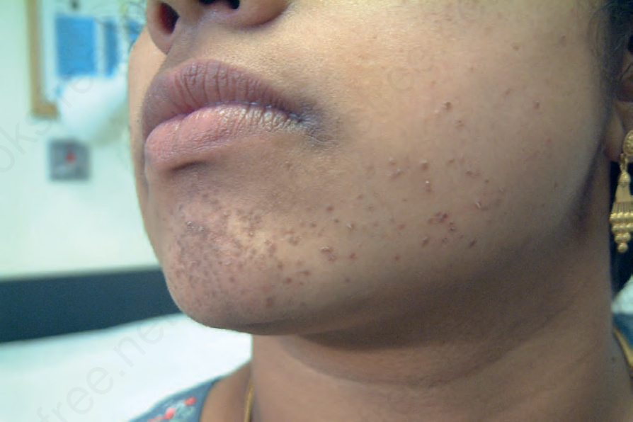
圖 18-17:扁平疣 (plane wart):注意典型扁平、膚色的丘疹,由於搔抓而呈線狀分布延伸(柯氏現象 [Koebner phenomenon])。By courtesy of B Al-Mahmoud, MD, Qatar, Oman.
Fig. 18.17 Plane wart: note the typical flat, flesh-colored papules, which have extended in a linear distribution due to scratching (Koebner phenomenon). By courtesy of B Al-Mahmoud, MD, Qatar, Oman.
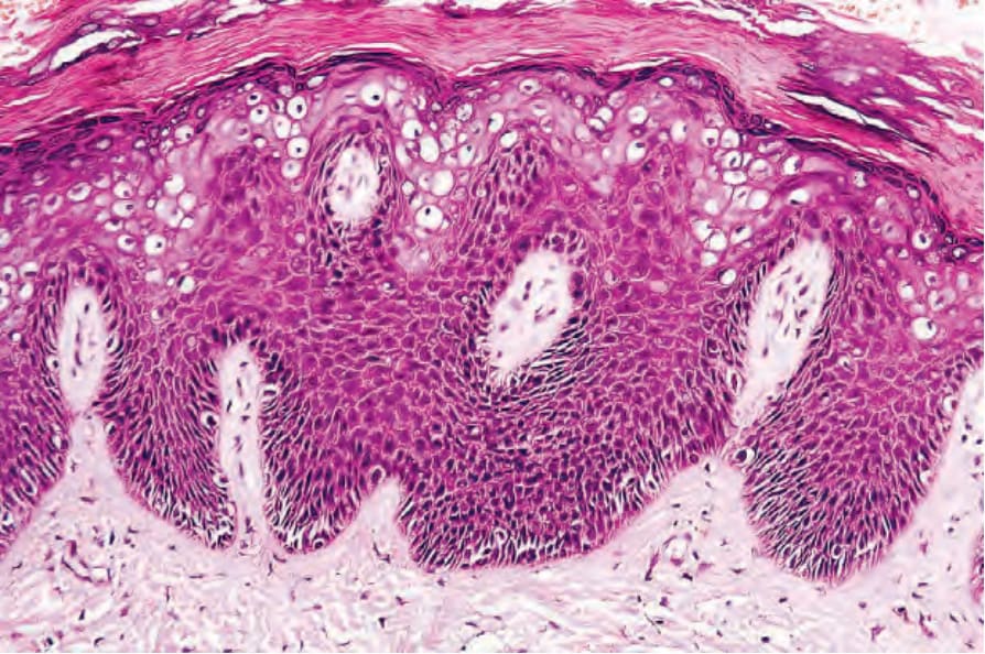
圖 18-18:扁平疣 (plane wart):可見過度角化 (hyperkeratosis) 與輕微規則性棘層肥厚 (regular acanthosis);乳頭瘤狀增生 (papillomatosis) 僅輕微。注意顯著的細胞質空泡化 (cytoplasmic vacuolation)。
Fig. 18.18 Plane wart: there is hyperkeratosis and slight regular acanthosis; papillomatosis is only mild. Note the prominent cytoplasmic vacuolation.
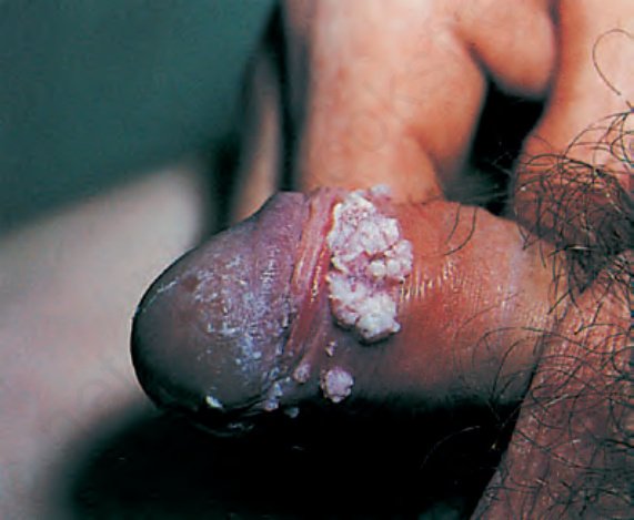
圖 18-19:尖型濕疣 (condyloma acuminatum):注意其典型的絲狀外觀。By courtesy of the Department of Genitourinary Medicine, St Thomas’ Hospital, London, UK.
Fig. 18.19 Condyloma acuminatum: note the typical filiform appearance. By courtesy of the Department of Genitourinary Medicine, St Thomas’ Hospital, London, UK.
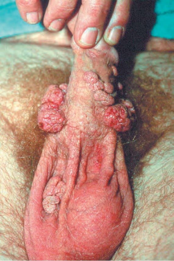
圖 18-20:尖型濕疣 (condyloma acuminatum):陰莖體與陰囊上有多發性病灶。By courtesy of the Department of Genitourinary Medicine, St Thomas’ Hospital, London, UK.
Fig. 18.20 Condyloma acuminatum: there are multiple lesions on the shaft of the penis and scrotum. By courtesy of the Department of Genitourinary Medicine, St Thomas’ Hospital, London, UK.
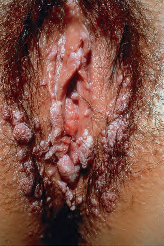
圖 18-21:尖型濕疣 (condyloma acuminatum):此病人有極為廣泛的外陰與會陰侵犯。此病人很可能合併子宮頸 HPV 感染。By courtesy of R.A. Marsden, MD, St George’s Hospital London, UK.
Fig. 18.21 Condyloma acuminatum: in this patient, there is very widespread involvement of the vulva and perineum. This patient is likely to have cervical HPV infection. By courtesy of R.A. Marsden, MD, St George’s Hospital London, UK.
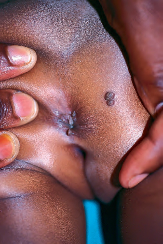
圖 18-22:尖型濕疣 (condyloma acuminatum):有極為廣泛的會陰侵犯。From the collection of the late N.P. Smith, MD, the Institute of Dermatology, London, UK.
Fig. 18.22 Condyloma acuminatum: there is very extensive involvement of the perineum. From the collection of the late N.P. Smith, MD, the Institute of Dermatology, London, UK.
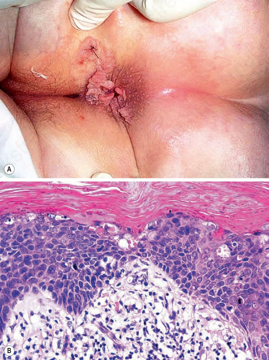
圖 18-23:(A, B) 尖型濕疣 (condyloma acuminatum):除多發性 condylomata 外,尚有原位鱗狀細胞癌 (in situ squamous cell carcinoma) 的組織學證據。By courtesy of P. Ngheim, MD, Dana Farber Cancer Institute and Harvard Medical School, Boston, USA.
Fig. 18.23 (A, B) Condyloma acuminatum: in addition to multiple condylomata, there was histologic evidence of in situ squamous cell carcinoma. By courtesy of P. Ngheim, MD, Dana Farber Cancer Institute and Harvard Medical School, Boston, USA.
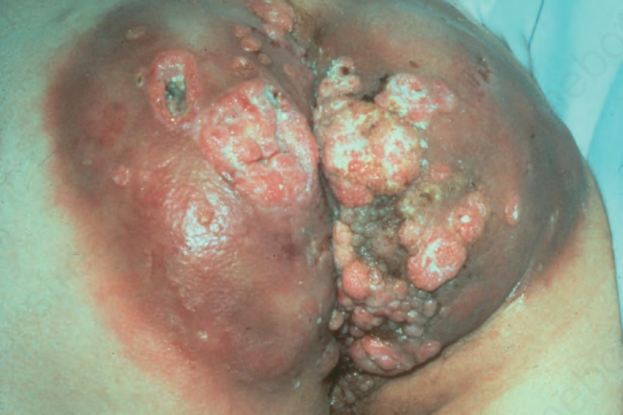
圖 18-24:Buschke-Löwenstein tumor:臀部與會陰有大量浸潤並伴有眾多竇道 (sinuses)。以 DNA 原位雜交 (DNA in situ hybridization) 及 Southern blot 分析鑑定出 HPV 第 6 型。By courtesy of A. Grassegger, MD, University of Innsbruk, Austria.
Fig. 18.24 Buschke-Löwenstein tumor: there is massive infiltration of the buttocks and perineum with numerous sinuses. HPV type 6 was identified by DNA in situ hybridization and Southern blot analysis. By courtesy of A. Grassegger, MD, University of Innsbruk, Austria.
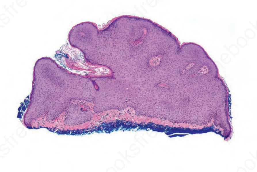
圖 18-25:尖型濕疣 (condyloma acuminatum):注意角化性、棘層肥厚的表皮並具有圓鈍的側緣。空凹細胞 (koilocytes) 出現於乳頭瘤狀上皮 (papillomatous epithelium) 的凹陷處 (declivities)。
Fig. 18.25 Condyloma acuminatum: note the keratotic acanthotic epidermis with rounded lateral borders. Koilocytes are present in the declivities of the papillomatous epithelium.
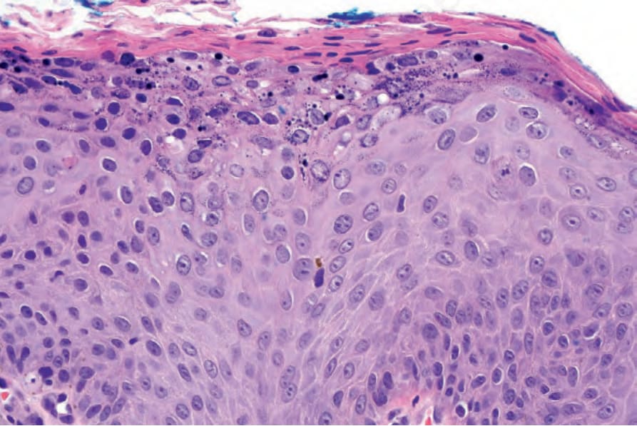
圖 18-26:尖型濕疣 (condyloma acuminatum):注意角化不全 (parakeratosis) 與表淺角質細胞 (superficial keratinocytes) 的空泡化。
Fig. 18.26 Condyloma acuminatum: note the parakeratosis and vacuolation of the superficial keratinocytes.