Gold
臨床特徵 (Clinical Features)
- Gold 治療可導致濕疹樣 (eczematous)、苔癬樣 (lichenoid)、糠疹樣 (pityriasiform) 與乾癬樣 (psoriasiform) 皮膚病變以及口腔炎 (stomatitis)。
- 由腸道外 (parenteral) 注射 gold salts 治療所造成的皮膚色素沉著稱為 chrysiasis(auriasis、chrysoderma、hautaurosis)。此為一種光依賴性 (photodependent)、不可逆 (irreversible) 的病況,最常見於類風濕性關節炎 (rheumatoid arthritis) 患者的紀錄。
- 一旦累積達到 50 mg/kg 的 gold 閾值,患者即處於風險之中。疾病嚴重度與 gold 的累積劑量 (cumulative dose) 相關。
- 顏色變化由淡紫/藍 (mauve/blue) 至藍 (blue) 至板岩灰 (slate-gray) 不等。臉部受日曬的皮膚特別容易受到影響。嚴重病例中,病灶可見於頸部、前胸,以及前臂與手部的背側 (Fig. 14.88)。
- 在禿頭患者中,有時可見頭皮受累。色素沉著也曾被描述於鞏膜 (sclera) 與頰黏膜 (buccal mucosa)。
致病機轉與組織學特徵 (Pathogenesis and Histologic Features)
- Chrysiasis 的致病機轉尚不明確。它可能與 UV 輻射對組織結合性 gold particles 的作用有關。支持此假說的觀察是:皮膚病灶可由 UVB 照射受日光防護的皮膚而誘發。同樣地,在接受 gold 治療的患者中,曾描述以 Q-switched ruby laser 治療後出現典型皮膚病灶。
鑑別診斷 (Differential Diagnosis)
- Gold pigment 必須與 silver deposits (argyria)、mercury 及刺青色素 (tattoo pigment) 區分。
- Silver pigment 主要沉積於基底膜 (basement membranes) 相關處,尤其是汗腺 (sweat glands) 的基底膜。它在交叉偏光 (cross-polarized light) 下不顯示橘紅色雙折射 (orange-red birefringence)。
- Mercury particles 體積大(直徑可達 340 µm),呈棕黑色 (brown-black)。
- 刺青通常由多種不同顏色的各式色素組成。在大多數病例中,臨床病史應可輕易確立診斷。
組織病理特徵 (Histopathology)
- Chrysiasis 的特徵為小型黑色、巨噬細胞結合性 (macrophage-bound) 顆粒沉積於較深層網狀真皮 (deeper reticular dermis) 的血管周圍以及汗腺管盤 (sweat gland coils) 周圍 (Fig. 14.89)。
- Perls Prussian blue (hemosiderin) 與針對 melanin 的 Masson-Fontana 染色皆為陰性。Gold particles 在交叉偏光下顯示橘紅色雙折射 (orange-red birefringence)。無發炎反應。表皮 melanin 色素沉著通常呈現正常。
- 亦曾報告一種局部型 chrysiasis,於注射部位 (injection sites) 伴有硬化性脂肪肉芽腫 (sclerosing lipogranulomas)。
- 在電子顯微鏡下,gold 呈現為顆粒狀 (granular)、微粒狀 (particulate) 與絲狀 (filamentous) 物質,有時在 phagolysosomes(aurosomes)內顯示星狀 (starlike) 形態。診斷可藉由 electron/X-ray probe microanalysis 加以確認。
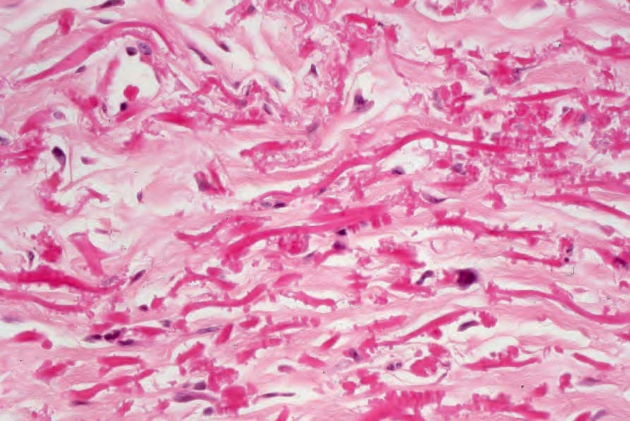
圖 14-86:Penicillamine dermopathy:鋸齒狀外觀 (serrated appearance) 為其特徵。
Fig. 14.86 Penicillamine dermopathy: the serrated appearance is characteristic.
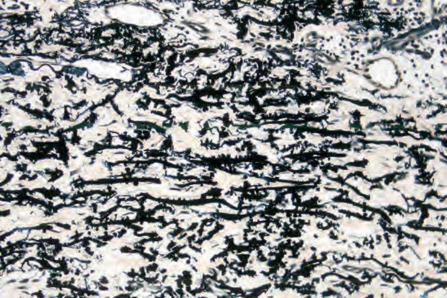
圖 14-87:Penicillamine dermopathy:此變化可藉由彈性組織染色 (elastic tissue stain) 加以凸顯。
Fig. 14.87 Penicillamine dermopathy: the changes can be highlighted by an elastic tissue stain.
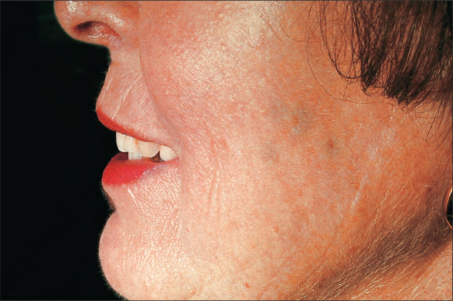
圖 14-88:Chrysiasis:臉頰上出現多處藍色變色 (blue discoloration) 病灶。By courtesy of J. Kerner, MD, Department of Dermatology, Harvard Medical School, Boston, USA.
Fig. 14.88 Chrysiasis: multiple foci of blue discoloration are present on the cheek. By courtesy of J. Kerner, MD, Department of Dermatology, Harvard Medical School, Boston, USA.
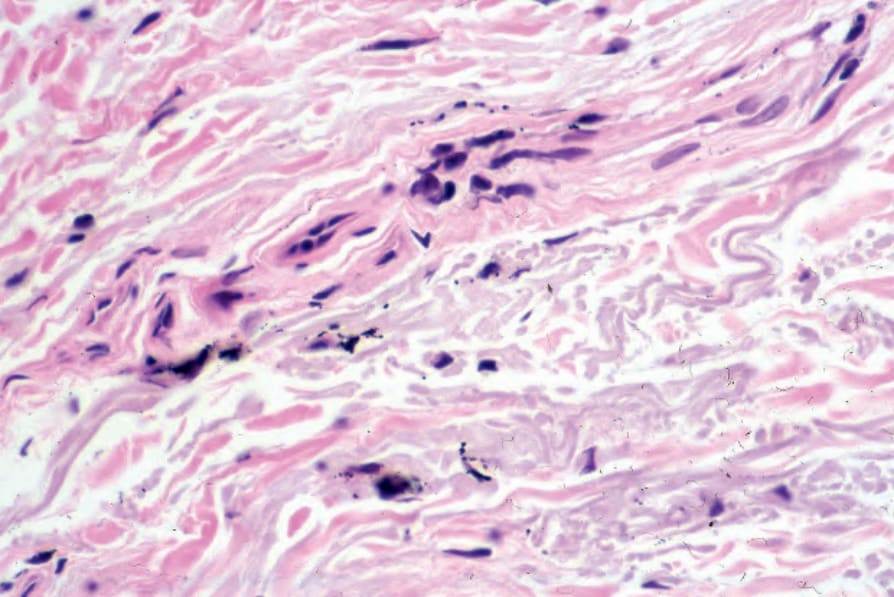
圖 14-89:Chrysiasis:在巨噬細胞 (macrophages) 內以及游離散布於淺層血管系統 (superficial vasculature) 周圍,皆可見細小的黑色顆粒。By courtesy of S. Lyle, MD, Beth Israel Deaconess Medical Center, Boston, USA.
Fig. 14.89 Chrysiasis: there are fine black granules both within macrophages and lying free around the superficial vasculature. By courtesy of S. Lyle, MD, Beth Israel Deaconess Medical Center, Boston, USA.