藥物誘發性色素沉著 (Drug-induced Hyperpigmentation)
臨床特徵 (Clinical Features)
-
皮膚色素沉著 (cutaneous hyperpigmentation) 是藥物治療常見的併發症。它可能源於黑色素合成增加,或藥物及其代謝物沉積於皮膚內。重金屬 (heavy metals) 亦可造成皮膚色素沉著。然而最常見的原因是發炎後色素沉著 (postinflammatory hyperpigmentation)。
-
長期以 minocycline 治療可能導致通常為可逆性(第 I 型與第 II 型)的皮膚色素沉著。一般將皮膚 minocycline 色素沉著區分為三種臨床變異型(圖 14.34–14.36):
- 第 I 型 (Type I):局限於瘢痕與發炎區域(例如臉部痤瘡瘢痕)的藍黑色斑 (blue-black macules)。
- 第 II 型 (Type II)(最常見):位於脛部、踝部與手臂的藍黑色、棕色或石板灰色 (slate-gray) 色素沉著。
- 第 III 型 (Type III):泛發性的污泥棕色 (muddy-brown) 色素沉著,可能在曝曬陽光的部位加重。另有報導一種第四變異型,影響嘴唇,可能代表固定型藥物疹 (fixed drug eruption)。
-
Minocycline 可能侵犯牙齒(造成綠灰色或藍灰色變色),主要影響牙冠的中間三分之一,偶爾影響切端三分之一。口腔黏膜病灶罕見,不過曾有報導色素沉著出現於頰黏膜、牙齦、舌頭與嘴唇。口腔下方的骨骼(黑骨病, black bone disease)是 minocycline 色素沉著最常侵犯的單一部位。觀察上頜與下頜前牙槽黏膜可最清楚地顯現此一變化。硬腭與舌側牙槽骨亦常受影響。
-
結膜、鞏膜、甲狀腺(黑甲狀腺, black thyroid)、主動脈、心內膜與動脈粥樣硬化斑塊 (atherosclerotic plaques) 亦可能在 minocycline 色素沉著中受到侵犯。Minocycline 色素沉著也可能影響外陰/陰道黏膜,臨床上可能類似黑色素細胞病變 (melanocytic lesion)。
-
許多其他四環素類藥物,包括 methacycline 與 tetracycline hydrochloride,也與皮膚色素沉著相關。
-
Amiodarone 主要用於治療心律不整 (cardiac arrhythmias),在高達百分之五十的病人中與光毒性/光敏感反應 (phototoxic/photosensitivity reaction) 相關。此外,皮膚的金棕色至石板灰色或藍/紫色色素沉著可能發生,主要影響曝露部位,包括臉部與手背,尤其是在長期接受高劑量者(圖 14.37)。色素沉著有時也見於鞏膜與角膜。
-
抗瘧疾藥 (Antimalarials) 也會導致異常的皮膚色素沉著。Mepacrine (quinacrine) 典型上產生黃色變色,不過曾有報導局限性的藍黑色皮膚黏膜病灶(圖 14.38 與 14.39)。Chloroquine 與 hydroxychloroquine 造成黃棕色至灰色的色素沉著。主要影響曝曬陽光的皮膚,不過黏膜色素沉著亦可能發生。
-
也曾有報導局限性 minocycline 色素沉著作為美容雷射術 (cosmetic laser procedures) 的併發症。
-
指甲色素沉著最常表現為近端甲床 (proximal nail bed) 的持續性石板灰色變色。其他特徵包括縱向黑甲 (longitudinal melanonychia)、瀰漫性指甲色素沉著與光甲剝離 (photo-onycholysis)。
-
除了造成光敏感與接觸性皮膚炎之外,chlorpromazine 治療(尤其在長期且高劑量時)可導致皮膚色素沉著,特別是在曝曬陽光的皮膚,例如臉部、手背與頸部。病人可能表現出金棕色、曬黑般的外觀,其他病人則出現石板灰色、偏藍或紫色的外觀。眼睛的角膜與水晶體 (lens) 亦可能受到侵犯。
-
長期以 imipramine 治療可能導致光分布性色素沉著 (photodistributed hyperpigmentation),影響臉部、頸部、胸部「V」形區域、手臂與手部(圖 14.40)。其色澤從金棕色到藍灰色或石板灰色不等。虹膜 (irises) 也可能變暗。
-
曾有文獻記載 desipramine 治療後出現光分布性藍灰色色素沉著。
-
重金屬 (heavy metals),包括金、銀與汞,皆可導致皮膚色素沉著(見下文)。
致病機轉與組織學特徵 (Pathogenesis and Histologic Features)
-
Minocycline 色素沉著的組織學特徵不一。在第 I 型與第 II 型變異型中,金棕色至棕黑色的顆粒 (granules) 主要見於巨噬細胞 (macrophages) 內,主要分布於血管周圍與汗腺腺體周圍(圖 14.41)。此色素在紫外光下發出黃色螢光,於第 II 型變異型中以 Masson-Fontana 與 Perls Prussian blue 反應皆呈陽性染色(圖 14.42 與 14.43)。該色素過碘酸希夫氏 (periodic acid-Schiff, PAS) 反應為陰性。相較之下,在第 I 型中,色素僅以 Perls 反應染色。一般認為它代表 minocycline 或其分解產物與血鐵質 (hemosiderin)、鐵蛋白 (ferritin) 或鐵 (iron) 螯合而成。鈣、硫與氯亦存在,但無黑色素 (melanin)。在第 I 型與第 II 型變異型中,黑色素細胞 (melanocytes) 與表皮並未顯示黑色素色素增加。第 III 型色素沉著的特徵則為表皮基底細胞黑色素色素增加。Perls 染色為陰性。曾有報導皮下脂肪 (subcutaneous fat) 的 minocycline 色素沉著出現於第 II 型疾病的臨床情境中。組織學上,皮下脂肪內的巨噬細胞與巨細胞 (giant cells) 中可見色素,Masson-Fontana 染色呈陽性,Perls 反應染色則不一。其中一項研究還描述了脂肪內巨噬細胞中的綠灰色非折射性 (nonrefractile) 球狀物。
-
這些顆粒為非折射性,Masson-Fontana 染色呈陽性,而鐵染色為陰性。
-
組織學上,chlorpromazine 色素沉著的特徵為環繞淺層血管的金棕色、巨噬細胞內 (macrophage-bound) 顆粒。這些顆粒以 Masson-Fontana 反應呈陽性,但不以 Perls Prussian blue 染色。超微結構上,此色素由溶酶體 (lysosome) 包覆,除巨噬細胞外,亦存在於內皮細胞、纖維母細胞、Schwann 細胞與平滑肌細胞中。黑色素增加亦促成皮膚色素沉著。
-
組織學上,amiodarone 色素沉著的特徵為巨噬細胞含有 PAS 陽性、黃棕色脂褐素樣 (lipofuscin-like) 顆粒,位於血管周圍分布(圖 14.44 與 14.45)。表皮的黑色素色素並未增加;事實上,近期已記載受侵犯皮膚中黑色素的缺乏。電子顯微鏡下,這些顆粒位於溶酶體內。亦可辨識出層狀髓鞘體 (lamellar myelin bodies)。類似的包涵體 (inclusions) 可見於肝細胞、Kupffer 細胞、肺部巨噬細胞與嗜中性球。
-
在 mepacrine (quinacrine) 色素沉著中,黃棕色色素見於整個真皮的組織球 (histiocytes) 細胞質內。此色素以 Perls Prussian blue 鐵反應呈弱陽性,Masson-Fontana 為陰性。Hydroxychloroquine 相關色素沉著的組織學表現被描述為巨噬細胞內與細胞外的黃棕色顆粒狀沉積物。
-
組織學上,imipramine 與 desipramine 色素沉著於上真皮含有 Masson-Fontana 陽性的金棕色顆粒,既游離存在,亦位於巨噬細胞內(圖 14.46)。Perls Prussian blue 為陰性。超微結構上,組織球除含有溶酶體包覆的電子緻密顆粒外,尚含有黑色素小體 (melanosomes)。
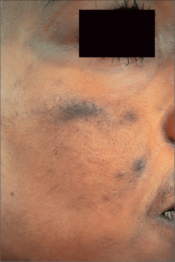
圖 14.34:Minocycline 色素沉著:侵犯臉頰與眼眶周圍區域的廣泛病灶。By courtesy of the Institute of Dermatology, London, UK.
Fig. 14.34 Minocycline pigmentation: extensive lesions involving the cheek and periorbital region. By courtesy of the Institute of Dermatology, London, UK.
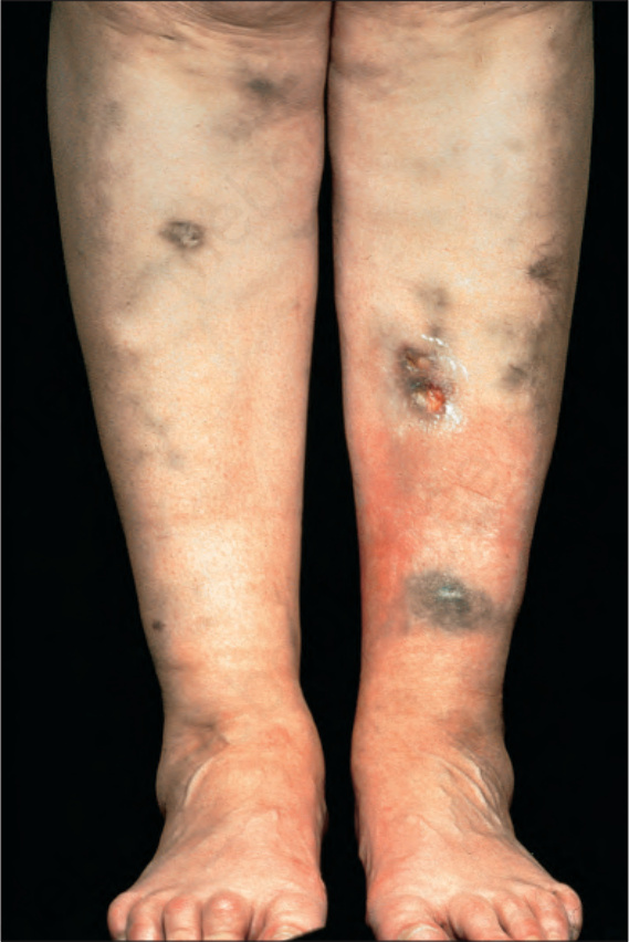
圖 14.35:Minocycline 色素沉著:這些藍黑色病灶發生於一位壞疽性膿皮症 (pyoderma gangrenosum) 病人。By courtesy of the Institute of Dermatology, London, UK.
Fig. 14.35 Minocycline pigmentation: these blue-black lesions have developed in a patient with pyoderma gangrenosum. By courtesy of the Institute of Dermatology, London, UK.
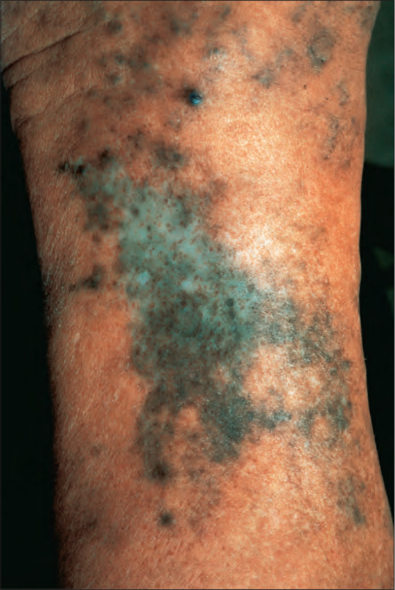
圖 14.36:Minocycline 色素沉著;侵犯脛部的典型色素沉著。By courtesy of the Institute of Dermatology, London, UK.
Fig. 14.36 Minocycline pigmentation; typical pigmentation affecting the shin. By courtesy of the Institute of Dermatology, London, UK.
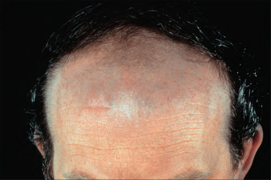
圖 14.37:Amiodarone 色素沉著:注意前額的石板灰色變色,此為特徵性部位。By courtesy of the Institute of Dermatology, London, UK.
Fig. 14.37 Amiodarone pigmentation: note the slate-gray discoloration on the forehead, a characteristic site. By courtesy of the Institute of Dermatology, London, UK.
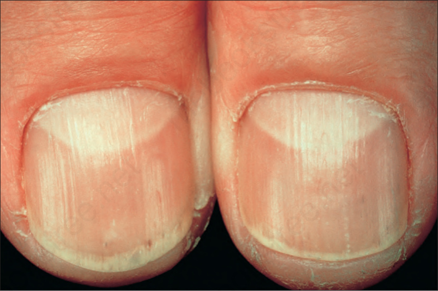
圖 14.38:Mepacrine 色素沉著:黃色變色為其特徵。By courtesy of the Institute of Dermatology, London, UK.
Fig. 14.38 Mepacrine pigmentation: a yellow discoloration is characteristic. By courtesy of the Institute of Dermatology, London, UK.
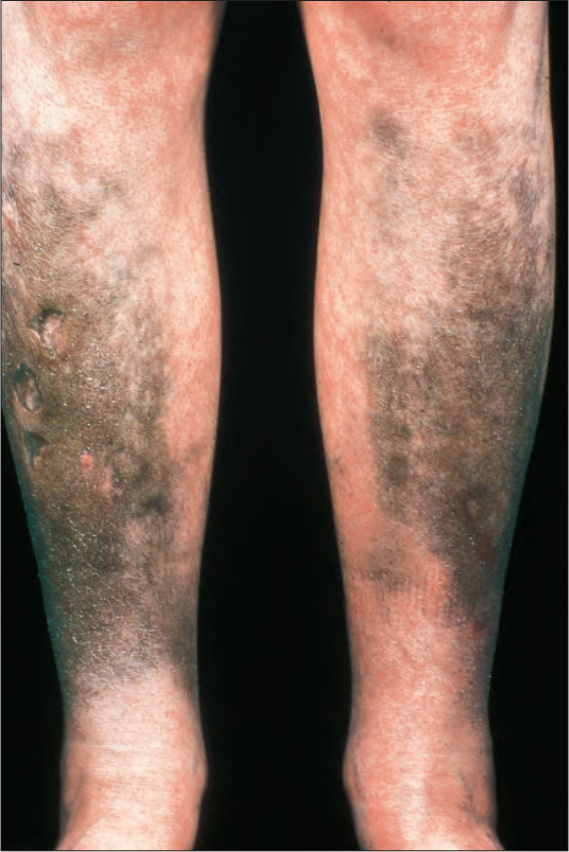
圖 14.39:Mepacrine 色素沉著:在此病人中,該藥物導致黑色病灶。By courtesy of the Institute of Dermatology, London, UK.
Fig. 14.39 Mepacrine pigmentation: in this patient, the drug resulted in black lesions. By courtesy of the Institute of Dermatology, London, UK.
圖 14.40:Imipramine 色素沉著:注意手部與前臂的濃烈棕色色素,相較於胸部更為明顯。By courtesy of L. Cohen, MD, Cohen Dermatopathology, Massachusetts, USA.
Fig. 14.40 Imipramine pigmentation: note the intense brown pigment of the hands and forearms in comparison with the chest. By courtesy of L. Cohen, MD, Cohen Dermatopathology, Massachusetts, USA.
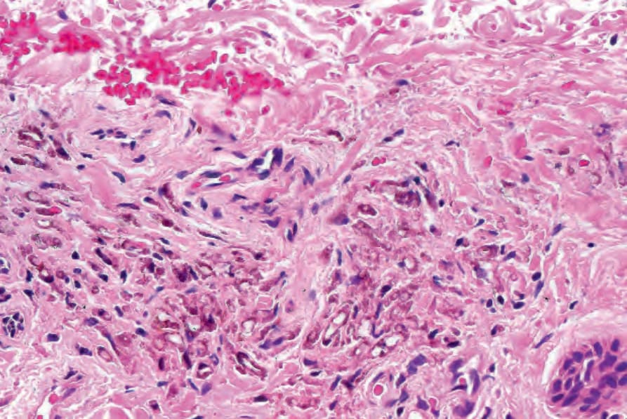
圖 14.41:Minocycline 色素沉著:注意血管周圍顆粒狀棕色色素的存在。
Fig. 14.41 Minocycline pigmentation: note the presence of perivascular granular brown pigment.
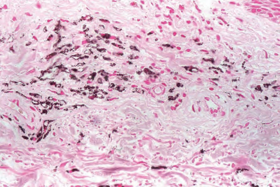
圖 14.42:Minocycline 色素沉著:色素以 Masson-Fontana 呈陽性染色。
Fig. 14.42 Minocycline pigmentation: the pigment stains positively with Masson-Fontana.
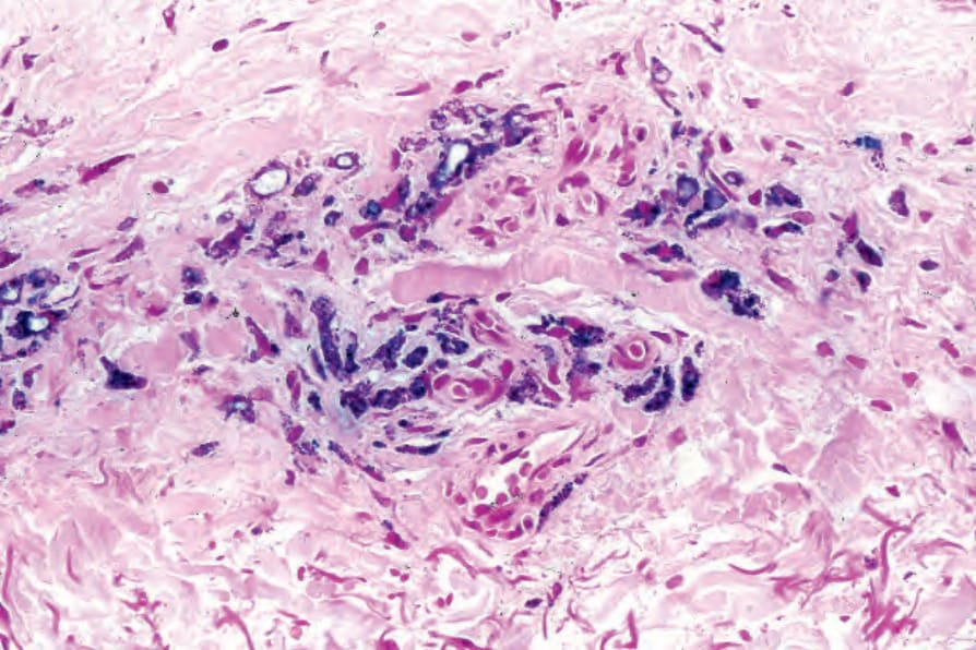
圖 14.43:Minocycline 色素沉著:色素亦以 Prussian blue 染色。
Fig. 14.43 Minocycline pigmentation: the pigment also stains with Prussian blue.
圖 14.44:Amiodarone 色素沉著:色素性巨噬細胞 (pigmented macrophages) 呈血管周圍分布。
Fig. 14.44 Amiodarone pigmentation: pigmented macrophages are present in a perivascular distribution.
圖 14.45:Amiodarone 色素沉著:高倍視野。
Fig. 14.45 Amiodarone pigmentation: high-power view.
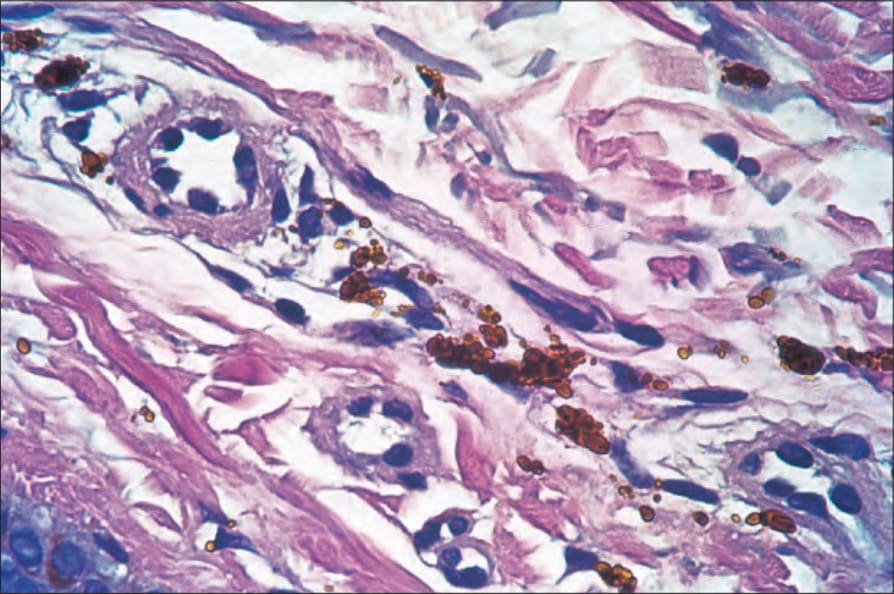
圖 14.46:Imipramine 色素沉著:典型的金棕色顆粒。注意 Prussian blue 反應為陰性。By courtesy of L. Cohen, MD, Cohen Dermatopathology, Massachusetts, USA.
Fig. 14.46 Imipramine pigmentation: typical golden-brown granules. Note that the Prussian blue reaction is negative. By courtesy of L. Cohen, MD, Cohen Dermatopathology, Massachusetts, USA.