臨床特徵 (Clinical Features)
-
局部性(脛前)黏液水腫 (localized (pretibial) myxedema) 最常與甲狀腺功能亢進 (hyperthyroidism) 相關,發生於 3–5% 的病例。
-
它是自體免疫性甲狀腺(Graves)疾病 (autoimmune thyroid (Graves) disease) 典型可見的三種病程之一,另外兩種為突眼 (exophthalmos) 與甲狀腺杵狀指 (thyroid acropachy,伴隨骨膜下新生骨形成的手指杵狀變化)。
-
脛前黏液水腫 (pretibial myxedema) 又稱為「Graves 皮膚病變」或「甲狀腺皮膚病變 (thyroid dermopathy)」,通常是 Graves disease 的晚期表現,且發生於 Graves 眼病變 (Graves ophthalmopathy) 出現之後。
-
它僅在極少數情況下被報告於 Graves disease 診斷之前出現,以及在無眼病變的情形下出現。
-
在 10% 的 Graves disease 病例中,病人臨床上並非甲狀腺功能亢進;他們可能為甲狀腺功能低下 (hypothyroid) 或甲狀腺功能正常 (euthyroid)。
-
脛前黏液水腫罕見地可與 Hashimoto 甲狀腺炎 (Hashimoto thyroiditis) 相關。
-
病灶為粉紅或黃色的蠟樣斑塊、結節,有時呈「腫瘤 (tumors)」狀,最常先出現於小腿前外側面 (anterolateral aspects of the lower legs)(圖 13.164)。
-
病灶典型為非凹陷性 (nonpitting)。部分病人有硬結 (induration) 並伴隨毛囊突出 (prominence of the follicles),形成橘皮樣外觀 (peau d’orange appearance),繼發性多毛症 (secondary hypertrichosis) 偶爾相當明顯。黏液水腫部位的局部多汗 (localized hyperhidrosis) 也可能罕見地發生。
-
此病可能進展至侵犯大部分的小腿,罕見地變成明顯的象皮病樣 (elephantiasiform)(圖 13.165)。足部與腳趾有時也可被侵犯。
-
小型病灶通常無症狀或輕微搔癢;較大的斑塊則常會疼痛。
-
局部性黏液水腫不常見地發生於其他部位,例如手臂、肩膀、腹部、頸部、臉部,甚至耳朵(圖 13.166)。結節性病灶罕見地發生於手部。
-
沉積於前臂者被描述為「橈側前黏液水腫 (preradial myxedema)」。
-
非典型部位之發生最可能與創傷 (trauma) 相關。例如,曾有局限於疤痕組織 (scar tissue) 的描述,後者包括天花疫苗接種疤痕 (smallpox vaccination scar) 的部位。亦曾報告出現於大腿供皮移植部位 (thigh donor graft site)。
-
罕見的脛前黏液水腫病人沒有甲狀腺疾病的證據。這些病人的切片傾向顯示與鬱滯 (stasis) 相關的變化,此特徵在組織學鑑別診斷上有用。其中一種此類變異型已被描述於合併淋巴水腫 (lymphedema) 的病態肥胖 (morbidly obese) 病人。病灶以丘疹、水疱及結節的形式出現於脛前表面。
-
脛前黏液水腫有時為自限性 (self-limiting),數年後出現消退 (involution)。完全緩解 (complete remission) 可發生於高達 26% 的病例,但這取決於疾病的嚴重程度。
-
一種異常型態的脛前黏液沉積,可能與 Graves disease 相關的脛前黏液水腫混淆,曾以「肢端魚鱗癬樣黏液沉積症 (acral ichthyosiform mucinosis)」之名被記錄於合併 Sjögren 症候群 (Sjögren syndrome) 的情形。在所描述的病例中,病人的甲狀腺功能檢查正常,且黏液沉積主要位於乳頭真皮 (papillary dermis)。
致病機轉與組織學特徵 (Pathogenesis and Histologic Features)
-
病因不確定;脛前黏液水腫的存在通常與血清中偵測到長效甲狀腺刺激物 (long-acting thyroid stimulator, LATS) 相關,但一般認為 LATS 並非致病因素。
-
1978 年曾提出,從脛前黏液水腫病人血清分離出的一種與致黏液性質 (mucigenic properties) 相關的纖維母細胞刺激因子 (fibroblast stimulating factor),可能在此病的致病機轉中扮演角色。
-
後續研究顯示,受侵犯病人脛前皮膚及眼眶 (orbit) 中的纖維母細胞,含有與甲狀腺刺激素受體 (thyroid stimulating hormone receptor) 完全相同的序列。
-
亦有人提出,這些纖維母細胞可能含有一種交叉反應蛋白 (cross-reacting protein),而非真正的受體,此蛋白與抗甲狀腺刺激因子受體 (thyroid stimulating factor receptor) 的自體抗體結合。
-
基於這些觀察,有人提出抗甲狀腺刺激素受體 (thyroid-stimulating hormone receptor) 的自體抗體與含有這些序列的纖維母細胞發生反應,導致細胞激素 (cytokines) 的產生,並誘發醣胺聚醣 (glycosaminoglycan) 分泌增加。
-
局限於下肢通常被認為是由於下垂性 (dependency) 與機械性因素所致。此外,菸草 (tobacco) 是脛前黏液水腫與 Graves 眼病變發生的已知危險因子;然而其確切機轉目前仍不明。
-
表皮常呈過度角化 (hyperkeratotic) 並伴隨毛囊角栓 (follicular plugging);在嚴重病例中,可能呈乳突瘤狀 (papillomatous) 與棘層肥厚 (acanthotic)。
-
網狀真皮 (reticular dermis) 顯示膠原束 (collagen bundles) 被大量黏液 (mucin) 分隔(圖 13.167 與 13.168)。可見膠原纖維 (collagen fibers) 的碎裂 (fragmentation)。
-
可見星狀纖維母細胞 (stellate fibroblasts),但其數量通常並未增加,或許僅在較象皮病樣的例子中例外。
-
在淋巴水腫與肥胖背景下所見的病灶,亦以小血管新生 (small vessel angiogenesis)、血管壁增厚、水腫及含鐵血黃素 (hemosiderin) 沉積為特徵。
-
免疫螢光 (immunofluorescent) 研究通常為陰性,雖然曾在表淺乳頭真皮 (superficial papillary dermis) 內辨識出 IgM 的顆粒狀沉積 (granular deposits)。
-
電子顯微鏡 (electron microscopic) 研究顯示,無定形顆粒狀物質 (amorphous granular material) 同時存在於纖維母細胞的內質網 (endoplasmic reticulum) 內、包覆纖維母細胞表面,以及在環繞著廣泛分隔之膠原與彈性纖維的間質 (interstitium) 中。曾在一例病人的內皮細胞 (endothelial cells) 細胞質中、以及另一例病人的真皮中辨識出管網狀結構 (tubuloreticular structures)。
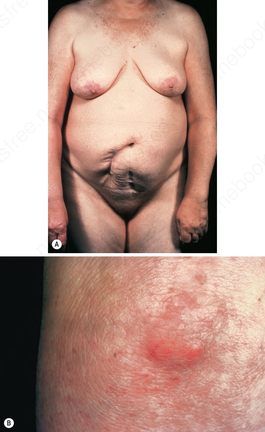
圖 13-163 (A, B):全身性黏液水腫 (Generalized myxedema):此病人有廣泛的黃色瘤 (xanthomata)。By courtesy of the Institute of Dermatology, London, UK.
Fig. 13.163 (A, B) Generalized myxedema: this patient has widespread xanthomata. By courtesy of the Institute of Dermatology, London, UK.
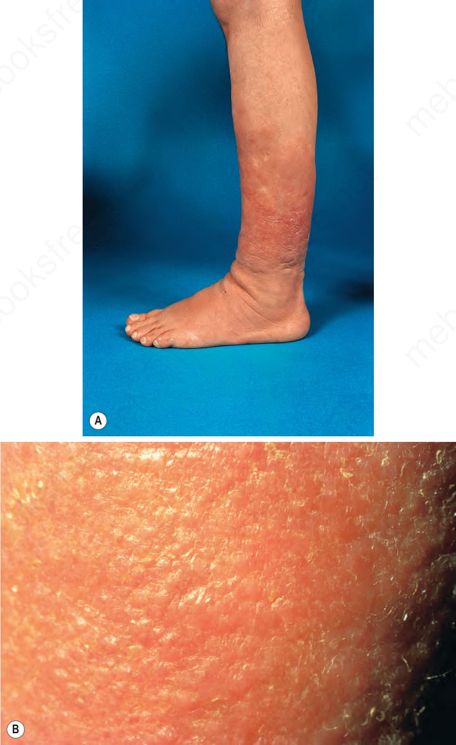
圖 13-164:脛前黏液水腫 (Pretibial myxedema):(A) 脛部出現紅斑性、略呈半透明的斑塊;(B) 近距離視野。By courtesy of R.A. Marsden, MD, St George’s Hospital, London, UK.
Fig. 13.164 Pretibial myxedema: (A) erythematous, somewhat translucent plaques are present over the shin; (B) close-up view. By courtesy of R.A. Marsden, MD, St George’s Hospital, London, UK.
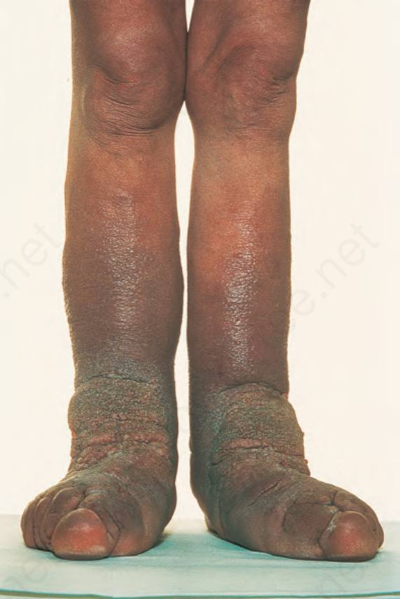
圖 13-165:脛前黏液水腫 (Pretibial myxedema):在此極端的例子中,其特徵類似象皮病 (elephantiasis)。By courtesy of R.A. Marsden, MD, St George’s Hospital, London, UK.
Fig. 13.165 Pretibial myxedema: in this extreme example, the features resemble elephantiasis. By courtesy of R.A. Marsden, MD, St George’s Hospital, London, UK.
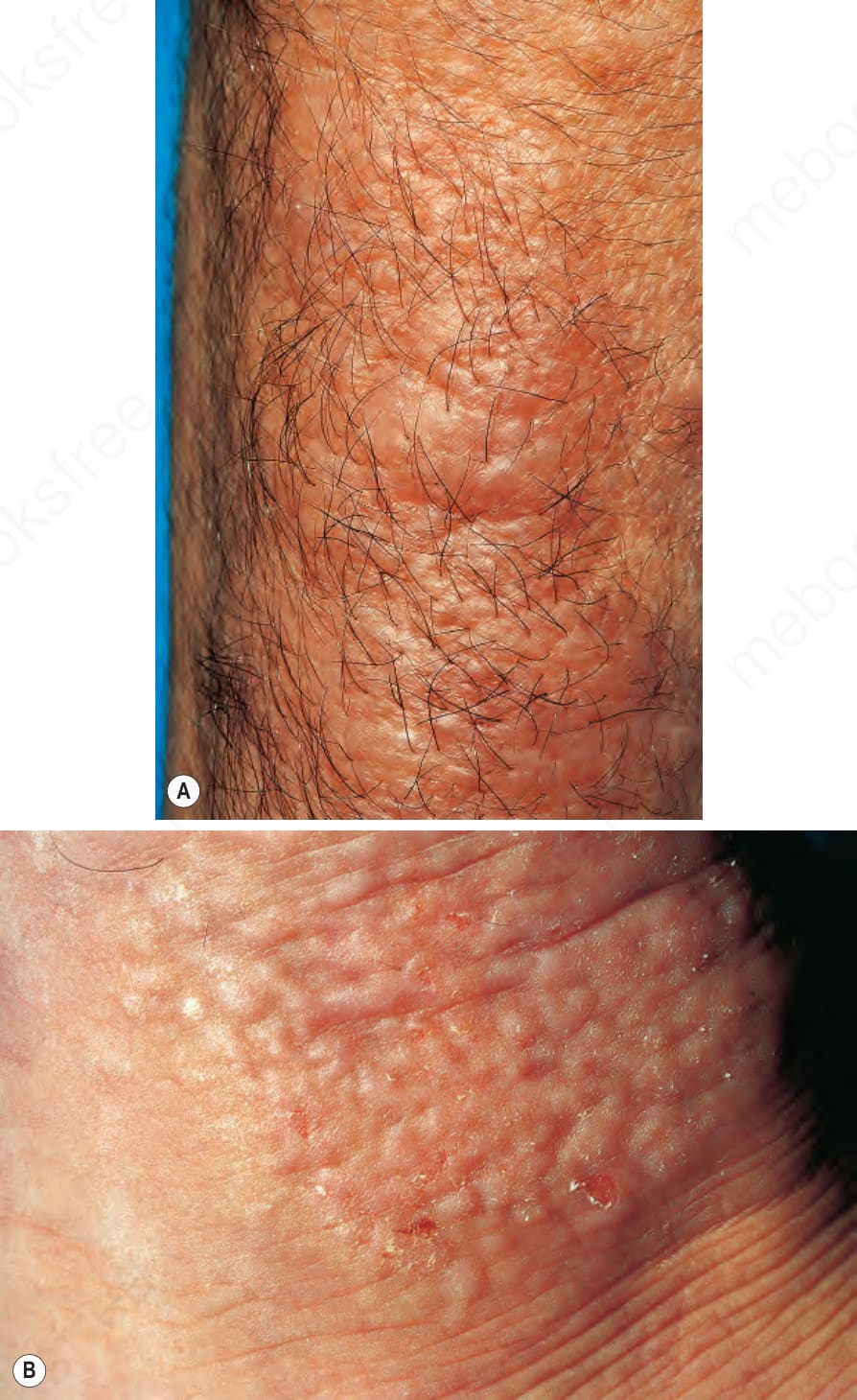
圖 13-166 (A, B):局部性黏液水腫 (Localized myxedema):這些圖片來自圖 13.162 所示的同一病人。在一次道路交通事故後,此病人於手臂靠近骨折部位處發生了額外的黏液性沉積。By courtesy of P.G. Goodwin, MD, The Royal Bournemouth Hospital, UK.
Fig. 13.166 (A, B) Localized myxedema: these pictures came from same patient shown in Fig. 13.162. Following a road traffic accident, the patient developed additional mucinous deposits on her arm close to the site of a fracture. By courtesy of P.G. Goodwin, MD, The Royal Bournemouth Hospital, UK.
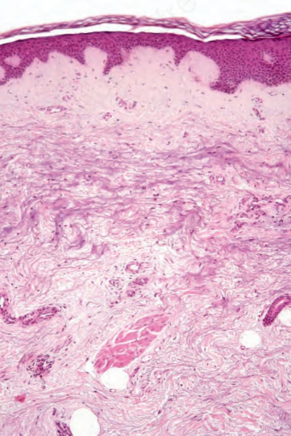
圖 13-167:脛前黏液水腫 (Pretibial myxedema):可見膠原纖維 (collagen fibers) 喪失並伴隨黏液 (mucin) 沉積。
Fig. 13.167 Pretibial myxedema: there is loss of collagen fibers associated with mucin deposition.