雜色卟啉症 (Variegate porphyria)
雜色卟啉症 (Variegate porphyria)
臨床特徵 (Clinical Features)
這種家族型卟啉症 (porphyria) 同時表現出遲發性皮膚卟啉症 (porphyria cutanea tarda) 的皮膚特徵,以及急性間歇性卟啉症 (acute intermittent porphyria) 的急性腹部與神經學發作,兩者通常在第二或第三個十年期間變得明顯 (Figs 13.106–13.109)。1,2 它在南非特別常見,可追溯至單一荷蘭家族的後裔。2,3 它是一種體染色體顯性遺傳 (autosomal dominantly inherited) 的疾病,且已辨識出受影響較嚴重的同型合子 (homozygotes) 個體 (Figs 13.110 and 13.111)。Variegate porphyria 與 protoporphyrinogen oxidase(血基質生合成途徑中倒數第二個酵素)活性降低有關。4 在位於染色體 1q22-23 的 protoporphyrin oxidase 基因上已證實有數種不同的突變。5–7 基因型並非臨床表現的重要決定因素。8–10 通常,該酵素的活性約降低 50%。10
……所描述的某些突變。2 此型卟啉症亦具異質性,UROD 基因中可能發生不同的突變。3–7 uroporphyrinogen decarboxylase 的活性遠低於 porphyria cutanea tarda。因此,疾病表現典型上較為嚴重。已有報告指出某些基因型 (genetic) 相關的輕度變異型。
急性發作可能由多種誘導肝臟微粒體 (hepatic microsomal) 活性的藥物所誘發,包括 barbiturates、酒精 (alcohol)、口服避孕藥 (oral contraceptives)、懷孕 (pregnancy)、抗痙攣藥 (anticonvulsants) 與 sulfonamides。11–14 急性 variegate porphyria 亦曾於病毒性肝炎 (viral hepatitis) 發作期間出現。11 在急性發作期間,皮膚表現有時輕微或缺如,因此此病可能被誤診為 acute intermittent porphyria。2
組織學特徵 (Histologic features of the porphyrias)
直接免疫螢光 (Direct immunofluorescence) 顯示免疫球蛋白(特別是 IgG,較少程度為 IgM)、纖維蛋白原 (fibrinogen) 與 C3 勾勒出真皮乳頭層 (papillary dermis) 內具特徵性的甜甜圈狀 (donut-shaped) 血管 (Fig. 13.112)。雖然這在紅血球生成性原卟啉症 (erythropoietic protoporphyria) 特別明顯,但它也是其他「皮膚型」變異型的特徵。1–3 免疫反應物 (Immunoreactants) 亦常見於真皮–表皮交界 (dermal–epidermal junction),並曾在外泌汗腺 (eccrine sweat glands) 與汗管 (ducts) 的基底膜區域 (basement membrane region) 內被辨識出。1–5 此發現被認為是由於血清成分的非特異性結合,而非免疫媒介的反應。4 此外,第四型膠原蛋白 (type IV collagen) 與層黏連蛋白 (laminin) 兩者皆以增多的量存在,因而促成血管壁增厚。6 類細胞體 (Cytoid bodies) 亦常見明顯。4 直接免疫螢光研究已證實,在 porphyria cutanea tarda 病人的淺層與中層真皮血管壁中有膜攻擊複合體 (membrane attack complex) C5b-9 的顆粒狀與均質性沉積。有人提出,紫外光 (UV light) 活化的尿卟啉 (uroporphyrins) 接續活化補體 (complement),可能扮演病理性角色。7 間接免疫螢光 (Indirect immunofluorescence) 對基底膜區自體抗體 (basement membrane zone autoantibodies) 一律為陰性。
所有類型卟啉症的組織學變化都非常相似。其特徵性表現為在受影響皮膚的血管周圍存在 PAS 陽性、抗澱粉酶 (diastase-resistant) 的玻璃樣 (hyaline) 物質 (Figs 13.113 and 13.114)。在輕度疾病中,沉積物纖細,通常侷限於真皮乳頭層血管,但在較嚴重的病例中,沉積物廣泛分布、出現於真皮更深處,並使血管壁呈現特徵性的層板狀 (lamellated) 外觀。這些外觀在 erythropoietic protoporphyria 特別顯著。1,8 在 porphyria cutanea tarda 與 erythropoietic protoporphyria 兩者中,有時可在血管周圍見到 Alcian blue 陽性的黏液素 (mucin),較少程度則見於真皮–表皮交界處。1 有時亦可顯示出脂質滴 (Lipid droplets)。在下層真皮可能出現針對澱粉樣物質 (amyloid) 的偽陽性 Congo red 染色。1
電子顯微鏡 (Electron microscopic) 觀察包括真皮血管周圍相當程度的基底膜重複 (basement membrane reduplication),較少程度則見於真皮–表皮交界處 (Fig. 13.115)。1,8 這與內皮細胞 (endothelial cell) 反覆損傷與再生、隨後形成新基底膜的效應一致。此外,典型上在血管周圍與表皮基底膜區域兩處皆存在細纖維狀 (finely fibrillar) 物質。亦可能出現不規則的電子緻密 (electron-dense) 無定形 (amorphous) 沉積物。1
可能有表皮下水疱 (subepidermal blisters),特徵性地伴隨輕微的單核發炎細胞浸潤 (mononuclear inflammatory cell infiltration) (Figs 13.116 and 13.117)。在 erythropoietic protoporphyria 的急性病灶中,曾描述呈現白血球破裂 (leukocytoclasis) 的嗜中性多形核白血球 (Neutrophil polymorphs),且有時可見紅血球外滲 (red cell extravasation)。9 真皮乳頭的花綵狀排列 (Festooning) 常見,但並非一律存在。裂隙平面 (plane of cleavage) 似乎是可變的。3,10 有些水疱在淺層真皮的緻密板 (lamina densa) 下方形成,類似後天性表皮鬆解水疱症 (epidermolysis bullosa acquisita)。在其他情況下,它們在重複形成的基底膜成分內發展。然而,最常見的情形,如抗原定位 (antigen mapping) 實驗所顯示,水疱形成始於透明板 (lamina lucida)。10,11 因此,第四型膠原蛋白與層黏連蛋白通常存在於水疱底部,而水疱性類天疱瘡抗原 (bullous pemphigoid antigen) 則明顯見於頂部。在 porphyria cutanea tarda 病人的水疱頂部,已辨識出由第四型膠原蛋白與層黏連蛋白組成的線狀分節結構。3,12 這些所謂的毛毛蟲體 (caterpillar bodies) 亦可見於 erythropoietic protoporphyria 與藥物誘發性偽卟啉症 (drug-induced pseudoporphyria) 病人的檢體中 (Fig. 13.117b)。13 它們為 PAS 陽性,並在覆蓋於表皮下水疱上方的表皮中以線狀排列的小球 (globules) 形式呈現。超微結構 (Ultrastructural) 研究顯示,這些小體代表退化中的角質細胞 (keratinocytes)、膠樣體 (colloid bodies) 與因反覆水疱形成及再上皮化所形成的基底膜片段三者的組合。14 毛毛蟲體存在於高達 43% 的 porphyria cutanea tarda 檢體中。15 「毛毛蟲體樣群集 (Caterpillar body-like clusters)」亦曾在 porphyria cutanea tarda、erythropoietic protoporphyria、水疱性類天疱瘡 (bullous pemphigoid) 以及交界型與營養不良型表皮鬆解水疱症 (junctional and dystrophic epidermolysis bullosa) 病人中被辨識出。這些群集在組織學上與典型毛毛蟲體完全相同,但不被第四型膠原蛋白或 PAS 染色所染色。15
罕見情況下,曾在 porphyria cutanea tarda 中記載有苔癬樣組織反應 (lichenoid tissue reaction)。16
在 variegate porphyria 所見水疱的組織學特徵,與 porphyria cutanea tarda 所描述者完全相同。17
在較慢性的病灶中常出現續發性硬皮病樣變化 (secondary sclerodermatous change),其特徵為膠原束 (collagen bundles) 增厚與皮膚附屬器 (cutaneous adnexae) 數量減少 (Fig. 13.118)。4,8,18 在整個受累真皮中可辨識出抗澱粉酶、PAS 陽性的物質。1 這在 porphyria cutanea tarda 特別顯著。要與硬皮病 (scleroderma) 的真皮變化作區分可能非常困難,但據稱卟啉症中膠原束的質地稍微較鬆散。1 硬皮病樣變化 (sclerodermoid changes) 在卟啉症中分布於日曬部位皮膚的臨床分布,有助於作此區分。19 由抗澱粉酶、PAS 陽性物質造成的基底膜增厚通常存在,尤其在 porphyria cutanea tarda。1,8 日光性彈力纖維變性 (Solar elastosis) 在後者情況中常見明顯,但這很可能主要是病人年齡的結果,不太可能是一個基本的病理過程 (Fig. 13.119)。它並非 erythropoietic protoporphyria 的特徵。1,8
在與 porphyria cutanea tarda 相關的禿髮 (alopecia) 中,初始變化為毛囊周圍結締組織鞘 (perifollicular connective tissue sheath) 的腫脹與均質化。20 之後,網狀真皮 (reticular dermis) 的硬皮病樣轉變特徵接續出現。面部中央丘疹性淋巴管擴張 (Centrofacial papular lymphangiectasis) 的特徵為淺層真皮中存在擴張的淋巴管。21
porphyria cutanea tarda 的肝臟變化是可變的,包括針狀尿卟啉結晶 (needle-shaped uroporphyrin crystals)、肝炎 (hepatitis)、肝細胞退化與再生、脂肪變性 (fatty change)、血鐵質沉著 (hemosiderosis) 與瘢痕化,有時達到肝硬化 (cirrhosis) 的程度 (Fig. 13.120)。20,22,23 肝細胞癌 (hepatocellular carcinoma) 的風險增加。23–27 erythropoietic protoporphyria 的肝臟變化包括肝細胞 (hepatocytes) 與庫佛細胞 (Kupffer cells) 內具雙折射性 (birefringent)、深棕色的原卟啉結晶 (protoporphyrin crystal) 沉積、肝細胞壞死、門脈與門脈周圍纖維化 (portal and periportal fibrosis)、膽汁鬱積 (cholestasis),以及較少見的肝硬化 (Fig. 13.121)。28
鑑別診斷 (Differential Diagnosis)
組織學上主要的鑑別診斷是在卟啉症 (porphyria)、偽卟啉症 (pseudoporphyria)、後天性與先天性表皮鬆解水疱症 (epidermolysis bullosa acquisita and congenita),以及水疱性澱粉樣變性 (bullous amyloidosis) 之間。這些全都產生細胞稀少 (cell-poor) 或無細胞 (cell-free) 的表皮下水疱。在多數病例中,藉由臨床資訊、免疫螢光研究與 Congo red 染色,可輕易作出區分。
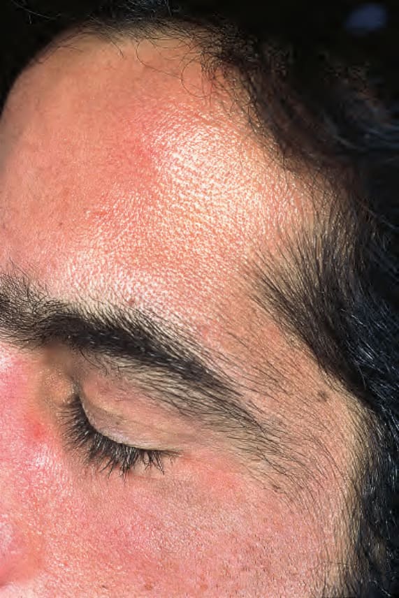
圖 13-105:遲發性皮膚卟啉症 (Porphyria cutanea tarda):如此病人所見的多毛症 (hypertrichosis) 是非常典型的特徵。承蒙英國倫敦 Institute of Dermatology 提供。
Fig. 13.105 Porphyria cutanea tarda: hypertrichosis as seen in this patient is a very typical feature. By courtesy of the Institute of Dermatology, London, UK.
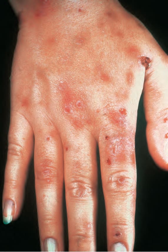
圖 13-106:雜色卟啉症 (Variegate porphyria):手背與手指上存在許多破裂的水疱 (ruptured vesicles)。承蒙愛爾蘭都柏林 Beaumont Hospital 之 G. Murphy, MD 提供。
Fig. 13.106 Variegate porphyria: numerous ruptured vesicles are present on the back of the hand and fingers. By courtesy of G. Murphy, MD, Beaumont Hospital, Dublin, Eire.
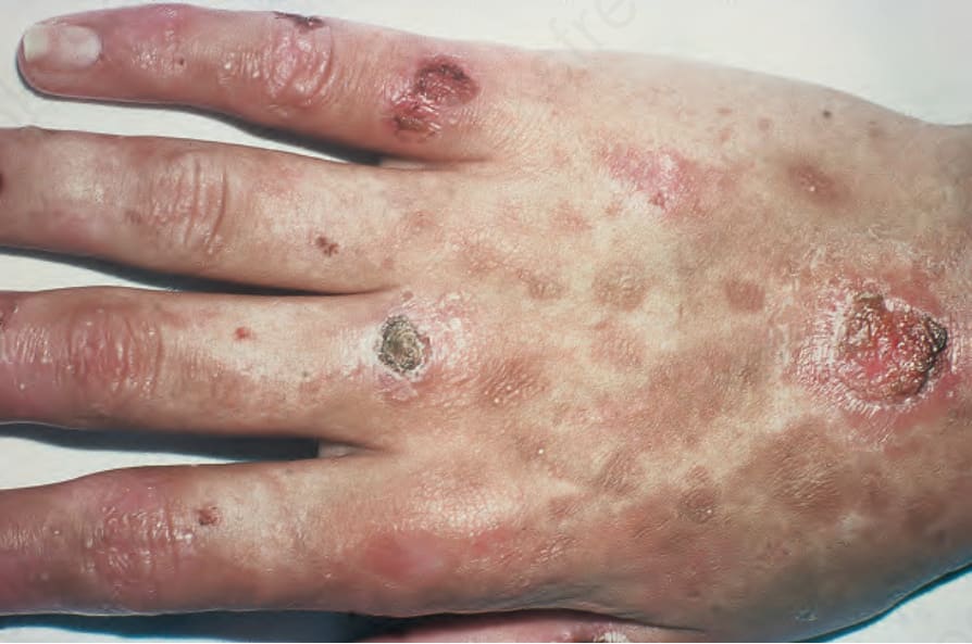
圖 13-107:雜色卟啉症 (Variegate porphyria):有破裂的水疱伴隨瘢痕化與粟粒疹 (milia)。承蒙英國倫敦 Institute of Dermatology 提供。
Fig. 13.107 Variegate porphyria: there are ruptured blisters with scarring and milia. By courtesy of the Institute of Dermatology, London, UK.
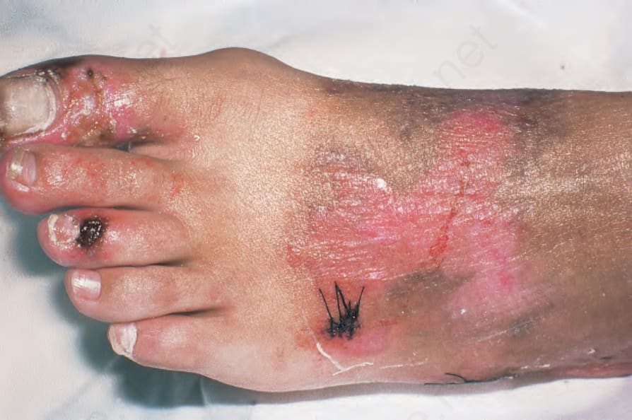
圖 13-108:雜色卟啉症 (Variegate porphyria):注意腳趾與足背上的水疱形成。承蒙英國倫敦 Institute of Dermatology 提供。
Fig. 13.108 Variegate porphyria: note the blistering over the toes and dorsum of the foot. By courtesy of the Institute of Dermatology, London, UK.
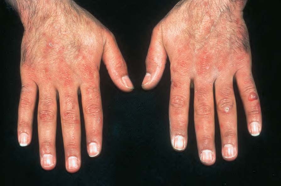
圖 13-109:雜色卟啉症 (Variegate porphyria):左小指上存在一個完整的水疱。其他部位則有明顯的瘢痕化並出現粟粒疹 (milia)。承蒙英國倫敦 Institute of Dermatology 提供。
Fig. 13.109 Variegate porphyria: an intact blister is present on the left little finger. Elsewhere, there is marked scarring and milia are present. By courtesy of the Institute of Dermatology, London, UK.
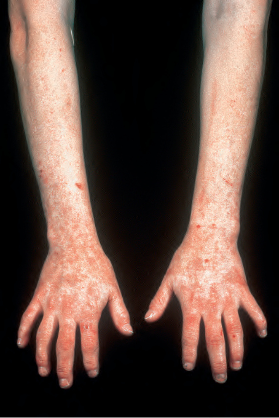
圖 13-110:同型合子雜色卟啉症 (Homozygous variegate porphyria):前臂、手部與手指背側表面有明顯的瘢痕化。承蒙英國倫敦 Institute of Dermatology 提供。
Fig. 13.110 Homozygous variegate porphyria: there is marked scarring of the dorsal surface of the forearms, hands, and fingers. By courtesy of the Institute of Dermatology, London, UK.
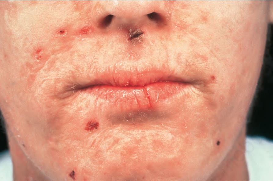
圖 13-111:同型合子雜色卟啉症 (Homozygous variegate porphyria):注意口周糜爛 (perioral erosions) 與瘢痕化。承蒙英國倫敦 Institute of Dermatology 提供。
Fig. 13.111 Homozygous variegate porphyria: note the perioral erosions and scarring. By courtesy of the Institute of Dermatology, London, UK.
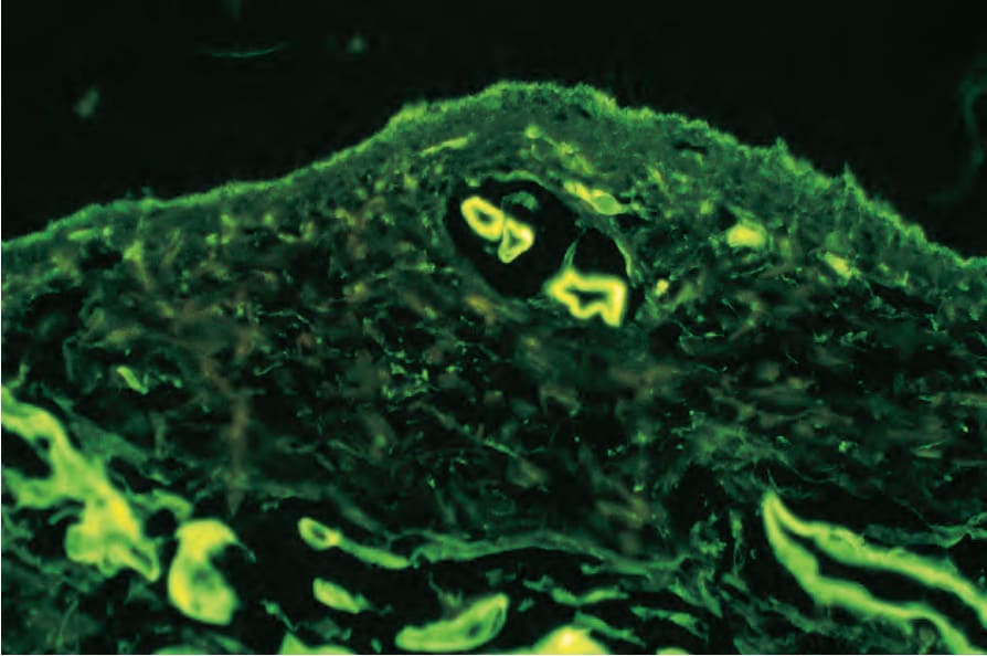
圖 13-112:遲發性皮膚卟啉症 (Porphyria cutanea tarda):淺層血管顯示顯著的 IgG 環周沉積 (circumferential deposition)。
Fig. 13.112 Porphyria cutanea tarda: the superficial blood vessels show striking IgG circumferential deposition.
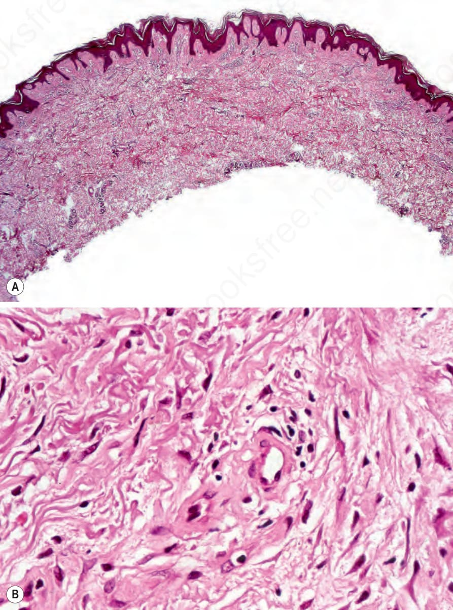
圖 13-113:(A, B) 遲發性皮膚卟啉症 (Porphyria cutanea tarda):淺層血管增厚並呈現玻璃樣化 (hyalinized)。
Fig. 13.113 (A, B) Porphyria cutanea tarda: the superficial vessels are thickened and appear hyalinized.
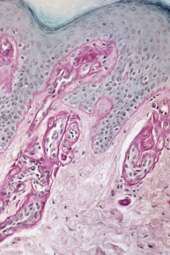
圖 13-114:紅血球生成性原卟啉症 (Erythropoietic protoporphyria):在此 periodic acid–Schiff (PAS) 染色切片中外觀更為戲劇性。
Fig. 13.114 Erythropoietic protoporphyria: the appearances are much more dramatic in this periodic acid–Schiff-stained section.
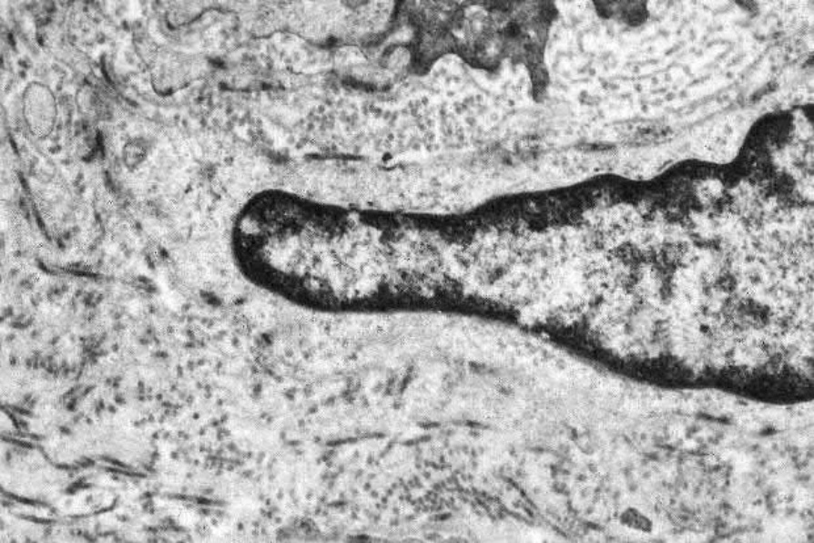
圖 13-115:遲發性皮膚卟啉症 (Porphyria cutanea tarda):此小型真皮血管周圍有顯著的基底膜重複 (basement membrane reduplication)。
Fig. 13.115 Porphyria cutanea tarda: there is striking basement membrane reduplication surrounding this small dermal vessel.
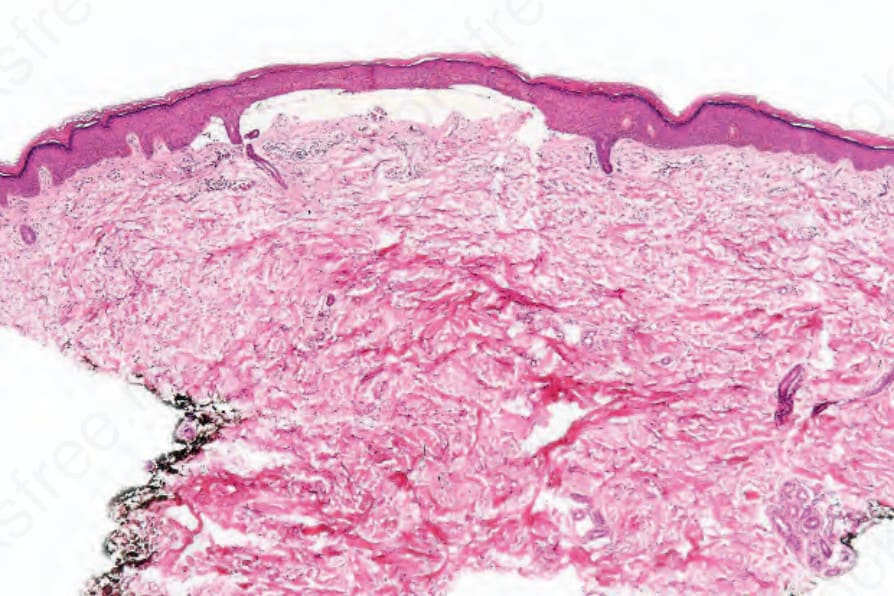
圖 13-116:遲發性皮膚卟啉症 (Porphyria cutanea tarda):存在一個平淡無奇的 (bland) 表皮下水疱。
Fig. 13.116 Porphyria cutanea tarda: a bland subepidermal blister is present.
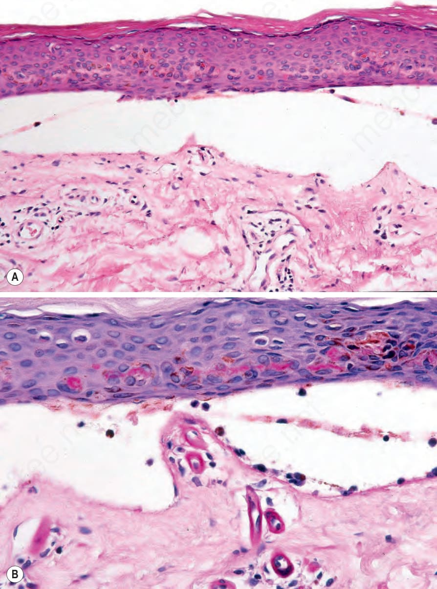
圖 13-117:遲發性皮膚卟啉症 (Porphyria cutanea tarda):(A) 水疱為無細胞 (cell free);(B) 淺層血管增厚 (periodic acid–Schiff)。注意覆蓋於上方表皮中的毛毛蟲體 (caterpillar bodies)。
Fig. 13.117 Porphyria cutanea tarda: (A) the blister is cell free; (B) the superficial vessels are thickened (periodic acid–Schiff). Note the caterpillar bodies in the overlying epidermis.
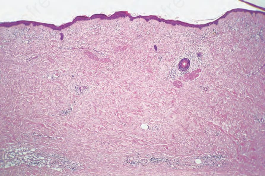
圖 13-118:遲發性皮膚卟啉症 (Porphyria cutanea tarda):整個真皮有強烈的瘢痕化。脂肪包陷 (fat entrapment) 令人聯想到硬皮病 (scleroderma)。
Fig. 13.118 Porphyria cutanea tarda: there is intense scarring of the entire dermis. The fat entrapment is reminiscent of scleroderma.
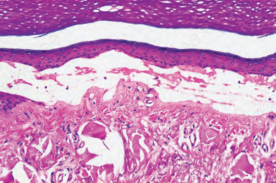
圖 13-119:遲發性皮膚卟啉症 (Porphyria cutanea tarda):此水疱深部有膠樣粟粒疹樣 (colloid milium-like) 的日光性彈力纖維變性 (solar elastosis)。
Fig. 13.119 Porphyria cutanea tarda: there is colloid milium-like solar elastosis deep to this blister.
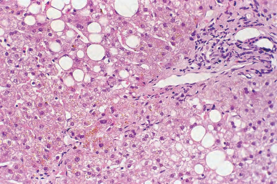
圖 13-120:遲發性皮膚卟啉症 (Porphyria cutanea tarda):除了脂肪變性 (fatty change) 與輕度慢性發炎外,可見棕色的尿卟啉結晶 (uroporphyrin crystals)。
Fig. 13.120 Porphyria cutanea tarda: in addition to fatty change and mild chronic inflammation, brown uroporphyrin crystals are evident.
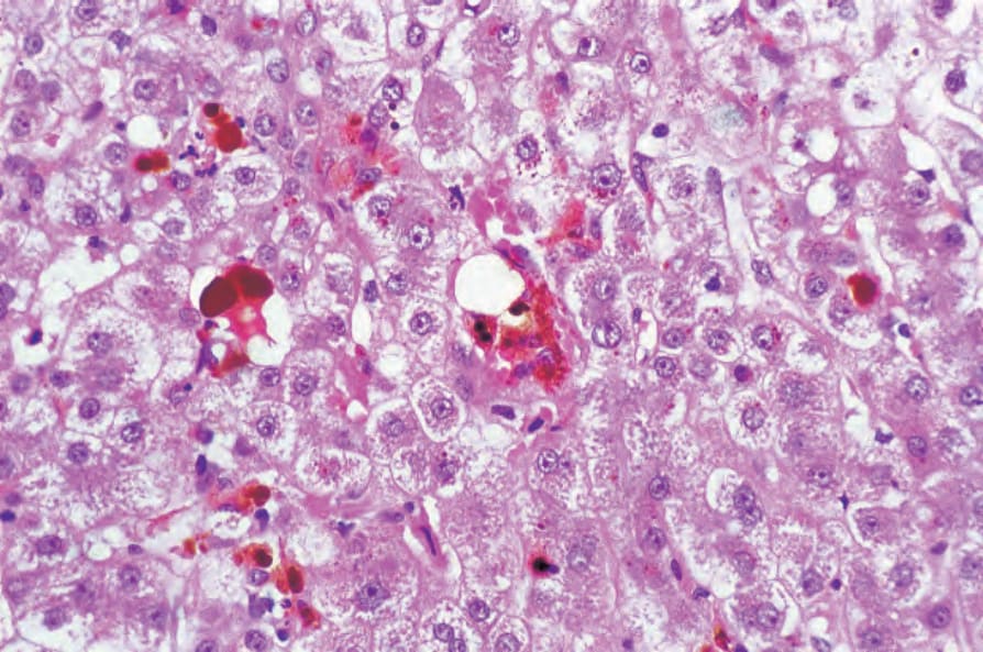
圖 13-121:紅血球生成性原卟啉症 (Erythropoietic protoporphyria):庫佛細胞 (Kupffer cells) 內含有豐富的棕色色素。承蒙英國倫敦 St Thomas’ Hospital 之 D.R. Davies, MD 提供。
Fig. 13.121 Erythropoietic protoporphyria: the Kupffer cells contain abundant brown pigment. By courtesy of D.R. Davies, MD, St Thomas’ Hospital, London, UK.