扁平黃色瘤 (Planar Xanthomata)
臨床特徵 (Clinical Features)
- 扁平黃色瘤 (planar xanthomata) 典型為柔軟的黃色真皮斑點 (macule) 或斑塊 (plaque),最常出現於眼周,在此部位稱為 xanthelasmata(瞼黃瘤)(Fig. 13.18)。
- 約 50% 的 xanthelasmata 病人合併有高脂血症 (hyperlipidemia)(hypercholesterolemia 或 HPL type III),且常伴隨膽固醇性角膜環 (cholesterol corneal arcus)。許多在常規檢驗上生化看似正常者,於更詳細的分析中卻顯示有細微的脂質代謝異常。較年輕的病人發生冠狀動脈粥狀硬化 (coronary artery atherosclerosis) 的風險特別增加。
- 當病灶非常廣泛(瀰漫性或泛發性扁平黃色瘤病 (diffuse or generalized plane xanthomatosis)),並合併頭頸部、偶爾上軀幹及上臂的橙黃色扁平黃色瘤時,可能伴隨某種全身性疾病,例如帶有副蛋白血症 (paraproteinemia) 的多發性骨髓瘤 (multiple myeloma)、冷凝球蛋白血症 (cryoglobulinemia)、良性副蛋白血症 (benign paraproteinemia),或較少見的白血病 (leukemia) 與類風濕性關節炎 (rheumatoid arthritis)(necrobiotic xanthogranuloma,壞死性黃色肉芽腫)(Fig. 13.19)。更為罕見的關聯包括特發性 Bence Jones 蛋白尿 (idiopathic Bence Jones proteinuria)、Sézary syndrome、Castleman disease、復發性多軟骨炎 (relapsing polychondritis)、後天性掌蹠角化症 (acquired palmoplantar keratoderma)、成人 T 細胞白血病/淋巴瘤 (adult T-cell leukemia/lymphoma) 與 Takayasu disease。曾有文獻記載一名單株免疫球蛋白血症 (monoclonal gammopathy) 病人,其皮膚病灶兼具 plane xanthoma 與類澱粉沉積症 (amyloidosis) 的特徵。後者該病例亦合併骨髓瘤 (myeloma)。
- 在骨髓瘤併扁平黃色瘤的病例中,已證實血清脂蛋白 (serum lipoproteins) 與副蛋白 (paraprotein) 之間會形成複合物,提示此交互作用可能誘發高脂血症與黃色瘤形成。瀰漫性扁平黃色瘤病人的血清脂質濃度可為正常或升高。扁平黃色瘤可出現於牙齦 (gingiva),在此部位通常與高脂血症有關。曾有一例罕見病例描述於一名嬰兒,以正常血脂 (normolipemic) 的丘疹及結節性病灶呈現,進展為扁平黃色瘤,並導致自發性消退。瀰漫性扁平正常血脂黃色瘤 (diffuse plane normolipemic xanthomata) 合併黏膜與結膜侵犯及主動脈瓣黃色瘤病 (aortic valve xanthomatosis) 偶可發生。曾有報告於一名全身性紅斑性狼瘡 (systemic lupus erythematosus) 病人出現臨床上類似扁平黃色瘤的病灶,但組織學上顯示膠原纖維束 (collagen bundles) 退化並繼發脂肪沉積。
- 見於 LDL 升高病人、且為同合子家族性高膽固醇血症 (homozygous familial hypercholesterolemia) 病徵性 (pathognomonic) 表現的皺褶間黃色瘤 (intertriginous xanthomata),呈現為黃色丘疹與斑塊,常具鵝卵石樣 (cobblestone) 外觀。這些病灶發生於指間蹼 (finger webspaces),較少見於腋窩、肘前窩與膕窩。它們與早發且嚴重的粥狀硬化有特別高的關聯。皺褶間黃色瘤也可罕見地見於異合子家族性高膽固醇血症 (heterozygous familial hypercholesterolemia)。
- 以掌部及手指皮膚皺紋處的黃橙色斑點呈現的扁平黃色瘤(xanthoma striatum palmare,掌紋黃色瘤),可診斷家族性異常β脂蛋白血症 (familial dysbetalipoproteinemia)(HPL type III,broad beta disease)(Fig. 13.20),其肇因於脂蛋白 ApoE 的異常(homozygous ApoE2/E2)。此導致肝臟與巨噬細胞對脂蛋白殘餘顆粒 (lipoprotein remnant particles) 的攝取受損,進而產生 HPL 與粥狀硬化增加。有趣的是,家族性異常β脂蛋白血症的傾向存在於 1% 的人口中,但似乎需要第二個脂質異常才會誘發症狀。
- 膽汁鬱積 (cholestasis) 性的扁平黃色瘤,例如因原發性膽汁性肝硬化 (primary biliary cirrhosis) 及膽道閉鎖 (biliary atresia) 所致者,呈現為界限分明的米色至橙色斑塊,特別常見於手與足,但亦可出現於他處。它們也可發生於糖尿病 (diabetes mellitus) 病人,並曾被描述於慢性移植物對抗宿主病 (chronic graft-versus-host disease) 所致膽汁鬱積的情境中。
- 扁平黃色瘤亦曾被描述為 HDL 缺乏 (HDL deficiency) 的一項特徵。
組織病理特徵 (Histologic Features)
- 在扁平黃色瘤中,特徵性的充滿脂質的泡沫細胞 (lipid-laden foam cells) 位於淺層真皮內 (Figs 13.21–13.23)。纖維化 (fibrosis) 極少。在罕見病例中,其組織學可能與 necrobiotic xanthogranuloma 的表現重疊。
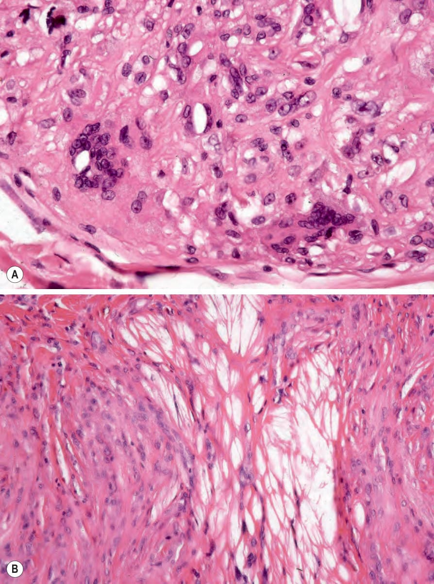
圖 13-17:(A, B) 結節性黃色瘤 (tuberous xanthoma):除黃色瘤細胞 (xanthoma cells) 外,偶可見含膽固醇裂隙 (cholesterol clefts) 的異物巨細胞 (foreign body giant cells)。脂質於組織處理過程中已被溶出。
Fig. 13.17 (A, B) Tuberous xanthoma: in addition to xanthoma cells, occasionally there are foreign body giant cells containing cholesterol clefts. The lipid has been dissolved out during processing.
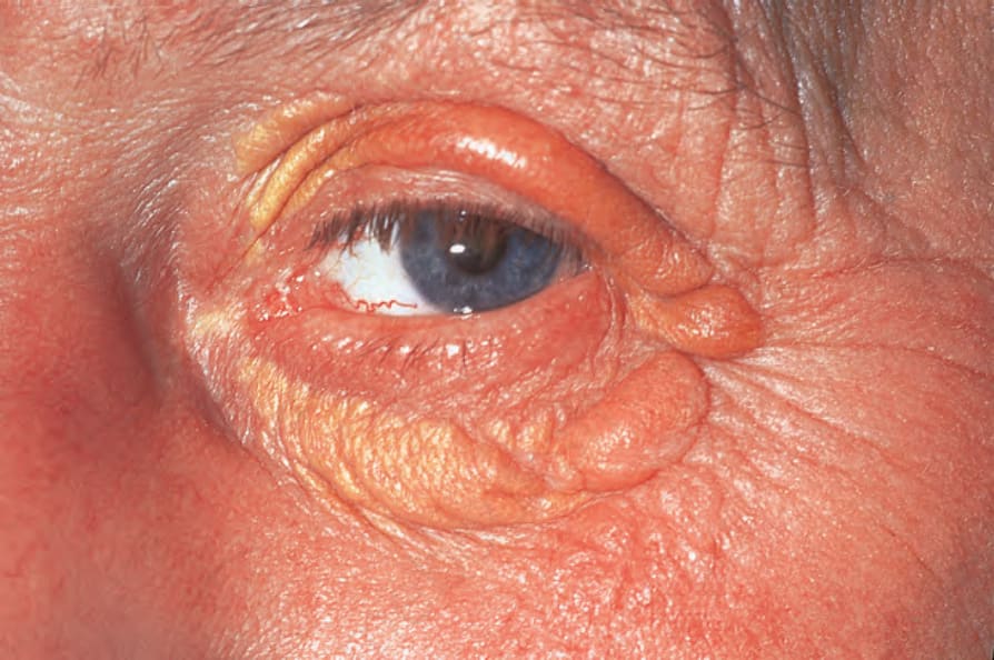
圖 13-18:瞼黃瘤 (xanthelasmata):注意黃色的眼周斑塊。這是高膽固醇血症 (hypercholesterolemia) 的常見表現。By courtesy of the Institute of Dermatology, London, UK.
Fig. 13.18 Xanthelasmata: note the yellow, periorbital plaques. These are a common manifestation of hypercholesterolemia. By courtesy of the Institute of Dermatology, London, UK.
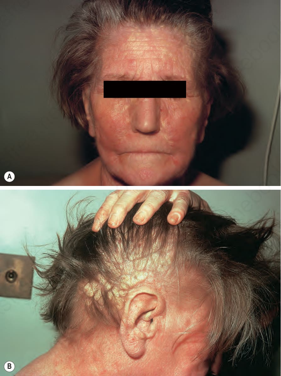
圖 13-19:扁平黃色瘤 (planar xanthoma):(A) 廣泛分布於前額、眼瞼與兩頰的病灶;(B) 頭皮上廣泛的黃色斑塊。此種外觀應促使尋找是否合併副蛋白血症 (paraproteinemia)。By courtesy of R.A. Marsden, MD, St George’s Hospital, London, UK.
Fig. 13.19 Planar xanthoma: (A) widely distributed lesions over the forehead, eyelids, and cheeks; (B) extensive yellow plaques on the scalp. This appearance should prompt a search for an associated paraproteinemia. By courtesy of R.A. Marsden, MD, St George’s Hospital, London, UK.
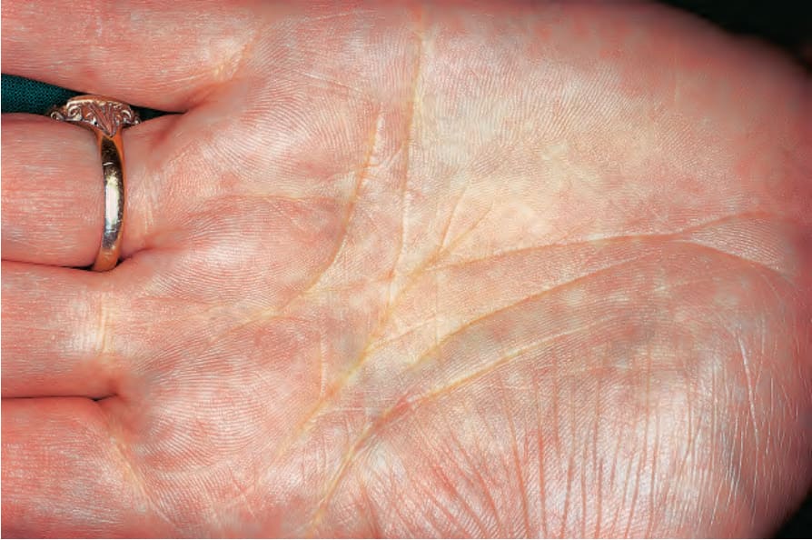
圖 13-20:扁平黃色瘤 (planar xanthoma):掌部病灶呈現為散在的斑點,於皮膚皺紋處增強。By courtesy of R.A. Marsden, MD, St George’s Hospital, London, UK.
Fig. 13.20 Planar xanthoma: palmar lesions presenting as discrete macules with accentuation in the skin creases. By courtesy of R.A. Marsden, MD, St George’s Hospital, London, UK.
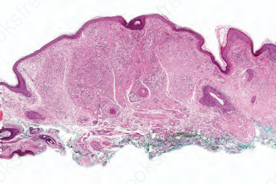
圖 13-21:扁平黃色瘤 (planar xanthoma):上層真皮內可見緻密的浸潤。
Fig. 13.21 Planar xanthoma: a dense infiltrate is present in the upper dermis.
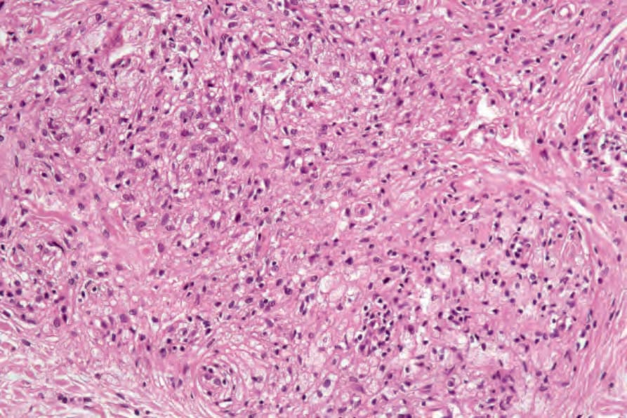
圖 13-22:扁平黃色瘤 (planar xanthoma):可見未脂化 (nonlipidized) 與脂化 (lipidized) 組織球 (histiocytes) 的混合存在。
Fig. 13.22 Planar xanthoma: there is an admixture on nonlipidized and lipidized histiocytes.