結節性黃色瘤 (Tuberous Xanthomata)
臨床特徵 (Clinical Features)
- 結節性黃色瘤 (tuberous xanthomata) 為堅實的黃—紅色丘疹與結節,最常見於膝、肘及臀部的伸側面 (extensor aspect)(Figs 13.11–13.13)。
- 病灶有時也發生於手部與手掌。少數病例則侵犯臉頰與鼻部。
- 最具特徵性者見於第 III 型家族性異常β脂蛋白血症 (familial dysbetalipoproteinemia type III),並有特別的周邊血管疾病 (peripheral vascular disease) 風險。另有四種疾病亦可能以結節性黃色瘤病 (tuberous xanthomatosis) 為特徵:
- 同型合子家族性高膽固醇血症 (homozygous familial hypercholesterolemia),
- 腦腱黃瘤病 (cerebrotendinous xanthomatosis),
- β-穀固醇血症 (β-sitosterolemia),
- 第 IV 型 HPL (type IV HPL)。
- tuberous xanthomata 也發生於次發性高脂血症 (secondary hyperlipidemia)(例如腎病症候群 nephrotic syndrome 或甲狀腺功能低下 hypothyroidism 所致)。蛋白酶抑制劑 (protease inhibitors) 可能引起高脂血症,並有報告指出 ritonavir 可誘發結節性與肌腱性黃色瘤 (tuberous and tendinous xanthoma) 病灶。
- 臨床上,tuberous xanthomata 偶爾與持久性隆起性紅斑 (erythema elevatum diutinum) 的病灶相似。文獻曾描述血脂正常的結節性與肌腱性黃色瘤 (tuberous and tendinous normolipemic xanthomata),但似乎在足夠的追蹤下,這些病人通常會發展出某種形式的高脂血症。
- 膽固醇沉積性纖維組織細胞瘤 (cholesterotic fibrous histiocytomas) 可能與高脂血症相關,且在臨床與組織學上常模擬 tuberous xanthoma。曾有文獻記載一例罕見的未分化多形性肉瘤 (undifferentiated pleomorphic sarcoma;惡性纖維組織細胞瘤 malignant fibrous histiocytoma),於一名第 IIA 型 HPL (type IIA HPL) 病人身上臨床上表現為 tuberous xanthoma。
組織病理特徵 (Histopathology)
- tuberous xanthomata 由位於網狀真皮 (reticular dermis) 內、有時也在皮下脂肪 (subcutaneous fat) 內的多發性結節構成(Fig. 13.14)。
- 其外觀依其演變階段而異(Fig. 13.15)。早期病灶以黃色瘤細胞 (xanthoma cells) 為主,但隨成熟而有纖維化 (fibrosis) 接續出現(Fig. 13.16)。
- 偶爾可見含膽固醇裂隙 (cholesterol clefts) 的異物巨細胞肉芽腫 (foreign body giant cell granulomata),有時也明顯可見血管周圍慢性發炎細胞浸潤 (perivascular chronic inflammatory cell infiltrate)(Fig. 13.17)。
鑑別診斷 (Differential Diagnosis)
- 主要發生於踝部周圍、脂質含量豐富的纖維組織細胞瘤 (heavily lipidized fibrous histiocytomas),在組織學上可能模擬 tuberous xanthoma。後者(tuberous xanthoma)缺乏纖維組織細胞瘤的結構,具有結節狀/多結節狀的生長型態及不等程度的纖維化,且缺乏表皮增生 (epidermal hyperplasia),以及缺乏見於纖維組織細胞瘤周邊的膠原蛋白玻璃樣變 (hyalinization of collagen) 型態。
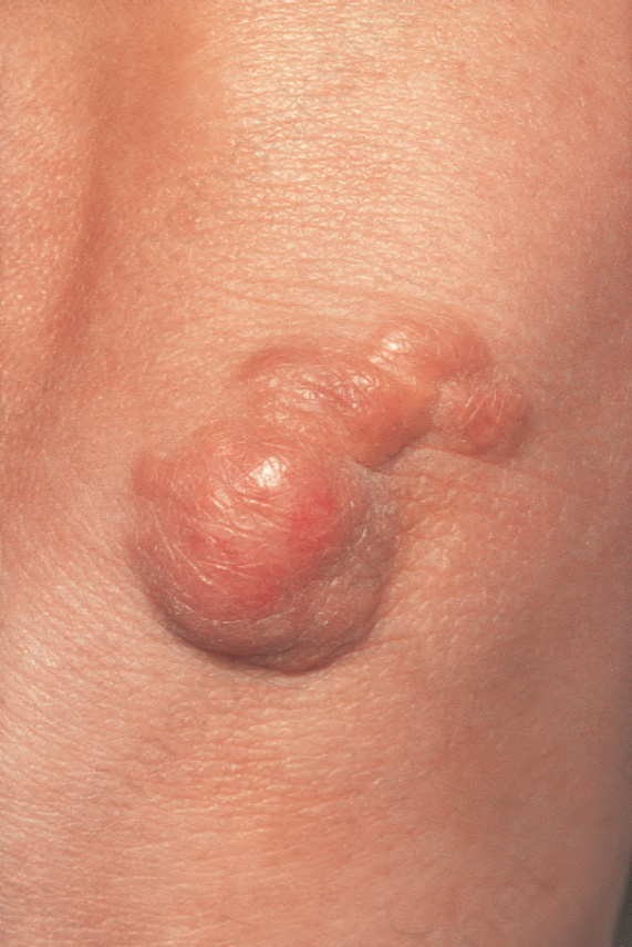
圖 13-11:結節性黃色瘤 (tuberous xanthoma):肘部上方堅實的紅斑性結節。By courtesy of R.A. Marsden, MD, St George’s Hospital, London, UK.
Fig. 13.11 Tuberous xanthoma: firm erythematous nodules over the elbow. By courtesy of R.A. Marsden, MD, St George’s Hospital, London, UK.
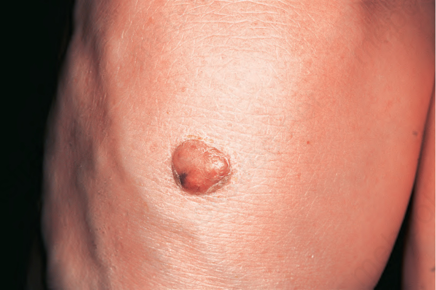
圖 13-12:結節性黃色瘤 (tuberous xanthoma):手臂背側的紅斑性結節。By courtesy of the Institute of Dermatology, London, UK.
Fig. 13.12 Tuberous xanthoma: erythematous nodule on the back of the arm. By courtesy of the Institute of Dermatology, London, UK.
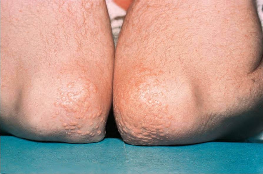
圖 13-13:結節性黃色瘤 (tuberous xanthoma):此例中,肘部出現噴發性 (eruptive) 病灶。By courtesy of the Institute of Dermatology, London, UK.
Fig. 13.13 Tuberous xanthoma: in this example, eruptive lesions are present on the elbows. By courtesy of the Institute of Dermatology, London, UK.
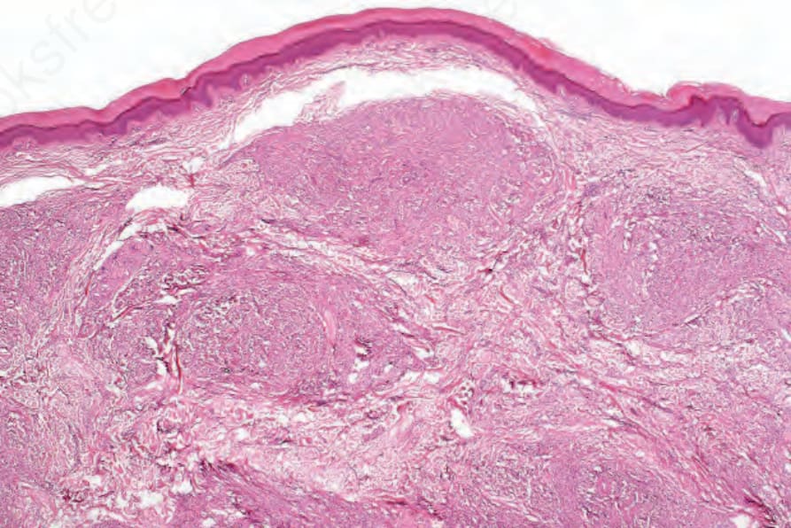
圖 13-14:結節性黃色瘤 (tuberous xanthoma):網狀真皮 (reticular dermis) 內可見數個結節。
Fig. 13.14 Tuberous xanthoma: several nodules are present in the reticular dermis.
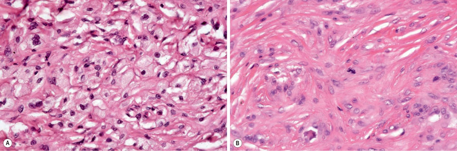
圖 13-15:結節性黃色瘤 (tuberous xanthoma):(A) 浸潤由均勻一致的黃色瘤細胞 (xanthoma cells) 組成,其特徵為淡染、泡沫狀的細胞質與小而位於中央的囊泡狀核 (vesicular nuclei);(B) 常可見到偶發的正常有絲分裂 (normal mitoses)。
Fig. 13.15 Tuberous xanthoma: (A) the infiltrate is composed of uniform xanthoma cells characterized by pale, foamy cytoplasm and small central vesicular nuclei; (B) occasional normal mitoses are commonly present.
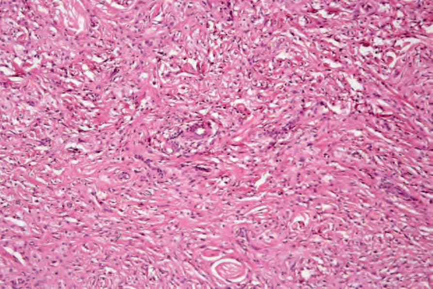
圖 13-16:結節性黃色瘤 (tuberous xanthoma):可見顯著的瘢痕形成 (scarring)。
Fig. 13.16 Tuberous xanthoma: there is marked scarring.
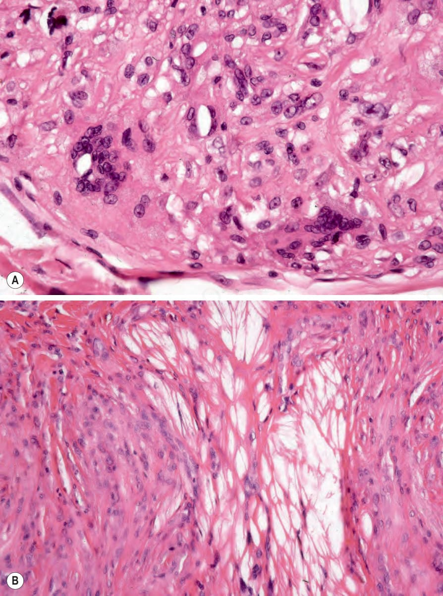
圖 13-17:(A, B) 結節性黃色瘤 (tuberous xanthoma):除黃色瘤細胞 (xanthoma cells) 外,偶爾可見含膽固醇裂隙 (cholesterol clefts) 的異物巨細胞 (foreign body giant cells)。脂質在組織處理 (processing) 過程中已被溶出。
Fig. 13.17 (A, B) Tuberous xanthoma: in addition to xanthoma cells, occasionally there are foreign body giant cells containing cholesterol clefts. The lipid has been dissolved out during processing.