Tuberous xanthomata
Tuberous xanthomata
Clinical features Tuberous xanthomata are firm yellow–red papules and nodules, which are found most frequently on the extensor aspect of the knees, elbows, and buttocks (Figs 13.11–13.13).1 Lesions sometimes also occur on the hands and palms.2 Rare cases involve cheeks and nose.3 They are most characteristically seen in familial dysbetalipoproteinemia type III, and there is a particular risk of peripheral vascular disease.4 Four other conditions may also be characterized by tuberous xanthomatosis:
• homozygous familial hypercholesterolemia,
• cerebrotendinous xanthomatosis,
• β-sitosterolemia,1
• type IV HPL.5
Tuberous xanthomata also occur in secondary hyperlipidemia (e.g., due to the nephrotic syndrome or hypothyroidism). Protease inhibitors may cause hyperlipidemia, and ritonavir has been reported to induce tuberous and tendinous xanthoma lesions.6 Clinically, tuberous xanthomata occasionally resemble the lesions of erythema elevatum diutinum. Tuberous and tendinous normolipemic xanthomata have been described but it seems that, with adequate follow-up, patients usually develop some form of hyperlipidemia.7
Cholesterotic fibrous histiocytomas may be associated with hyperlipidemia and often simulate a tuberous xanthoma clinically and histologically.8 A rare case of undifferentiated pleomorphic sarcoma (malignant fibrous histiocytoma) clinically presenting as a tuberous xanthoma in a patient with type IIA HPL has been documented.9
Histologic features Tuberous xanthomata consist of multiple nodules in the reticular dermis and sometimes the subcutaneous fat (Fig. 13.14). Their appearance varies, depending upon their stage of evolution (Fig. 13.15). Xanthoma cells predominate in early lesions, but with maturity fibrosis supervenes (Fig. 13.16). On occasion, foreign body giant cell granulomata containing cholesterol
565 The hyperlipidemias
A
B
clefts are seen and a perivascular chronic inflammatory cell infiltrate is sometimes evident (Fig. 13.17).
Differential diagnosis Heavily lipidized fibrous histiocytomas that tend to occur mainly around the ankle may histologically mimic tuberous xanthoma.10 The latter lesions lack the architecture of fibrous histiocytomas, have a nodular/multinodular growth pattern with variable fibrosis and lack epidermal hyperplasia and the hyalinization of collagen pattern seen at the periphery of fibrous histiocytomas.
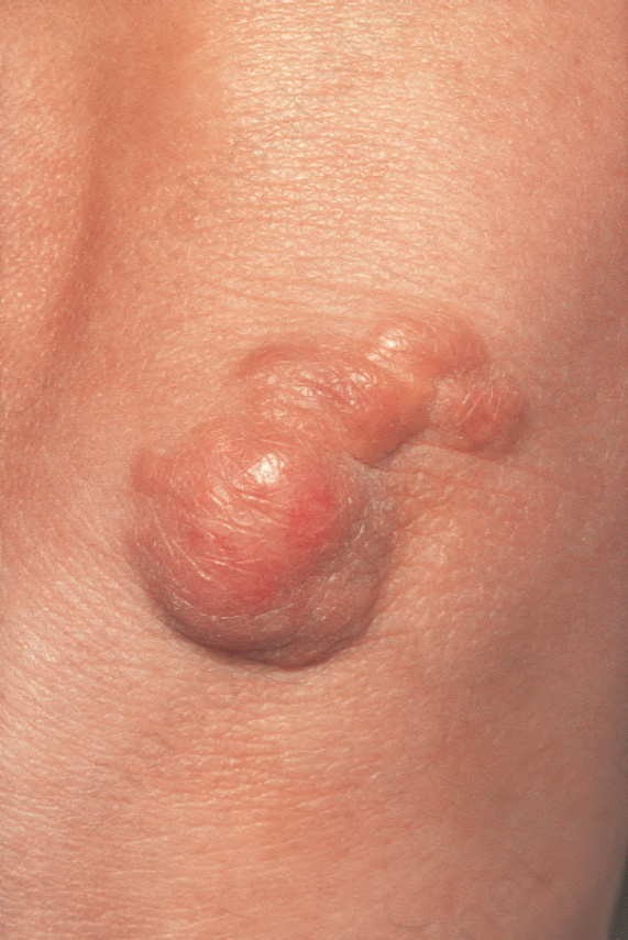
Fig. 13.11 Tuberous xanthoma: firm erythematous nodules over the elbow. By courtesy of R.A. Marsden, MD, St George’s Hospital, London, UK.
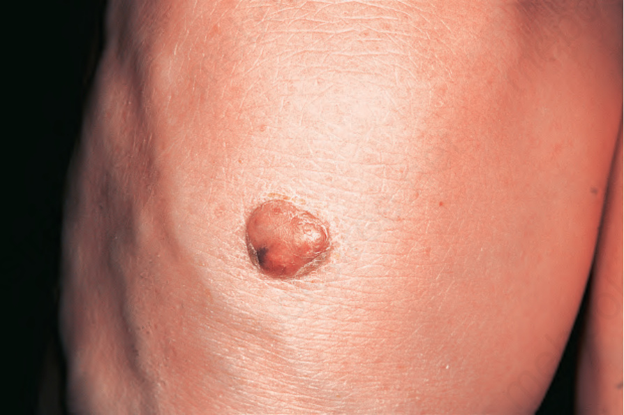
Fig. 13.12 Tuberous xanthoma: erythematous nodule on the back of the arm. By courtesy of the Institute of Dermatology, London, UK.
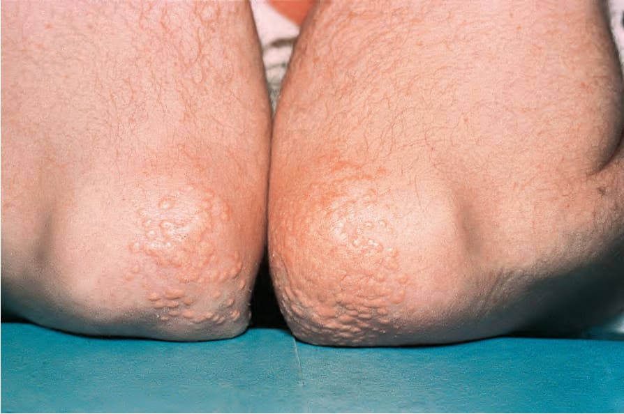
Fig. 13.13 Tuberous xanthoma: in this example, eruptive lesions are present on the elbows. By courtesy of the Institute of Dermatology, London, UK.
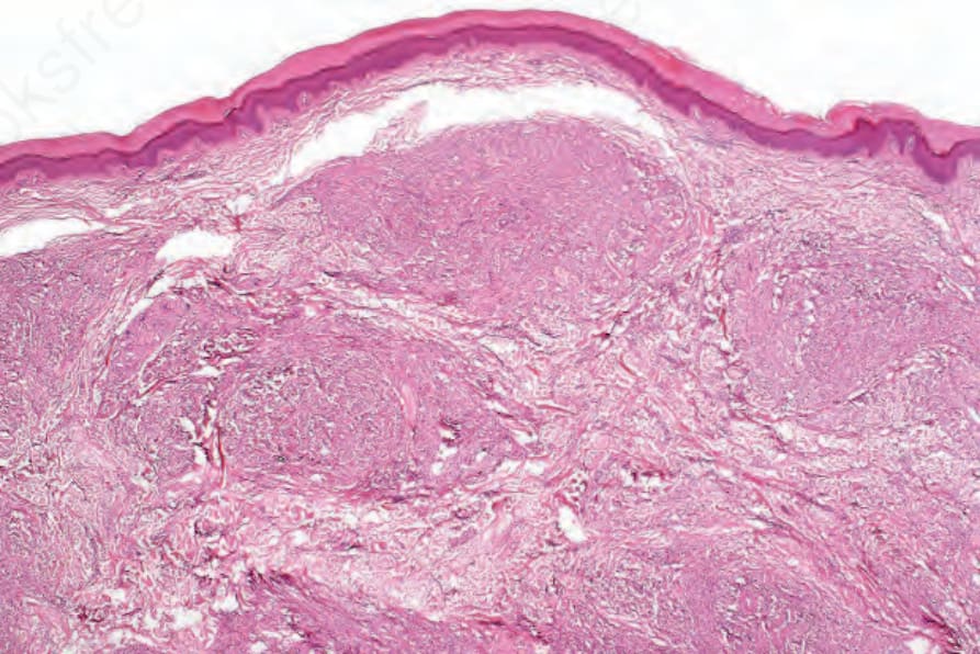
Fig. 13.14 Tuberous xanthoma: several nodules are present in the reticular dermis.
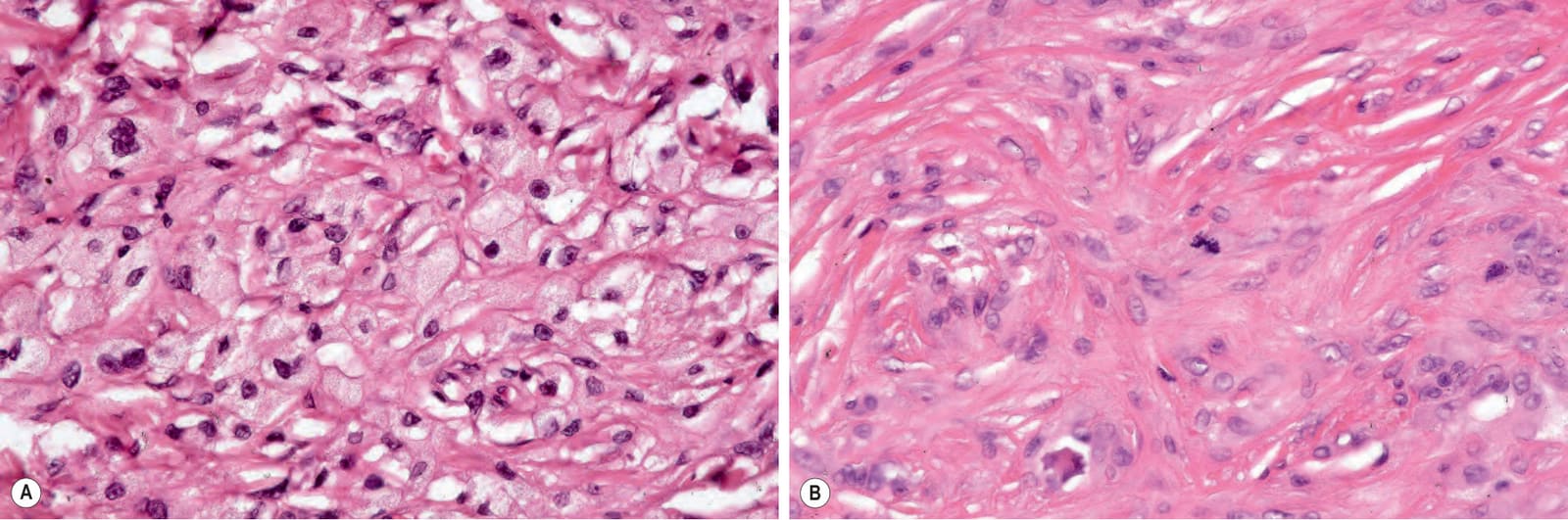
Fig. 13.15 Tuberous xanthoma: (A) the infiltrate is composed of uniform xanthoma cells characterized by pale, foamy cytoplasm and small central vesicular nuclei; (B) occasional normal mitoses are commonly present.
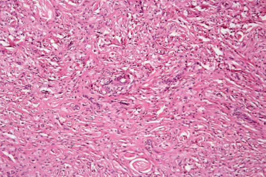
Fig. 13.16 Tuberous xanthoma: there is marked scarring.
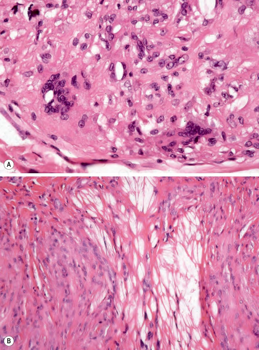
Fig. 13.17 (A, B) Tuberous xanthoma: in addition to xanthoma cells, occasionally there are foreign body giant cells containing cholesterol clefts. The lipid has been dissolved out during processing.