生殖器黑色素沉著症 (Genital Melanosis)
組織病理特徵 (Histopathology)
- 生殖器黑色素沉著症 (genital melanosis) 的特徵為基底層角質細胞 (basal keratinocytes) 與黑色素細胞 (melanocytes) 的色素增加 (Fig. 12.155)。
- 依定義,黑色素細胞數量並未增加。然而,若黑色素細胞數量確實增加,則「生殖器雀斑樣痣 (genital lentiginosis)」一詞或許更為恰當。在實際情況中,此差異僅屬學術性質。沒有交界活性 (junctional activity) 的證據,且在多數病例中,並無細胞學異型性 (cytological atypia)。
- 然而,有人提出,在黑色素細胞數量增加且具任何程度細胞學異型性的病灶中,可能與皮膚其他部位的黑色素瘤 (melanomas) 有關聯。其下方真皮內可能明顯可見載有色素的巨噬細胞 (pigment-laden macrophages)。一個重要的陷阱是合併存在的 LS(lichen sclerosus),因為其真皮變化可能類似一個完全消退的黑色素瘤 (melanoma)。
- Dowling-Degos disease 的組織學特徵具有特異性:中度正角化 (orthokeratosis) 或角化過度 (hyperkeratosis)、乳頭上皮 (suprapapillary epithelium) 變薄、表皮突 (rete ridges) 呈「鹿角狀 (antler-like)」分支、基底層色素沉著但黑色素細胞數量未增加(S100 正常)。
臨床特徵 (Clinical Features)
- 生殖器黑色素沉著症的特徵為色素沉著,而無明顯的先前發炎性皮膚病 (inflammatory dermatosis) 證據。然而,在男性,臨床上通常會懷疑為過去或慢性低度的 LS(lichen sclerosus)、LP(lichen planus)、ZB(Zoon balanitis)或非特異性龜頭包皮炎 (balanoposthitis)。
- 色素沉著的強度可能不一,且通常呈不規則。此問題通常侵犯數個部位,包括皮膚與黏膜表面。色素沉著緩慢發展,且範圍可能非常廣泛。在多數生殖器黑色素沉著症的病例中,病灶會趨於穩定且可能消退。
- 有時也可能發生單一病灶 (unifocal lesions)。小型而界線分明的單發或多發病灶通常被描述為生殖器黑色素斑 (genital melanotic macules)。最常見的部位為陰莖龜頭與陰莖體,以及外陰內側面(包括前庭 vestibule)(Figs 12.153 and 12.154)。病灶也可能侵犯陰道與子宮頸。
- 此疾病被認為是良性的,但有罕見的零星報告指出黑色素瘤 (melanoma) 在黑色素沉著區域繼發發生。黑色素瘤在外陰黑色素沉著症 (vulval melanosis) 的情境下罕見發生,這可能是因為在後者中黑色素細胞數量並未增加,僅有基底細胞層的色素過度沉著 (basal cell layer hyperpigmentation)。另一方面,陰莖病灶則常顯示出……(按:原文於此處中斷)
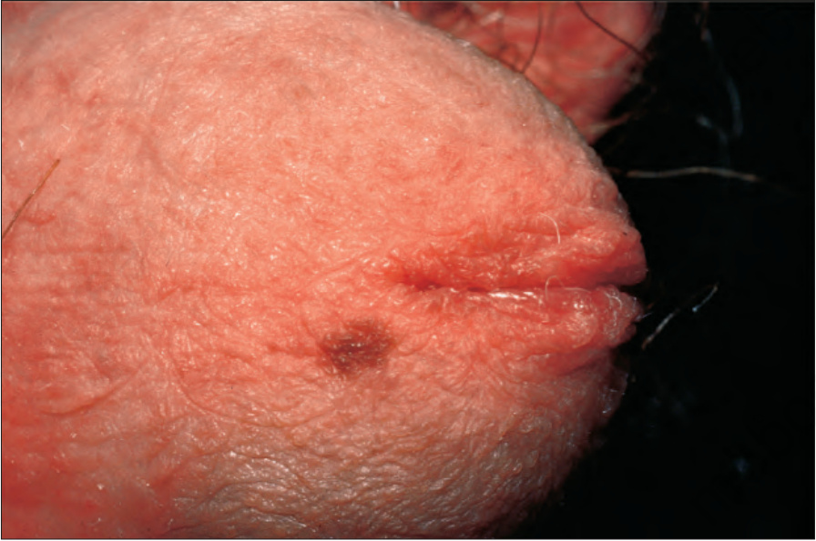
圖 12-153:陰莖黑色素斑 (Penile melanotic macule):陰莖龜頭上有一個小型不規則的色素性斑 (pigmented macule)。承蒙英國倫敦皮膚科研究所 (Institute of Dermatology, London, UK) 提供。
Fig. 12.153 Penile melanotic macule: there is a small irregular pigmented macule on the glans penis. By courtesy of the Institute of Dermatology, London, UK.
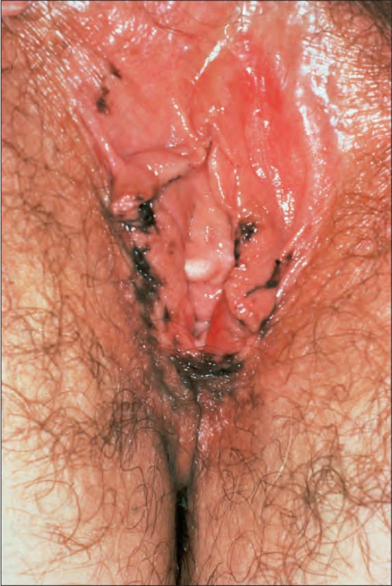
圖 12-154:外陰黑色素沉著症 (Vulval melanosis):外陰及鄰近皮膚上有多發不規則的色素性斑 (pigmented macules)。承蒙英國倫敦皮膚科研究所 (Institute of Dermatology, London, UK) 提供。
Fig. 12.154 Vulval melanosis: there are multiple irregular pigmented macules on the vulva and adjacent skin. By courtesy of the Institute of Dermatology, London, UK.
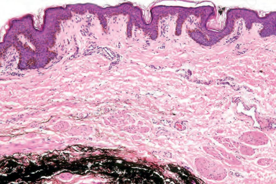
圖 12-155:外陰黑色素沉著症 (Vulval melanosis):可見明顯的基底細胞色素沉著 (basal cell pigmentation)。
Fig. 12.155 Vulval melanosis: there is marked basal cell pigmentation.
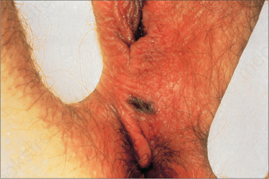
圖 12-156:非典型外陰痣 (Atypical vulval nevus):會陰部 (perineum) 有一個不規則且深度色素沉著的病灶。承蒙英國倫敦皮膚科研究所 (Institute of Dermatology, London, UK) 提供。
Fig. 12.156 Atypical vulval nevus: there is an irregular darkly pigmented lesion on the perineum. By courtesy of the Institute of Dermatology, London, UK.
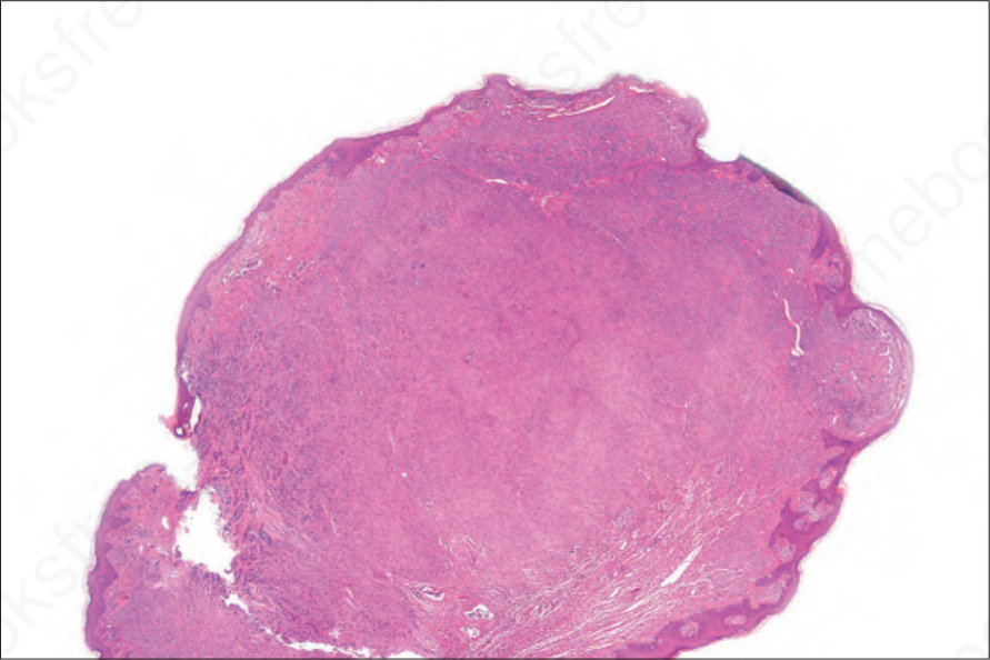
圖 12-157:非典型外陰痣 (Atypical vulval nevus):一名 17 歲女性息肉樣病灶 (polypoid lesion) 的掃描視野。注意視野上方有大量色素沉著的巢狀結構 (heavily pigmented nests)。承蒙英國倫敦皮膚科研究所 (Institute of Dermatology, London, UK) 提供。
Fig. 12.157 Atypical vulval nevus: scanning view of a polypoid lesion from a 17-year-old female. Note the heavily pigmented nests at the top of the field. By courtesy of the Institute of Dermatology, London, UK.