臨床特徵 (Clinical Features)
- 軟斑症 (malacoplakia,意為「軟斑塊」) 是巨噬細胞 (macrophage) 在對細菌病原體的發炎反應中功能受損的結果,最常見的病原為大腸桿菌 (Escherichia coli),但其他菌種亦可參與,包括 S. aureus、Proteus、Pseudomonas、Klebsiella 與 mycobacteria。
- 在 40% 的患者中發現有原發性或後天性免疫缺陷 (primary or acquired immunodeficiency)。後者包括 HIV 與實體器官移植 (solid organ transplantation),主要為腎臟移植,極罕見為心臟移植。
- 最常被描述侵犯泌尿道 (urinary tract),但也可侵犯許多其他器官,包括胃腸系統、淋巴結、下生殖道 (lower genital tract)、腦、骨、肺、腎上腺與皮膚。
- 皮膚病灶可位於真皮 (dermal) 與/或皮下 (subcutaneous),約 41% 的病例最常見於生殖器周圍 (尤其是外陰 vulva) 或會陰 (perineum),但也可見於其他部位,包括 20% 患者的軀幹、20% 的頭頸部、10% 的四肢與 10% 的腋窩。
- 也有罕見報告侵犯 Bartholin gland。可能發生陰道出血。曾有一位患者被描述合併子宮頸的腹股溝肉芽腫 (granuloma inguinale)。
- 可侵犯多個皮膚部位,在一個特殊案例中,皮膚受累並延伸至顱蓋骨 (calvarium) 與腦實質 (brain parenchyma)。皮膚表現多變,包括丘疹 (papules)、斑塊 (plaques)、息肉 (polyps)、潰瘍 (ulcers) 與竇道 (sinuses)。
- 潛在或相關疾病通常與免疫抑制 (immunosuppression) 相關,包括癌 (carcinoma)、類風濕性關節炎 (rheumatoid arthritis)、系統性紅斑狼瘡 (systemic lupus erythematosus)、C 型肝炎 (hepatitis C)、結節病 (sarcoidosis)、白血病 (leukemia)、淋巴瘤 (lymphoma) 與移植 (transplantation)。皮膚病灶為非進行性 (nonprogressive),但典型為持續性 (persistent)。
致病機轉與組織學特徵 (Pathogenesis and Histologic Features)
- 軟斑症的特徵為融合成片的組織球 (confluent sheets of histiocytes),具有嗜伊紅顆粒狀細胞質 (eosinophilic granular cytoplasm) 與小型、通常偏心 (eccentric) 的細胞核。
- 具有特徵性的細胞質內、鈣化、von Kossa 陽性的包涵體,稱為 Michaelis-Gutmann bodies(圖 12.146 與 12.147)。這些包涵體有時呈層狀 (laminated),並可用 PAS 染色加以強化。它們在以 Perl’s reaction 染鐵時也可能呈陽性。
- Michaelis-Gutmann body 具有足夠的特殊性,可在皮膚刮取 (skin scraping) 製備的標本中對軟斑症作出細胞學上的區別。
- 組織球浸潤可能混雜有嗜中性球 (neutrophils)、淋巴球 (lymphocytes) 與漿細胞 (plasma cells),並伴隨肉芽組織 (granulation tissue)。
- 軟斑症的電子顯微鏡 (electron microscopy) 顯示組織球內含大量吞噬溶酶體 (phagolysosomes),有時帶有完整與/或部分消化的細菌。吞噬溶酶體似乎是為了回應慢性細菌感染而累積。
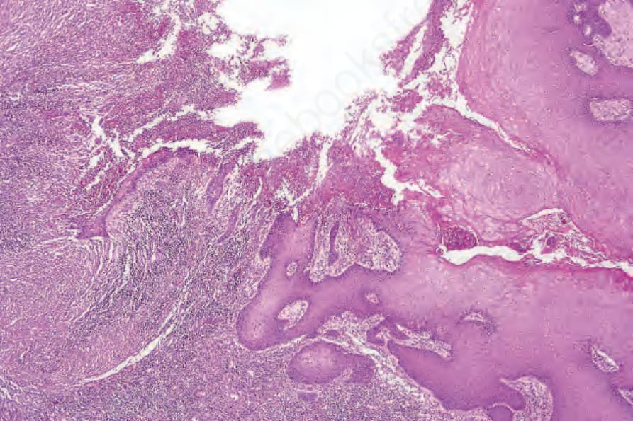
圖 12-143:阿米巴病 (amebiasis):取自外陰潰瘍 (vulval ulcer) 的切片,該潰瘍是由肛門直接蔓延而形成。
Fig. 12.143 Amebiasis: biopsy from a vulval ulcer, which developed as a result of direct spread from the anus.
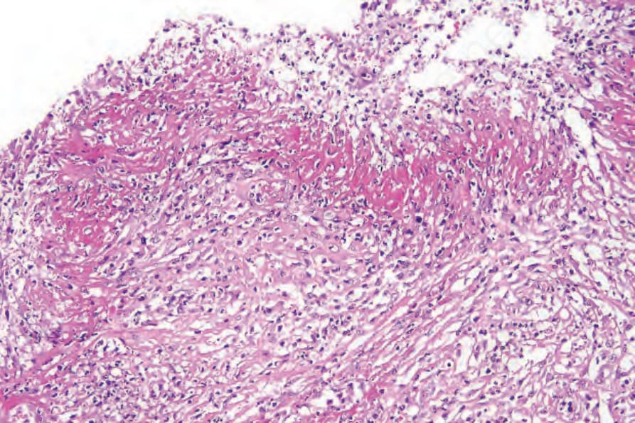
圖 12-144:阿米巴病 (amebiasis):潰瘍底部覆蓋著緻密的纖維蛋白性滲出物 (fibrinous exudate)。
Fig. 12.144 Amebiasis: the floor of the ulcer is covered by a dense fibrinous exudate.
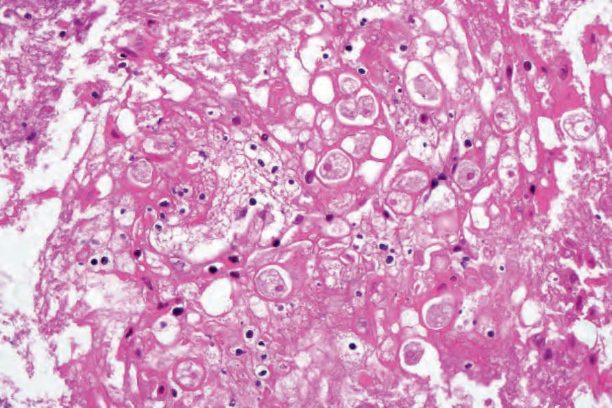
圖 12-145:阿米巴病 (amebiasis):可見大量滋養體 (trophozoites)。注意被吞噬的紅血球 (ingested red blood cells)。
Fig. 12.145 Amebiasis: there are numerous trophozoites present. Note the ingested red blood cells.
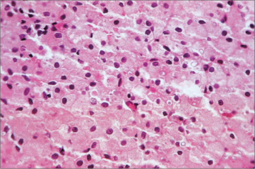
圖 12-146:軟斑症 (malacoplakia):浸潤由具嗜伊紅細胞質 (eosinophilic cytoplasm) 的組織球組成。注意淡藍色、層狀的 Michaelis-Gutmann bodies。
Fig. 12.146 Malacoplakia: the infiltrate consists of histiocytes with eosinophilic cytoplasm. Note the pale blue, laminated Michaelis-Gutmann bodies.