臨床特徵 (Clinical Features)
- 腹股溝肉芽腫 (granuloma inguinale,又稱杜諾凡病 donovanosis) 發生於熱帶地區衛生條件不佳的族群,主要見於印度、巴西、西印度群島、中國與西非;過去曾見於美國南部,但現已罕見。早年曾於美國南部出現,現已少見。澳洲的原住民族群中仍可見此病。
- 此微生物感染力低,推測經由性接觸傳播,可能透過擦傷的皮膚。最常發生於 30 至 50 歲(第三至第五個十年)。潛伏期不確定,可能為 2 至 3 週至數個月不等。
- 在女性,初始表現通常為一個或多個位於大陰唇內側、陰唇繫帶 (fourchette) 或陰蒂周圍的硬結性丘疹或結節 (Fig. 12.127)。在男性,則侵犯龜頭 (glans)、包皮 (prepuce)、冠狀溝 (coronal sulcus) 或陰莖體 (shaft)(Fig. 12.128)。包皮的背側穿孔 (dorsal perforation) 可作為晚期併發症發生。
- 會陰部、腹股溝區與子宮頸亦可受侵犯。在一例侵犯子宮頸的病例中,合併有軟斑症 (malacoplakia)。丘疹會不規則潰瘍,若未治療則廣泛擴展。潰瘍底部呈「牛肉狀 (beefy)」,邊緣則被掘空 (undermined) 且硬結。有時可擴散至鄰接的「相吻 (kissing)」部位。變異型包括疣狀 (verrucous)、壞死性 (necrotic) 與瘢痕性 (scarring) 病灶。淋巴結原發感染並不會發生,但因續發性感染而出現疼痛性淋巴結病變則常見。罕見情況下可見原發性生殖器外病灶(主要見於口腔或唇部,但亦可見於足部等不尋常部位)。極為例外的情況下,曾有以腰大肌膿瘍 (psoas abscess) 及以模擬卵巢癌之骨盆腫塊作為表現的描述。
- 極偶爾,會出現侵犯多個器官(包括肝臟)的全身性感染,並於骨骼出現溶骨性病灶 (osteolytic lesions)。後者可能特別與原發性子宮頸病灶有關。亦曾有脊椎壓迫 (spinal compression) 之報告。晚期併發症包括尿道、陰道或肛門的狹窄,陰莖體破壞並伴自截 (autoamputation),以及假性象皮病 (pseudoelephantiasis)。
- 與其他性傳染病相同,granuloma inguinale 的病人常為 HIV 陽性,並可能同時罹患梅毒 (syphilis)。生殖器鱗狀細胞癌 (genital SCC) 是不常見但重要的併發症。兒童感染罕有報告,係由受感染母親於出生時傳染所致。此病在兒童的罕見表現包括頸部腫塊、中耳炎 (otitis media) 與乳突炎 (mastoiditis)。
致病機轉與組織學特徵 (Pathogenesis and Histologic Features)
- 腹股溝肉芽腫由肉芽腫莢膜桿菌 (Calymmatobacterium granulomatis,舊稱 Donovania granulomatis) 引起,此為一種有莢膜的短小 (1–2 µm)、革蘭氏陰性桿菌,具特徵性的雙極染色 (bipolar staining)。經由性接觸傳播,但感染力低。此微生物可見於糞便中,這可能作為感染力的儲存庫,或在偶發病例中成為生殖器感染的來源。同性戀者較高的感染發生率可能支持此一概念。
- 病灶以極為密集的發炎浸潤為特徵,其中以漿細胞 (plasma cells) 為主 (Fig. 12.129)。局灶性嗜中性球微膿瘍 (neutrophilic microabscesses) 的形成常見。具病理特徵性 (pathognomonic) 的巨噬細胞含有胞質內囊腫狀空泡,其中的細菌可藉由 Giemsa 染色或 Warthin-Starry 反應顯示出來 (Figs 12.130 and 12.131)。細菌亦可見於細胞外。多數病例中伴有棘層肥厚 (acanthosis),有時可達假上皮瘤性增生 (pseudoepitheliomatous hyperplasia) 的程度。潰瘍常見。一項大型研究顯示頻繁的微生物經表皮排除 (transepidermal elimination)。此現象很可能是此病擴散的重要機轉之一。
- 診斷係藉由在潰瘍刮取物或切片(以 Giemsa 或 Warthin-Starry 染色)中辨認典型微生物(杜諾凡小體 Donovan bodies)來確認。PCR 也已成功用於確認診斷。應進行暗視野顯微鏡 (dark-field illumination microscopy) 以排除 syphilis。在電子顯微鏡下,可於巨噬細胞的吞噬體 (phagosomes) 內顯示出有莢膜的微生物。
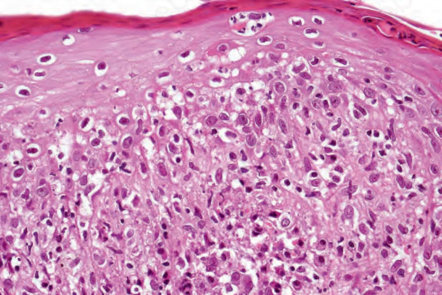
圖 12-122:續發性梅毒 (secondary syphilis):在此視野中可見明顯的介面變化 (interface change)。
Fig. 12.122 Secondary syphilis: in this field, there is marked interface change.
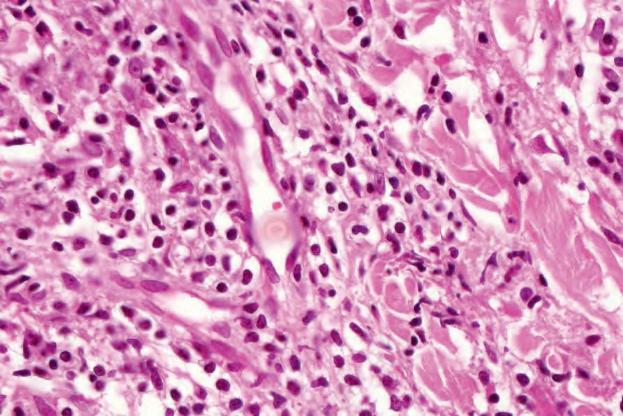
圖 12-123:續發性梅毒 (secondary syphilis):注意明顯的內皮細胞腫脹 (endothelial cell swelling)。
Fig. 12.123 Secondary syphilis: note the marked endothelial cell swelling.
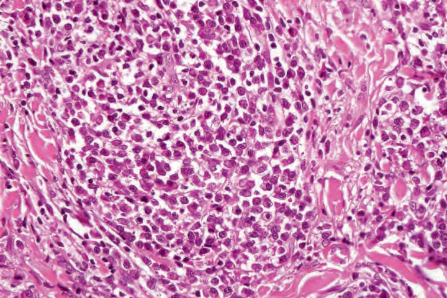
圖 12-124:續發性梅毒 (secondary syphilis):可見無數的漿細胞 (plasma cells)。
Fig. 12.124 Secondary syphilis: innumerable plasma cells are present.
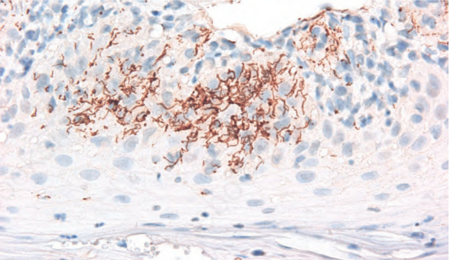
圖 12-125:續發性梅毒 (secondary syphilis):在一位續發性梅毒病人中,藉由免疫組織化學 (immunohistochemistry) 可見表皮內有眾多螺旋體 (spirochetes)。
Fig. 12.125 Secondary syphilis: numerous spirochetes are seen by immunohistochemistry within the epidermis in a patient with secondary syphilis.
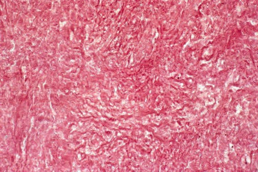
圖 12-126:梅毒瘤 (gumma):高倍視野顯示細胞與結締組織的鬼影輪廓 (ghost outlines)。
Fig. 12.126 Gumma: high-power view reveals ghost outlines of cells and connective tissue.
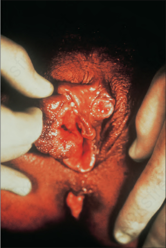
圖 12-127:腹股溝肉芽腫 (granuloma inguinale):早期病灶,顯示緊鄰陰蒂 (clitoris) 的潰瘍性丘疹。承蒙英國 University of Newcastle-upon-Tyne 之 J. Lawson, MD 提供。
Fig. 12.127 Granuloma inguinale: early lesion showing an ulcerated papule adjacent to the clitoris. By courtesy of J. Lawson, MD, University of Newcastleupon-Tyne, UK.
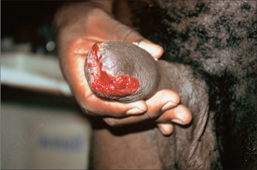
圖 12-128:腹股溝肉芽腫 (granuloma inguinale):此病人有龜頭 (glans penis) 的廣泛潰瘍。注意其典型的「牛肉狀 (beefy)」外觀。承蒙千里達 Port of Spain 之 C. Furlonge, MD 提供。
Fig. 12.128 Granuloma inguinale: in this patient, there is extensive ulceration of the glans penis. Note the typical ‘beefy’ appearance. By courtesy of C. Furlonge, MD, Port of Spain, Trinidad.
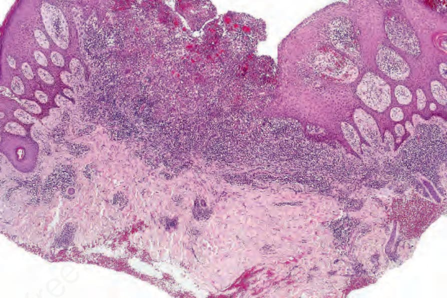
圖 12-129:腹股溝肉芽腫 (granuloma inguinale):取自陰莖的切片。注意假上皮瘤性增生 (pseudoepitheliomatous hyperplasia)。有密集的發炎變化。
Fig. 12.129 Granuloma inguinale: biopsy from the penis. Note the pseudoepitheliomatous hyperplasia. There are intense inflammatory changes.
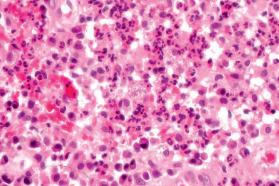
圖 12-130:腹股溝肉芽腫 (granuloma inguinale):浸潤由淋巴球、嗜中性球、漿細胞及明顯的淡染組織球 (pale-staining histiocytes) 組成。
Fig. 12.130 Granuloma inguinale: the infiltrate consists of lymphocytes, neutrophils, plasma cells, and conspicuous pale-staining histiocytes.
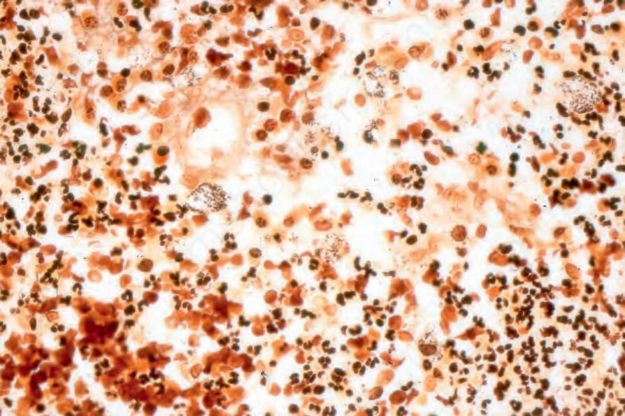
圖 12-131:腹股溝肉芽腫 (granuloma inguinale):組織球內含有特徵性的杜諾凡小體 (Donovan bodies)(Warthin-Starry stain)。
Fig. 12.131 Granuloma inguinale: the histiocytes contain characteristic Donovan bodies (Warthin-Starry stain).