尖圭濕疣 (Condyloma acuminatum, genital warts, HPV infection)
臨床特徵 (Clinical Features)
- 生殖器疣(尖圭濕疣,genital warts, condyloma acuminatum)通常由 HPV 第 6、11、16、18、30–32、42–44 與 51–55 型所引起。HPV-7 通常與屠夫的疣 (warts in butchers) 相關,偶爾也可能造成濕疣。HPV-6 與 11 約佔此類病灶的 90%。然而,從單一病灶中可分離出一種以上的 HPV 型別。
- 病灶發生於陰莖龜頭 (glans penis) 與包皮 (prepuce) 或陰莖體 (shaft),呈柔軟、肉質(有時呈絲狀,filiform)的斑塊,並可延伸至尿道口 (meatus)(圖 12.86 與 12.87)。在陰莖體上,其外生性 (exophytic) 較不明顯。外陰部 (vulval) 病灶可能體積龐大且浸軟 (macerated),並可延伸至陰道口 (introitus)(圖 12.88–12.90)。病灶在臨床檢查時可能難以察覺。外陰部濕疣通常侵犯小陰唇 (labia minora)、陰唇間溝 (interlabial sulcus) 或陰道口周圍區域。
- 類似的肉質與絲狀柔軟腫塊也可能發生於肛周 (perianally) 與肛門 (anus) 內,較常見於男性(圖 12.91)。兒童的生殖器疣總是會引起對性虐待 (sexual abuse) 可能性的考量,但也可能因密切的非性接觸而發生。然而,主要由 HPV 第 2 型引起的非性病性病毒疣(尋常疣,verruca vulgaris)可發生於年幼女孩,以及較少見於成年女性,因此 HPV 分型 (HPV typing) 可能非常重要。最常受影響的是性活躍的年輕成人(第二與第三個十年),且常合併其他經由性行為獲得的感染。
- 罕見情況下,尖圭濕疣可表現於口腔 (oral cavity)。亦曾有報告指出,少數肥胖病人的腹部脂肪垂皺褶 (abdominal pannus fold) 中發生濕疣。
- 必須認識到,相當比例的生殖器 HPV 感染是無症狀的。已證實,患有生殖器疣的男性病人的女性伴侶,發生子宮頸 HPV 感染與上皮內贅瘤 (intraepithelial neoplasia) 的風險增加。與既存外陰疣相關的子宮頸贅瘤 (cervical neoplasia),至少在某些病人中亦與免疫抑制 (immunosuppression) 有關。多達 80% 的侵襲性子宮頸鱗狀癌 (invasive cervical squamous carcinomas) 已被證實含有 HPV DNA。第 16、18、31–33、35、39、42 與 51–54 型最常與子宮頸、外陰與陰莖的癌症相關。
- 生殖器疣的惡性轉化 (malignant transformation) 罕見,但可能與陰莖鮑文樣丘疹病 (penile bowenoid papulosis) 及普通型(未分化)VIN(VIN usual type, undifferentiated)相關。在最新的 VIN 分類中,尖圭濕疣被歸類為低度鱗狀上皮內病變 (low-grade squamous intraepithelial lesions)。
- 罕見情況下,可能遇到一種巨大、增生旺盛且局部破壞性的濕疣變異型(Buschke-Löwenstein tumor)。此型與 HPV-6、11 或 16 相關。許多人認為它是疣狀癌 (verrucous carcinoma) 的一種變異型,即分化良好鱗狀癌 (well-differentiated squamous carcinoma) 的一個亞型(見下文)。然而此議題仍具爭議,其他作者則視其為一個獨特的疾病實體。若接受放射治療 (irradiated),它們可能出現惡性進展 (malignant progression)。此病況曾在一位 HIV 陽性病人中被描述。
- 含有 HPV-6 與 11 的幼年型喉乳頭瘤 (juvenile laryngeal papillomata),可見於母親患有尖圭濕疣所生的兒童。
組織病理特徵 (Histopathology)
- 尖圭濕疣的特徵為顯著的棘層肥厚 (acanthosis),呈實心或小樑狀 (trabecular) 模式,並有寬廣、圓鈍的外生性 (exophytic) 生長(圖 12.92 與 12.93)。其深部邊緣銳利且相當規則。病灶表面呈過度角化 (hyperkeratotic) 與角化不全 (parakeratotic)。表淺空泡化的角質形成細胞(挖空細胞,koilocytes)為其特徵(圖 12.94)。空泡化的上皮通常在凹陷處 (declivities) 最為明顯。必須小心,勿將挖空細胞與黏膜上皮中空泡化、含肝醣 (glycogenated) 的角質形成細胞,或與人工假象造成空泡化的角質形成細胞混淆。由於挖空細胞具有增大、皺縮、深染 (hyperchromatic) 的細胞核,故可相當容易地加以區分。
- 對於先前曾以 podophyllin 治療過的病灶,在組織學判讀時應特別小心(雖然自從 imiquimod 問世後,此療法現已甚少使用)。這些病灶可顯現出明顯的退化性變化,伴隨細胞質空泡化 (cytoplasmic vacuolation)、細胞核增大與中期停滯 (metaphase arrest)。然而這些變化往往較為局灶性,且不會見到異常的有絲分裂像 (abnormal mitotic figures)。針對乳頭瘤病毒共同抗原 (papillomavirus common antigen) 的免疫組織化學染色曾被用於確認診斷,但僅約 60% 的病例呈陽性。近期,石蠟包埋組織 (paraffin-embedded tissue) 中的廣效原位雜交 (broad-spectrum in situ hybridization) 已可使用。
- 巨大尖圭濕疣 (Giant condyloma acuminatum, Buschke-Löwenstein tumor) 最常發生於生殖器,較大且更呈花椰菜狀 (cauliflower-like)。它顯示某種程度的內生性 (endophytic) 生長傾向,但不帶有任何明顯浸潤 (frank infiltration) 的跡象。它可局部復發。肛門濕疣 (anal condylomata) 可能發展出鮑文樣 (bowenoid) 特徵,偶爾會繼發侵襲性腫瘤。
- 在最新的 VIN 分類中,尖圭濕疣被歸類為低度鱗狀上皮內病變 (low-grade squamous intraepithelial lesions)。
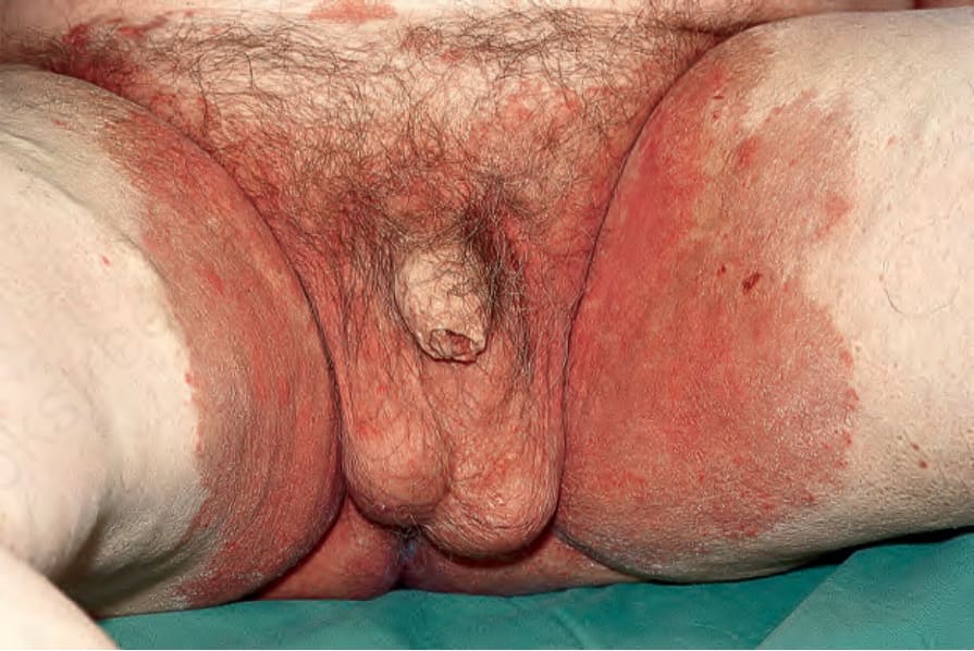
圖 12.85:隱匿性癬 (Tinea incognito):腹部、腹股溝、大腿與陰囊有廣泛侵犯。此係不當使用外用類固醇 (topical steroids) 之後發生。摘自 Bunker C. Male Genital Skin Disease. Saunders Ltd./Elsevier 2004。
Fig. 12.85 Tinea incognito: there is extensive involvement of the abdomen, groins, thighs and scrotum. This followed injudicious use of topical steroids. From Bunker C. Male Genital Skin Disease. Saunders Ltd./Elsevier 2004.
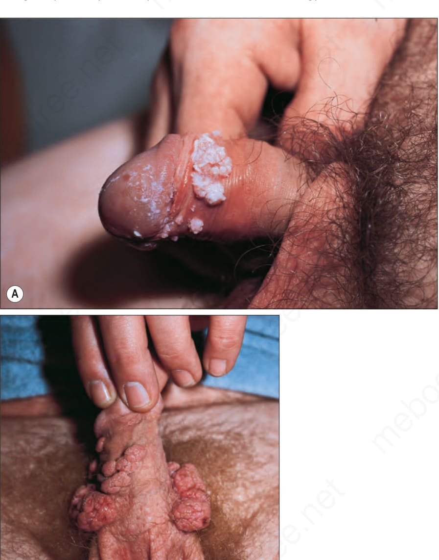
圖 12.86:尖圭濕疣 (Condyloma acuminatum):陰莖龜頭上有多個紅斑性、絲絨樣 (velvety) 斑塊。承蒙英國倫敦 Institute of Dermatology 惠予提供。
Fig. 12.86 Condyloma acuminatum: multiple erythematous, velvety plaques are present on the glans penis. By courtesy of the Institute of Dermatology, London, UK.
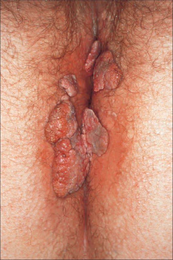
圖 12.87:尖圭濕疣 (Condyloma acuminatum):(A) 此病人的病灶呈典型的絲狀 (filiform) 外觀;(B) 陰莖與陰囊上有多個濕疣。承蒙英國倫敦 St Thomas’ Hospital, Department of Genitourinary Medicine 惠予提供。
Fig. 12.87 Condyloma acuminatum: (A) in this patient, the lesions have a typical filiform appearance; (B) multiple condylomata are present on penis and scrotum. By courtesy of the Department of Genitourinary Medicine, St Thomas’ Hospital, London, UK.
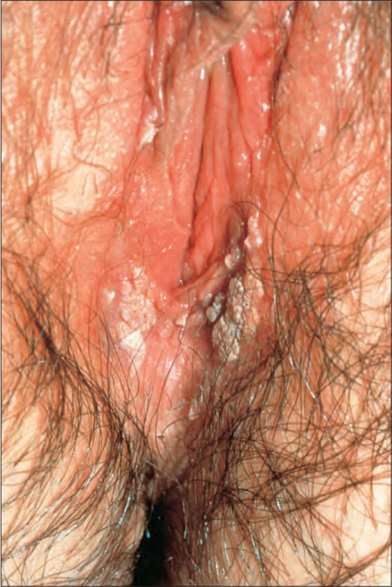
圖 12.88:尖圭濕疣 (Condyloma acuminatum):小陰唇 (labia minora) 與前庭 (vestibule) 周圍可見多個灰色病灶。承蒙英國倫敦 Institute of Dermatology 惠予提供。
Fig. 12.88 Condyloma acuminatum: multiple gray lesions are evident on the labia minora and around the vestibule. By courtesy of the Institute of Dermatology, London, UK.
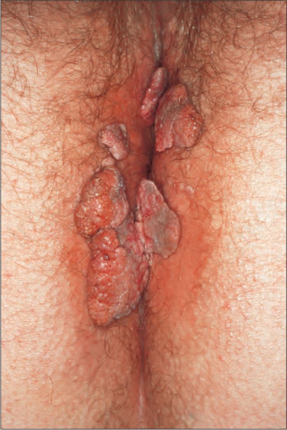
圖 12.89:尖圭濕疣 (Condyloma acuminatum):此病人的濕疣呈有蒂狀 (pedunculated),並已延伸至大腿。承蒙英國倫敦 Institute of Dermatology 惠予提供。
Fig. 12.89 Condyloma acuminatum: in this patient, the condylomata are pedunculated and have extended onto the thighs. By courtesy of the Institute of Dermatology, London, UK.
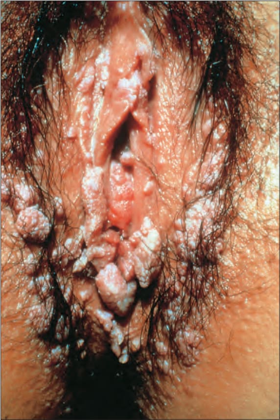
圖 12.90:尖圭濕疣 (Condyloma acuminatum):病變非常廣泛。此病人有相當大的風險發展出子宮頸疾病。承蒙英國倫敦 St George’s Hospital 的 R.A. Marsden, MD 惠予提供。
Fig. 12.90 Condyloma acuminatum: there is very extensive disease. The patient is at considerable risk of developing cervical disease. By courtesy of R.A. Marsden, MD, St George’s Hospital, London, UK.
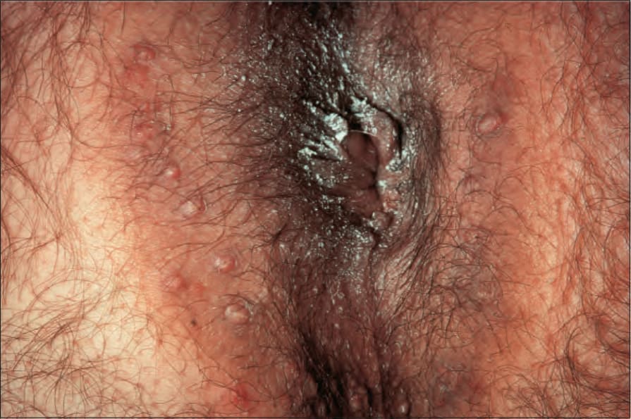
圖 12.91:尖圭濕疣 (Condyloma acuminatum):肛周侵犯很可能與同性戀性行為 (homosexual activity) 相關。承蒙英國倫敦 St George’s Hospital 的 R.A. Marsden, MD 惠予提供。
Fig. 12.91 Condyloma acuminatum: perianal involvement is likely to be associated with homosexual activity. By courtesy of R.A. Marsden, MD, St George’s Hospital, London, UK.
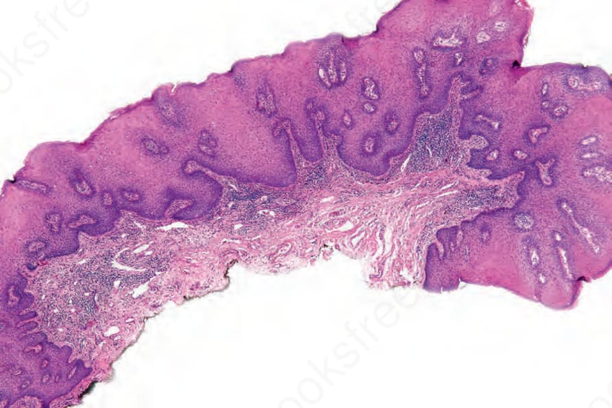
圖 12.92:尖圭濕疣 (Condyloma acuminatum):有局灶性角化不全 (parakeratosis)、輕度乳頭瘤狀增生 (papillomatosis) 與非常顯著的棘層肥厚 (acanthosis)。下緣界線分明。
Fig. 12.92 Condyloma acuminatum: there is focal parakeratosis, slight papillomatosis and very marked acanthosis. The lower border is sharply demarcated.
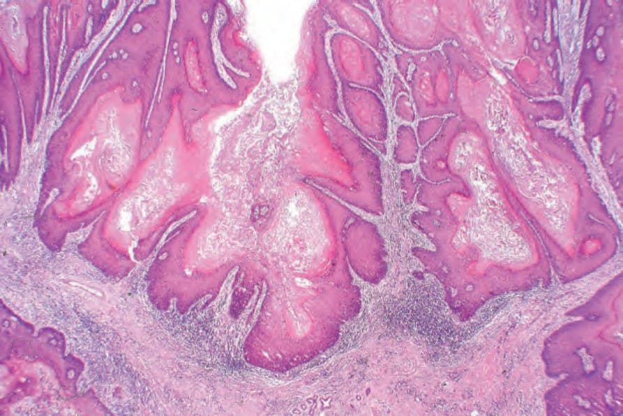
圖 12.93:尖圭濕疣 (Condyloma acuminatum):此為一個更為旺盛 (florid) 的範例。注意明顯的乳頭瘤狀增生 (papillomatosis) 與非常顯著的棘層肥厚 (acanthosis)。
Fig. 12.93 Condyloma acuminatum: this is a much more florid example. Note the gross papillomatosis and very marked acanthosis.
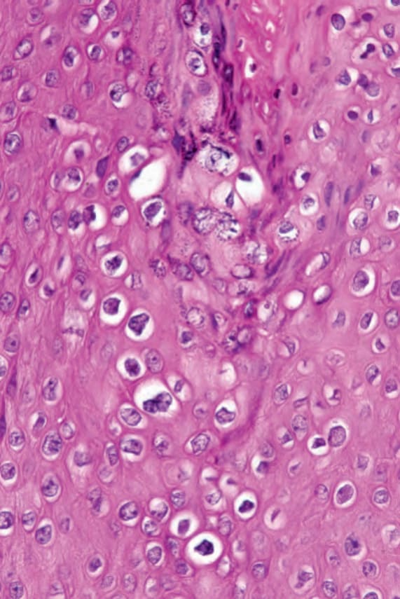
圖 12.94:尖圭濕疣 (Condyloma acuminatum):可見明顯的挖空細胞 (koilocytes),具有不規則的細胞核與空泡化的細胞質。
Fig. 12.94 Condyloma acuminatum: there are conspicuous koilocytes with irregular nuclei and vacuolated cytoplasm.
圖 12.95:原發性硬下疳 (Primary chancre):硬下疳是一種無痛性潰瘍,具有硬化 (indurated) 的邊緣。基底呈黃色,並藏有大量螺旋體 (spirochetes)。承蒙英國倫敦 King’s College Hospital 的 F. Lim, MD 惠予提供。
Fig. 12.95 Primary chancre: the chancre is a painless ulcer with an indurated edge. The base is yellow and harbors large numbers of spirochetes. By courtesy of F. Lim, MD, King’s College Hospital, London, UK.