臨床特徵 (Clinical Features)
- 在全身性疾病的患者中,肛門生殖器 (anogenital) 病灶可見於高達 40% 的病人。然而在部分病人,疾病侷限於下生殖道與/或肛周 (perianal) 區域。扁平苔癬 (lichen planus, LP) 會表現 Koebner 現象 (Koebner phenomenon),這或可部分解釋其口腔–生殖器 (orogenital) 好發傾向。兒童的生殖器 LP 屬例外罕見。¹ 患有口腔 LP 的女性常合併生殖器疾病,而此生殖器疾病可能無症狀。²,³
- 表皮呈棘層肥厚 (acanthotic),伴表皮突 (epidermal ridges) 的延長與肥大,以及角化不全 (parakeratosis)。乳頭上板 (suprapapillary plates) 變薄,且表皮有嗜中性球 (neutrophils) 浸潤,伴隨表淺角質細胞 (superficial keratinocytes) 的空泡化,並形成海綿狀膿疱 (spongiform pustules) 與微膿瘍 (microabscesses)(圖 12-32)。發炎延伸至鄰近的下方真皮,該處浸潤以單核細胞 (mononuclear) 為主。其組織學基本上與膿疱性乾癬 (pustular psoriasis) 所見者完全相同。² 因此,緊密的臨床病理對照 (clinicopathological correlation) 對於建立診斷至關重要。反應性關節炎 (reactive arthritis) 患者典型病灶的偶發切片,可能會顯示其下存在白血球破碎性血管炎 (leukocytoclastic vasculitis)。⁴¹
- 已有少數病人在臨床上以皮膚病灶表現,且組織學顯示無菌性嗜中性球性毛囊炎 (sterile neutrophilic folliculitis) 合併毛囊周圍血管病變 (perifollicular vasculopathy)。⁴² 作者認為此組織學型態可能是全身性疾病的標記。相關疾病可包括反應性關節炎、發炎性腸道疾病 (inflammatory bowel disease)、Behçet 病 (Behçet disease)、B 型肝炎感染 (hepatitis B infection)、瘰癧性皮膚結核 (scrofuloderma)、結締組織疾病 (connective tissue diseases),以及血液惡質病 (hematological dyscrasias)。
- 病灶為典型的紫羅蘭色 (violaceous) 或白色斑塊,或為紅斑與糜爛區域。Wickham 紋 (Wickham striae)(常見於口腔受侵犯時)雖有時可見,但在肛門生殖器皮膚較少出現(圖 12-33)。侵犯肛門生殖器皮膚的 LP 臨床變異型包括鱗屑丘疹型 (squamo-papular)、糜爛型 (erosive)、肥厚型 (hypertrophic),以及色素過度沉著的皺褶部位疾病 (hyperpigmented flexural disease)(圖 12-34 至 12-41)。外陰也曾描述有毛髮扁平苔癬 (lichen planopilaris)。⁴
- LP 的糜爛型在肛門生殖器部位較為常見,並可導致瘢痕形成與構造扭曲。⁵ 外陰前庭 (vulval vestibule)、陰道及子宮頸亦可能受侵犯,有時則單獨影響陰道與/或子宮頸。⁶,⁷ 女性還有一種不尋常的糜爛型 LP 變異型,侵犯口腔牙齦 (oral gingivae)、外陰前庭與陰道,稱為外陰陰道–牙齦症候群 (vulvovaginal-gingival syndrome)(圖 12-42 至 12-44)。⁸,⁹ 此可導致嚴重的外陰與陰道瘢痕,伴陰道沾黏 (vaginal adhesions)、束帶 (constriction bands),且在部分病例造成完全狹窄 (complete stenosis)。¹⁰ 男性也曾描述有相當於 Hewitt 外陰陰道症候群的對應病症,表現為慢性糜爛性牙齦與生殖器病灶(生殖器–牙齦症候群 (genito-gingival syndrome))。¹¹,¹² 有生殖器病灶的病人可能合併口腔、耳部 (aural)、結膜 (conjunctival) 與食道 (esophageal) 受侵犯。¹³⁻¹⁷
- 肛周疾病可造成深部、疼痛的皸裂 (fissuring),且常是肥厚型變異侵犯此部位。
- 在男性生殖器,LP 可表現為包莖 (phimosis)。¹⁸,¹⁹ 在未割包皮 (uncircumcised) 的男性可見沾黏,包括冠狀溝橫向 (transcoronal) 與冠狀溝下 (subcoronal) 的沾黏。
- 肛門生殖器 LP 帶有些微增加的惡性風險,通常為鱗狀細胞癌 (squamous cell carcinoma, SCC)(圖 12-45)。²⁰⁻²³ 龜頭 (glans penis) 的肥厚型 LP 應視為具有潛在不良生物學行為。²⁴
致病機轉與組織學特徵 (Pathogenesis and Histologic Features)
- 對口腔 LP 的研究支持其有免疫學基礎,即活化的 T 細胞對某種未明的抗原刺激 (antigenic stimulus) 起反應。²⁵ 肛門生殖器 LP 的組織學特徵,往往比表現於非黏膜表面的 LP 更難辨認。表皮可能變平 (effaced) 或增厚,且在真皮–表皮交界 (dermal–epidermal junction) 處有緊貼的、緻密的帶狀浸潤 (band-like infiltrate)(圖 12-46)。許多生殖器病灶屬黏膜性,其發炎細胞浸潤常富含漿細胞 (plasma cells),這與其他部位以淋巴球 (lymphocytes) 及組織球 (histiocytes) 為主的 LP 病灶形成對比(圖 12-47)。基底層 (basal layer) 常因再生 (regeneration) 進行而被破壞,並出現某些細胞學異型性 (cytological atypia)。可見類細胞體 (cytoid bodies),但傾向不顯著。此伴隨角化不全 (parakeratosis)。局部次發性海綿水腫變化 (secondary spongiotic changes) 並不少見,尤其在黏膜表面。在病程久遠的疾病中,緻密的帶狀浸潤可能被斑片狀、稀疏的浸潤所取代,僅見小灶的苔癬樣發炎 (lichenoid inflammation)。許多男性生殖器 LP 病例被誤診為 ZB 或 LS。
鑑別診斷 (Differential Diagnosis)
- 臨床鑑別診斷包括乾癬 (psoriasis)、男性的 ZB、LS、病毒疣 (viral warts)、鮑恩樣丘疹病 (bowenoid papulosis),以及汗孔角化症 (porokeratosis)。LP 是肛門搔癢症 (pruritus ani) 的成因之一。切片檢查常為診斷目的所必需,但更重要的是用於追蹤罕見的慢性肛門生殖器疾病病例,因為當出現潰瘍–糜爛 (ulcero-erosive) 或疣狀 (verrucous) 特徵時,會引發對 SCC 形成的疑慮。LP 常與 LS 的特徵重疊,且在部分病人中兩種疾病可能並存。乳頭真皮 (papillary dermis) 或表淺固有層 (superficial lamina propria) 的玻璃樣變性 (hyalinization) 提示後者 (LS) 之存在。對於罹患此種慢性重疊症候群 (chronic overlap syndrome) 的病人,應特別小心辨認異型增生 (dysplastic) 區域或 SCC。臨床上以黏膜疾病為主的病人會類似黏膜類天疱瘡 (mucous membrane pemphigoid),但免疫螢光 (immunofluorescence) 研究始終為陰性。曾有一例伴有口腔生殖器侵犯與瘢痕性結膜炎 (cicatrizing conjunctivitis)、並與胸腺瘤 (thymoma) 相關的副腫瘤性 LP (paraneoplastic LP) 被描述。²⁷
- 免疫螢光研究可顯示沿基底膜帶 (basement membrane zone) 有 fibrinogen 與 IgM,較罕見有 IgG 或 IgA。²⁶ 類細胞體亦可能被標記。²⁶
- 有時藥物可誘發全身性苔癬樣疹 (generalized lichenoid eruption)。曾報告一例因 propranolol 所致、侷限於陰莖的苔癬樣藥物疹 (lichenoid drug eruption)。²⁸
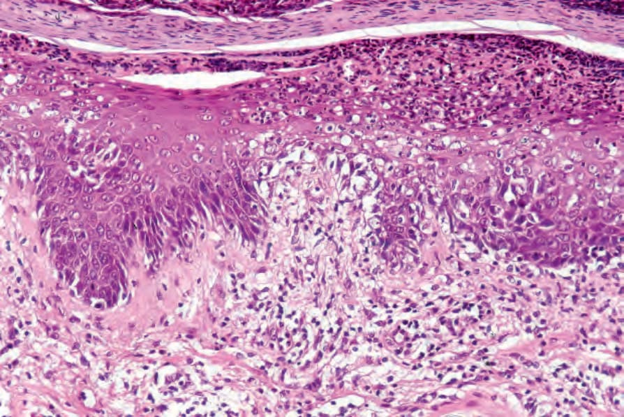
圖 12-32:反應性關節炎 (reactive arthritis):高倍視野顯示角化不全 (parakeratosis) 與一處膿疱 (pustule)。
Fig. 12.32 Reactive arthritis: high-power view showing parakeratosis and a pustule.
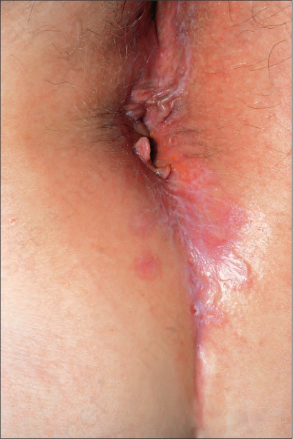
圖 12-33:扁平苔癬 (lichen planus):會陰 (perineal) 病灶顯示明顯的條紋 (striae)。蒙倫敦皮膚科研究所 (Institute of Dermatology, London, UK) 惠允。
Fig. 12.33 Lichen planus: perineal lesions showing conspicuous striae. By courtesy of the Institute of Dermatology, London, UK.
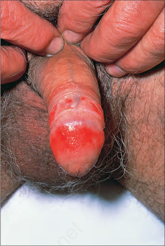
圖 12-34:糜爛型扁平苔癬 (erosive lichen planus):龜頭 (glans penis) 有廣泛糜爛。蒙倫敦皮膚科研究所 (Institute of Dermatology, London, UK) 惠允。
Fig. 12.34 Erosive lichen planus: there is extensive erosion of the glans penis. By courtesy of the Institute of Dermatology, London, UK.
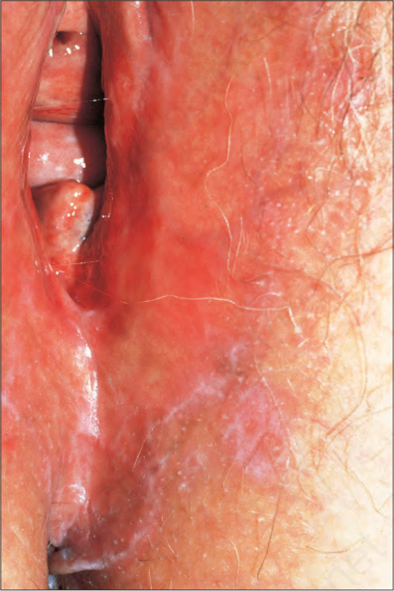
圖 12-35:外陰扁平苔癬 (vulval lichen planus):扁平苔癬的網狀 (reticulated) 病灶延伸至會陰 (perineum)。蒙倫敦皮膚科研究所 (Institute of Dermatology, London, UK) 惠允。
Fig. 12.35 Vulval lichen planus: reticulated lesions of lichen planus extending into the perineum. By courtesy of the Institute of Dermatology, London, UK.
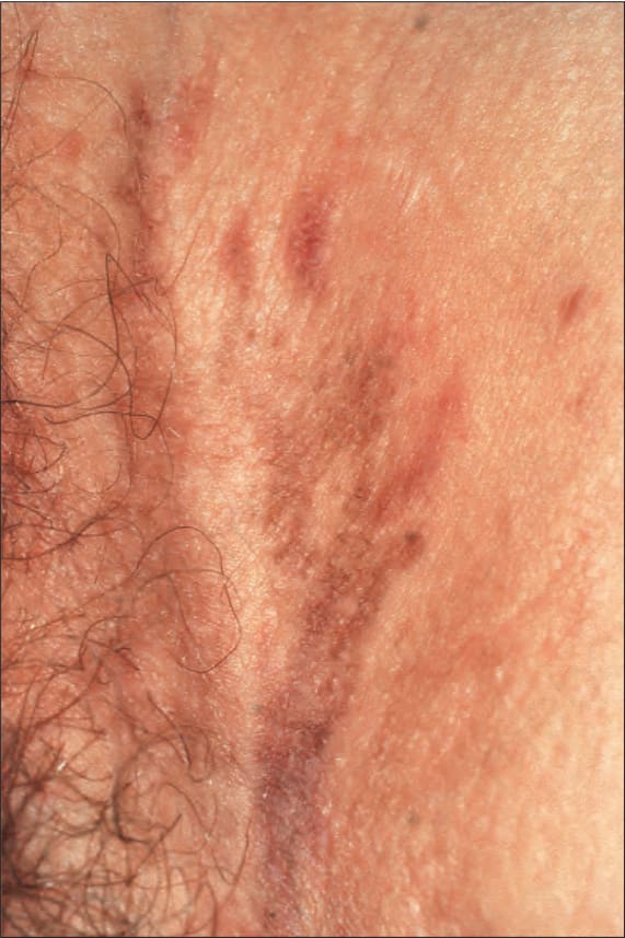
圖 12-36:外陰扁平苔癬 (vulval lichen planus):在此消退中疾病的例子,可見線狀色素過度沉著 (hyperpigmented) 病灶。蒙倫敦皮膚科研究所 (Institute of Dermatology, London, UK) 惠允。
Fig. 12.36 Vulval lichen planus: in this example of resolving disease, there are linear hyperpigmented lesions. By courtesy of the Institute of Dermatology, London, UK.
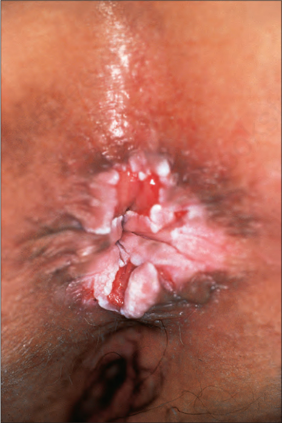
圖 12-37:會陰扁平苔癬 (perineal lichen planus):可見伴有 Wickham 紋 (Wickham striae) 的典型丘疹 (papules)。蒙倫敦皮膚科研究所 (Institute of Dermatology, London, UK) 惠允。
Fig. 12.37 Perineal lichen planus: typical papules with Wickham striae are present. By courtesy of the Institute of Dermatology, London, UK.
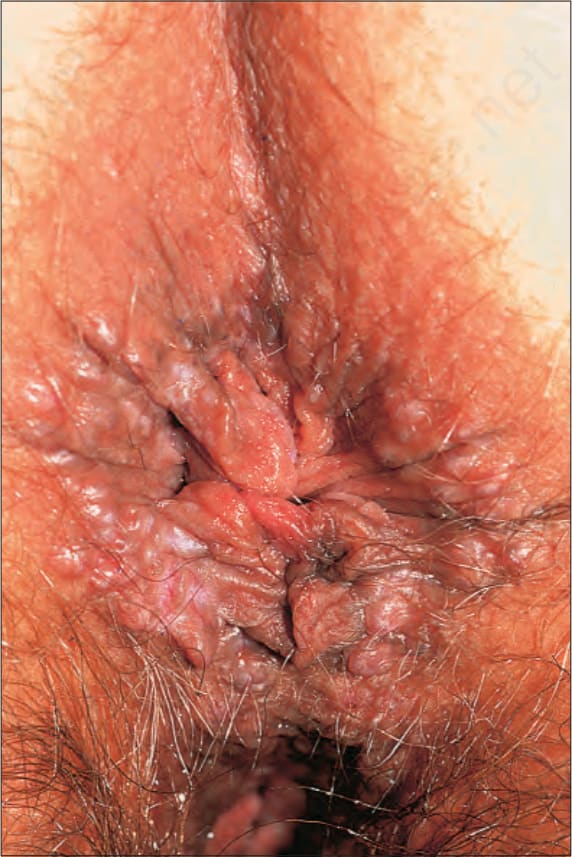
圖 12-38:肥厚型肛周扁平苔癬 (hypertrophic perianal lichen planus):慢性搔抓導致疊加的苔癬化 (lichenification)。蒙倫敦皮膚科研究所 (Institute of Dermatology, London, UK) 惠允。
Fig. 12.38 Hypertrophic perianal lichen planus: chronic scratching has resulted in superimposed lichenification. By courtesy of the Institute of Dermatology, London, UK.
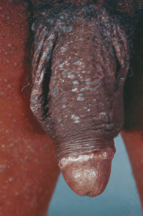
圖 12-39:陰莖扁平苔癬 (penile lichen planus):有陰莖體 (shaft) 與龜頭 (glans) 的受侵犯。摘自 Bunker C. Male Genital Skin Disease. Saunders Ltd./Elsevier 2004.
Fig. 12.39 Penile lichen planus: there is involvement of the shaft and glans. From Bunker C. Male Genital Skin Disease. Saunders Ltd./Elsevier 2004.
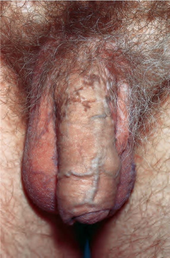
圖 12-40:陰莖扁平苔癬 (penile lichen planus):近端陰莖體 (proximal shaft) 顯示發炎後色素過度沉著 (post inflammatory hyperpigmentation)。摘自 Bunker C. Male Genital Skin Disease. Saunders Ltd./Elsevier 2004.
Fig. 12.40 Penile lichen planus: the proximal shaft shows post inflammatory hyperpigmentation. From Bunker C. Male Genital Skin Disease. Saunders Ltd./Elsevier 2004.
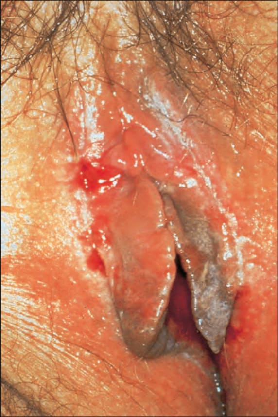
圖 12-41:糜爛型扁平苔癬 (erosive lichen planus):可見雙側糜爛 (bilateral erosions)。蒙倫敦皮膚科研究所 (Institute of Dermatology, London, UK) 惠允。
Fig. 12.41 Erosive lichen planus: bilateral erosions are present. By courtesy of the Institute of Dermatology, London, UK.
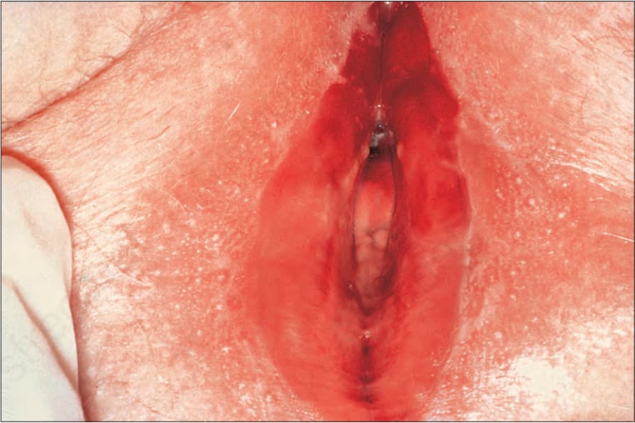
圖 12-42:外陰陰道–牙齦症候群 (vulvovaginal-gingival syndrome):有廣泛的前庭 (vestibular) 紅斑與糜爛,周圍伴有細緻的白色鱗屑。蒙倫敦皮膚科研究所 (Institute of Dermatology, London, UK) 惠允。
Fig. 12.42 Vulvovaginal-gingival syndrome: there is extensive vestibular erythema and erosion with a surrounding delicate white scale. By courtesy of the Institute of Dermatology, London, UK.
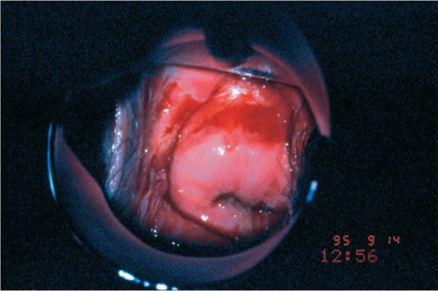
圖 12-43:外陰陰道–牙齦症候群 (vulvovaginal-gingival syndrome):有陰道與子宮頸的潰瘍 (ulceration)。蒙倫敦皮膚科研究所 (Institute of Dermatology, London, UK) 惠允。
Fig. 12.43 Vulvovaginal-gingival syndrome: there is ulceration of the vagina and cervix. By courtesy of the Institute of Dermatology, London, UK.
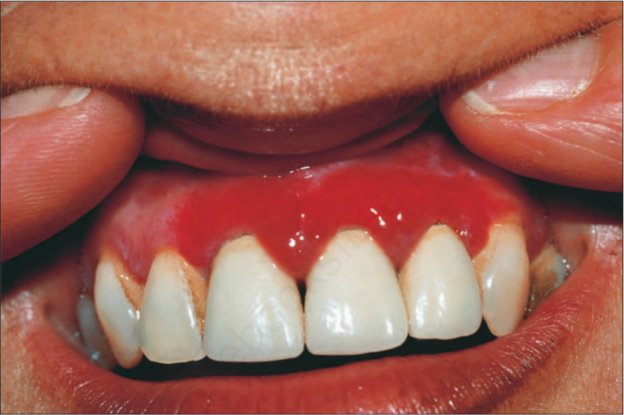
圖 12-44:外陰陰道–牙齦症候群 (vulvovaginal-gingival syndrome):注意伴有牙齦糜爛 (erosion of the gum) 的強烈紅斑。蒙倫敦皮膚科研究所 (Institute of Dermatology, London, UK) 惠允。
Fig. 12.44 Vulvovaginal-gingival syndrome: note the intense erythema with erosion of the gum. By courtesy of the Institute of Dermatology, London, UK.
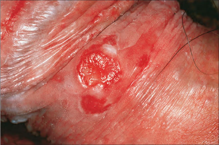
圖 12-45:扁平苔癬 (lichen planus):慢性陰莖病灶併發潰瘍性鱗狀細胞癌 (ulcerated squamous cell carcinoma)。蒙倫敦皮膚科研究所 (Institute of Dermatology, London, UK) 惠允。
Fig. 12.45 Lichen planus: chronic penile lesion complicated by an ulcerated squamous cell carcinoma. By courtesy of the Institute of Dermatology, London, UK.
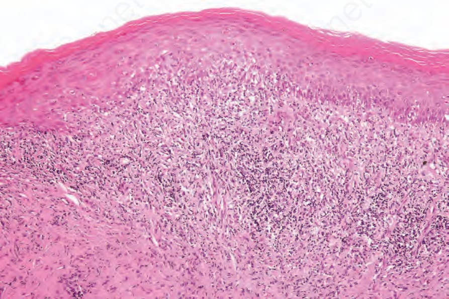
圖 12-46:扁平苔癬 (lichen planus):有角化過度 (hyperkeratosis)、棘層肥厚 (acanthosis) 與帶狀發炎細胞浸潤 (bandlike inflammatory cell infiltrate)。
Fig. 12.46 Lichen planus: there is hyperkeratosis, acanthosis, and a bandlike inflammatory cell infiltrate.
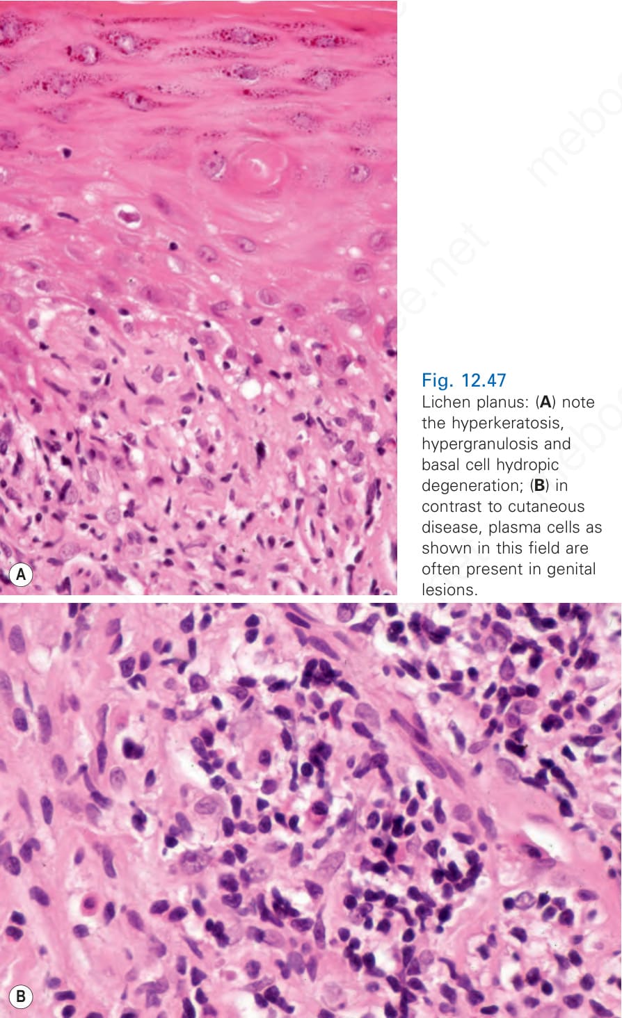
圖 12-47:扁平苔癬 (lichen planus):(A) 注意角化過度 (hyperkeratosis)、顆粒層增厚 (hypergranulosis) 與基底細胞水樣變性 (basal cell hydropic degeneration);(B) 與皮膚 (cutaneous) 疾病相反,如本視野所示的漿細胞 (plasma cells) 常出現於生殖器病灶。
Fig. 12.47 Lichen planus: (A) note the hyperkeratosis, hypergranulosis and basal cell hydropic degeneration; (B) in contrast to cutaneous disease, plasma cells as shown in this field are often present in genital lesions.