臨床特徵 (Clinical Features)
- 細胞性血管纖維瘤 (cellular angiofibroma) 是一種特殊的腫瘤,主要發生於中年女性的外陰 (vulva),極少數發生於陰道 (vagina)。
- 在男性較不常見,病灶發生於腹股溝陰囊 (inguinoscrotal) 區,罕見於肛周 (perianal) 區。
- 例外個案曾報告發生於後腹膜 (retroperitoneum),以及胸部、腹部、膝部、後腹膜、尿道、肛門、前列腺、上眼瞼、口腔黏膜及肘部的皮下組織。
- 腫瘤傾向於體積小且行為為良性,幾乎無局部復發傾向。極大的病灶屬例外。曾描述過兩處同時出現的睪丸病灶。曾報告肉瘤性變化 (sarcomatous change),但目前為止此變化並未與侵襲性行為相關聯。
組織病理特徵 (Histopathology)
- 腫瘤邊界清楚但無包膜 (unencapsulated),僅偶爾向周圍軟組織延伸 (Figs 12.291–12.293)。浸潤性型態 (infiltrative pattern) 罕見。
- 大多數病灶相當富細胞 (cellular),由短而溫和的梭形細胞 (spindle-shaped cells) 組成,其胞質界限不清、呈淡嗜伊紅性 (pale eosinophilic),並具空泡狀核 (vesicular nuclei)。核溝 (nuclear grooves) 與核內包涵體 (intranuclear inclusions) 常見。
- 有絲分裂相 (mitotic figures) 數目不一,但有時可能很明顯。除了纖細的膠原束 (collagen bundles) 與肥大細胞 (mast cells) 之外,常見厚壁、中等大小的玻璃樣變 (hyalinized) 血管。有時可見假血管腔隙 (pseudovascular spaces)。
- 常出現成熟脂肪細胞 (mature adipocytes)(多達 30% 的個案)。
- 曾描述局灶性細胞學異型 (focal cytological atypia),類似其他腫瘤中所見的合胞體樣變化 (symplastic changes),且可辨認出肉瘤性轉化 (sarcomatous transformation)。惡性區域通常顯示高細胞密度、細胞學異型及多核細胞 (multinucleated cells)。腫瘤罕見可顯示多形性脂肪肉瘤 (pleomorphic liposarcoma) 或非典型脂肪瘤樣腫瘤 (atypical lipomatous tumor) 的特徵。
致病機轉與組織學特徵 (Pathogenesis and Histologic Features)
- 細胞性血管纖維瘤的細胞遺傳學分析 (cytogenetic analysis) 顯示第 13q14 號染色體 (chromosome 13q14) 的缺失,此特徵亦見於梭形細胞脂肪瘤 (spindle cell lipoma) 與乳腺型肌纖維母細胞瘤 (mammary-type myofibroblastoma)。結合組織學上的相似性,此進一步支持這些腫瘤之間的關聯。
- 所描述的細胞遺傳學異常導致 Rb protein 核內表現的喪失,此特徵有助於與組織學上的模擬病灶 (histologic mimics) 區分。
- 腫瘤細胞對 vimentin 呈陽性,並在多達 50% 的個案中對 CD34 呈陽性。actin、desmin、caldesmon、S100 protein 及上皮性標記 (epithelial markers) 染色為陰性。
鑑別診斷 (Differential Diagnosis)
- 主要與血管肌纖維母細胞瘤 (angiomyofibroblastoma) 區分。後者由較具上皮樣 (epithelioid) 且 desmin 陽性的細胞組成,呈巢狀型態 (nested pattern) 並傾向於血管周圍分布 (perivascular distribution)。細胞性血管纖維瘤對 desmin 為陰性,且常對 CD34 呈陽性。
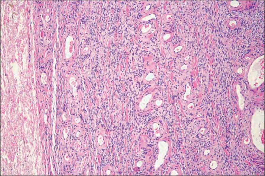
圖 12-291:細胞性血管纖維瘤 (cellular angiofibroma):腫瘤以厚壁、玻璃樣變 (hyalinized) 血管為特徵,並伴隨緻密富細胞的間質 (densely cellular stroma)。By courtesy of M. Nucci, MD, Brigham and Women’s Hospital and Harvard Medical School, Boston, USA.
Fig. 12.291 Cellular angiofibroma: the tumor is characterized by thick-walled, hyalinized blood vessels associated with a densely cellular stroma. By courtesy of M. Nucci, MD, Brigham and Women’s Hospital and Harvard Medical School, Boston, USA.
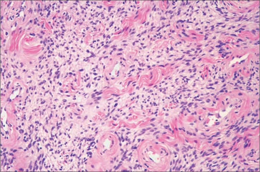
圖 12-292:細胞性血管纖維瘤 (cellular angiofibroma):注意伴隨的膠原纖維 (collagen fibers)。By courtesy of M. Nucci, MD, Brigham and Women’s Hospital and Harvard Medical School, Boston, USA.
Fig. 12.292 Cellular angiofibroma: note the associated collagen fibers. By courtesy of M. Nucci, MD, Brigham and Women’s Hospital and Harvard Medical School, Boston, USA.
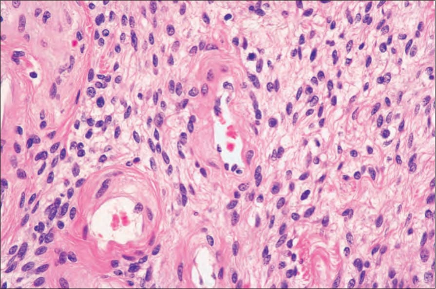
圖 12-293:細胞性血管纖維瘤 (cellular angiofibroma):腫瘤細胞小、均勻一致,具圓形至卵圓形的空泡狀核 (vesicular nuclei)。By courtesy of M. Nucci, MD, Brigham and Women’s Hospital and Harvard Medical School, Boston, USA.
Fig. 12.293 Cellular angiofibroma: the tumor cells are small, uniform and have round to oval vesicular nuclei. By courtesy of M. Nucci, MD, Brigham and Women’s Hospital and Harvard Medical School, Boston, USA.
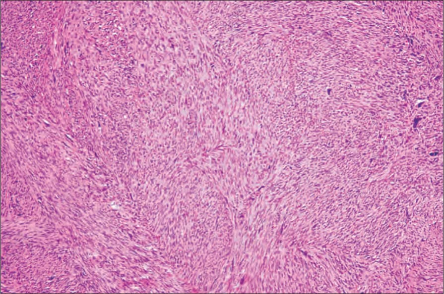
圖 12-294:外陰平滑肌肉瘤 (vulval leiomyosarcoma):此低倍視野顯示具嗜伊紅性胞質 (eosinophilic cytoplasm) 的腫瘤細胞束 (fascicles)。By courtesy of C. Crum, MD, Brigham and Women’s Hospital and Harvard Medical School, Boston, USA.
Fig. 12.294 Vulval leiomyosarcoma: this low-power view shows fascicles of tumor cells with eosinophilic cytoplasm. By courtesy of C. Crum, MD, Brigham and Women’s Hospital and Harvard Medical School, Boston, USA.