Cellular angiofibroma
Cellular angiofibroma
Clinical features Cellular angiofibroma is a distinctive tumor that occurs mainly on the vulva of middle-aged women,1–7 and very rarely in the vagina.6,7 Presentation in males is less common, with lesions occurring in the inguinoscrotal region or rarely in the perianal region.1,2,6,7 Exceptional cases have been reported in the retroperitoneum and in the subcutaneous tissue of the chest, abdomen, knee, retroperitoneum, urethra, anus, prostate, upper eyelid, oral mucosa, and elbow.2,7–12 Tumors tend to be small and behavior is benign, with almost no tendency for local recurrence.1,7,13 Very large lesions are exceptional.14,15 Two testicular lesions presenting simultaneously have been described.16 Sarcomatous change has been reported, but so far this has not been associated with aggressive behavior.7,17–19
Tumors are well circumscribed but unencapsulated, with only occasional extension into the surrounding soft tissues (Figs 12.291–12.293).1 An infiltrative pattern is rare.7 Most lesions are fairly cellular and composed of short, bland, spindle-shaped cells with poorly defined pale eosinophilic cytoplasm and vesicular nuclei. Nuclear grooves and intranuclear inclusions are common. The number of mitotic figures varies, but sometimes they may be prominent. Thick-walled, medium-sized hyalinized blood vessels are frequent in addition to slender collagen bundles and mast cells. Pseudovascular spaces are sometimes seen. Mature adipocytes are frequently present (in up to 30% of cases). Focal cytological atypia resembling symplastic changes seen in other tumors are described, and sarcomatous transformation can be identified.7,17–19,24,25 Malignant areas usually show high cellularity, cytologic atypia, and multinucleated cells.7 Tumors may rarely show features of a pleomorphic liposarcoma or of an atypical lipomatous tumor.6,18,19
Pathogenesis and histologic features Cytogenetic analysis of cellular angiofibroma has shown loss of chromosome 13q14, a feature also seen in spindle cell lipoma and mammary-type myofibroblastoma.7,20–22 In conjunction with histologic similarities, this gives further support to a link between these neoplasms. The cytogenetic abnormality described results in loss of nuclear expression of the Rb protein, a feature that is useful in distinction from histologic mimics.23
Tumor cells are positive for vimentin and are positive for CD34 in up to 50% of cases. Staining for actin, desmin, caldesmon, S100 protein, and epithelial markers is negative.1,2
556 Diseases of the anogenital skin
Differential diagnosis Distinction is mainly with angiomyofibroblastoma. The latter consists of more epithelioid desmin-positive cells with a nested pattern and a tendency for perivascular distribution. Cellular angiofibroma is negative for desmin and is often positive for CD34.
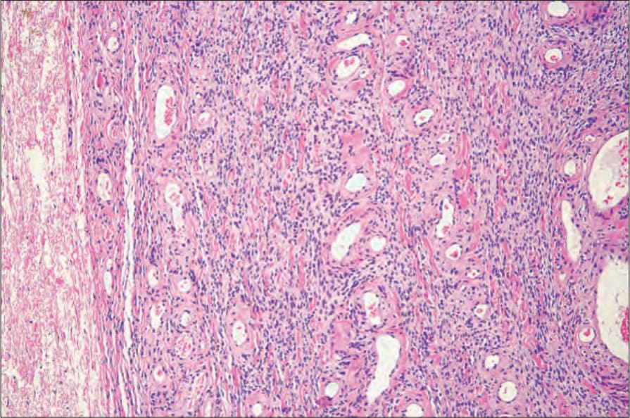
Fig. 12.291 Cellular angiofibroma: the tumor is characterized by thick-walled, hyalinized blood vessels associated with a densely cellular stroma. By courtesy of M. Nucci, MD, Brigham and Women’s Hospital and Harvard Medical School, Boston, USA.
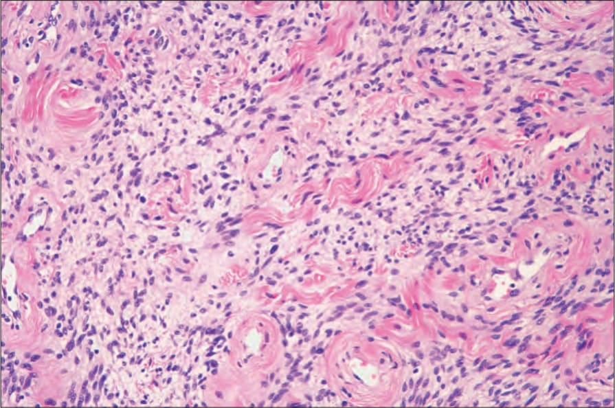
Fig. 12.292 Cellular angiofibroma: note the associated collagen fibers. By courtesy of M. Nucci, MD, Brigham and Women’s Hospital and Harvard Medical School, Boston, USA.
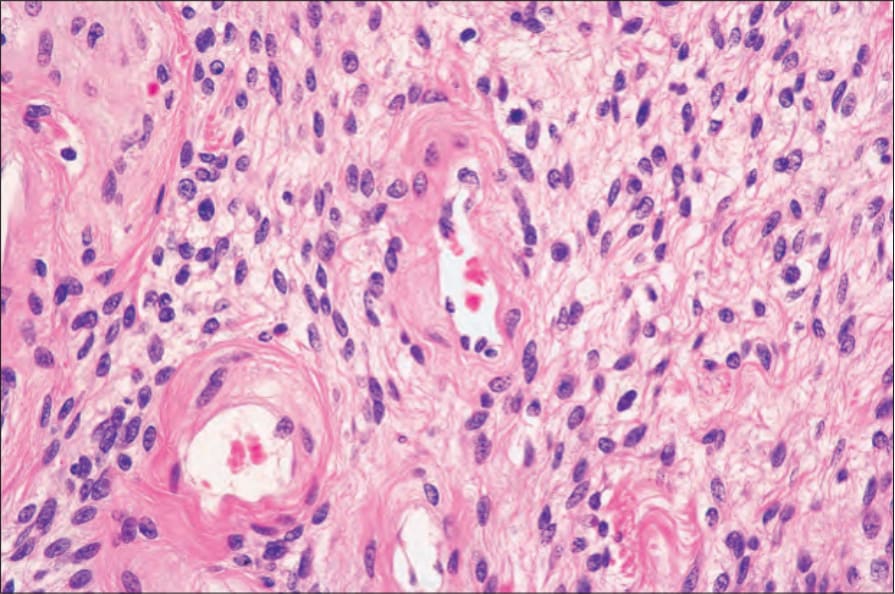
Fig. 12.293 Cellular angiofibroma: the tumor cells are small, uniform and have round to oval vesicular nuclei. By courtesy of M. Nucci, MD, Brigham and Women’s Hospital and Harvard Medical School, Boston, USA.
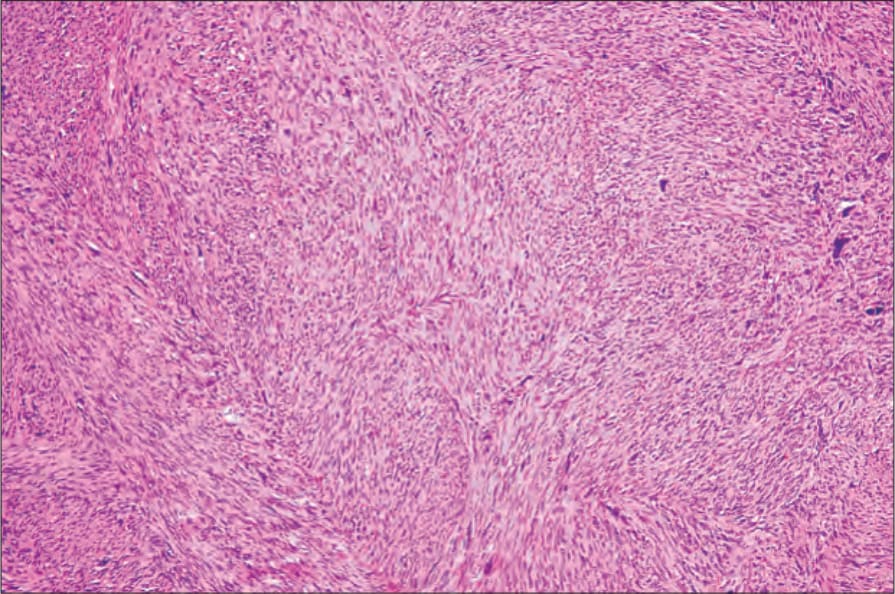
Fig. 12.294 Vulval leiomyosarcoma: this low-power view shows fascicles of tumor cells with eosinophilic cytoplasm. By courtesy of C. Crum, MD, Brigham and Women’s Hospital and Harvard Medical School, Boston, USA.