侵襲性血管黏液瘤 (Aggressive Angiomyxoma)
組織病理特徵 (Histopathology)
- 肉眼檢查可見一質軟、界線不清、呈分葉狀並帶有黏液樣變化 (myxoid change) 的腫瘤。
- 顯微鏡下,病灶呈浸潤性 (infiltrative),於黏液樣間質 (myxoid stroma) 中可見眾多小型與中型血管,以及少量腫瘤細胞 (Figs 12.288–12.290)。
- 血管壁厚,常呈玻璃樣變 (hyalinized)。
- 腫瘤細胞為小型、紡錘狀 (spindle-shaped) 或星狀 (stellate),具界線不清的淡粉紅色細胞質與泡狀核 (vesicular nuclei)。
- 細胞學上無異型性 (cytological atypia),有絲分裂象 (mitotic figures) 罕見。
- 血管旁常可見平滑肌束 (bundles of smooth muscle),此發現可以 desmin 染色凸顯。10
- 腫瘤常包陷殘存的正常結構,包括腺體與平滑肌。
- 散在的肥大細胞 (mast cells) 常見。
- 偶可見類似間質性息肉 (stromal polyps) 中所見的多核巨細胞 (multinucleated giant cells)。
- 部分病例在組織學上與 angiomyofibroblastoma 有重疊 (見前文)。
臨床特徵 (Clinical Features)
-
此腫瘤表現為一緩慢生長、無症狀的腫塊,侵犯骨盆與會陰部。1–6
-
罕見情況下,病灶可出現於後腹膜 (retroperitoneum)。7
-
主要影響第三或第四個十年 (third or fourth decade) 的女性,在兒童中極為罕見。8,9
-
不到 5% 的病例發生於男性,好發於陰囊 (scrotum)、會陰 (perineum) 或腹股溝 (groin)。10–12
-
在男性中,病灶可能類似陰囊水腫 (hydrocele) 或腹股溝疝氣 (inguinal hernia)。13,14
-
腫瘤通常直徑達 10 cm 或更大,有時可達非常大的體積。
-
因腫瘤外部壓迫,通常會繼發泌尿生殖系統 (genitourinary) 與肛門直腸 (anorectal) 症狀。
-
在女性中,病灶主要出現於外陰 (vulva) 或會陰,其次為陰道 (vagina) 與骨盆。
-
由於其廣泛的浸潤性生長,完整的手術切除往往困難;因此局部復發 (local recurrences) 頻繁,可見於高達 30% 的病例。
-
轉移 (metastasis) 極為罕見。15,16
-
已有罕見病例報告指出,腫瘤在以 gonadotrophin releasing hormone agonists 治療後體積顯著縮小。17,18
免疫組化與分子 (Immunohistochemistry & Molecular)
-
免疫組化方面,腫瘤細胞對 smooth-muscle actin 與 desmin 呈陽性。
-
亦可見 estrogen 與 progesterone receptors 陽性,而在男性中 androgen receptors 呈陽性。5,7,19
-
對多個侵襲性血管黏液瘤所做的細胞遺傳學分析 (cytogenetic analysis) 常顯示 chromosome 12q13–15 的重排 (rearrangements)。
-
後者導致 HMGA2 (高遷移率群蛋白家族 high-mobility-group protein family 的成員,先前稱為 HMGIC) 的異常表現。20–24
-
有趣的是,所涉及的區域 (12q14–15) 與其他數種腫瘤所報導的區域相同,包括 leiomyoma 與脂肪性腫瘤 (lipomatous neoplasms)。
-
HMGA2 染色可能有助於將 aggressive angiomyxoma 與其潛在的相似病灶 (主要為 angiomyofibroblastoma,見該節) 區別開來,但此標記雖然敏感,特異性並不高。25,26
-
電子顯微鏡 (electron microscopy) 顯示細胞具有纖維母細胞 (fibroblasts) 與肌纖維母細胞 (myofibroblasts) 的特徵。5
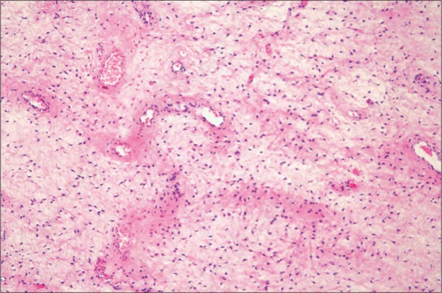
圖 12-288:侵襲性血管黏液瘤 (aggressive angiomyxoma):黏液樣間質 (myxoid stroma) 中散布著明顯的血管。By courtesy of M. Nucci, MD, Brigham and Women’s Hospital and Harvard Medical School, Boston, USA.
Fig. 12.288 Aggressive angiomyxoma: there are conspicuous blood vessels dispersed in a myxoid stroma. By courtesy of M. Nucci, MD, Brigham and Women’s Hospital and Harvard Medical School, Boston, USA.
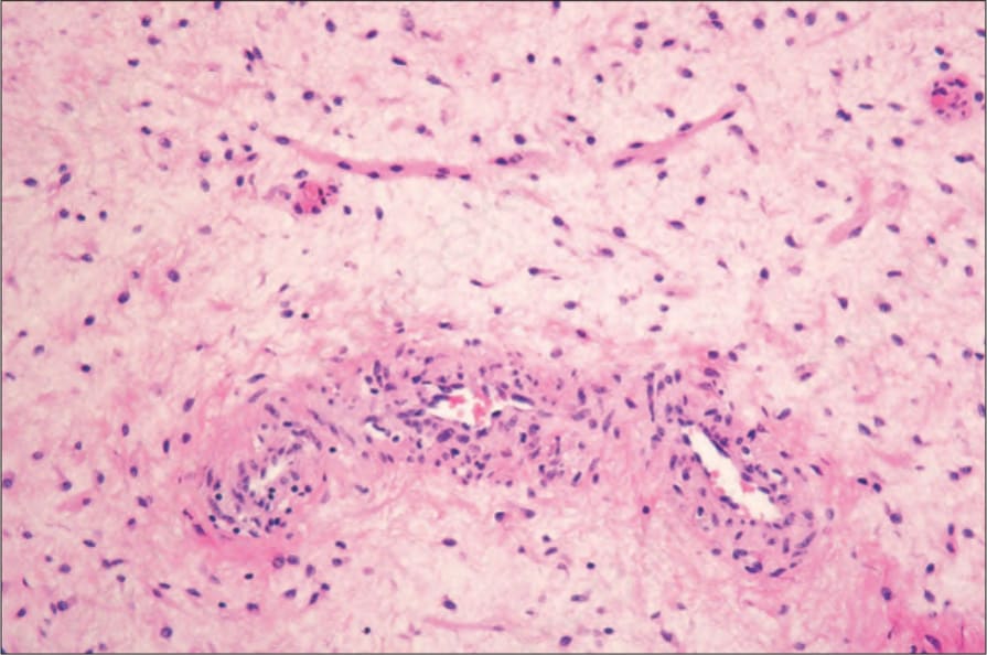
圖 12-289:侵襲性血管黏液瘤 (aggressive angiomyxoma):此視野中,上方可見明顯的平滑肌束 (smooth muscle bundle)。By courtesy of M. Nucci, MD, Brigham and Women’s Hospital and Harvard Medical School, Boston, USA.
Fig. 12.289 Aggressive angiomyxoma: in this view, a smooth muscle bundle is evident in the upper field. By courtesy of M. Nucci, MD, Brigham and Women’s Hospital and Harvard Medical School, Boston, USA.
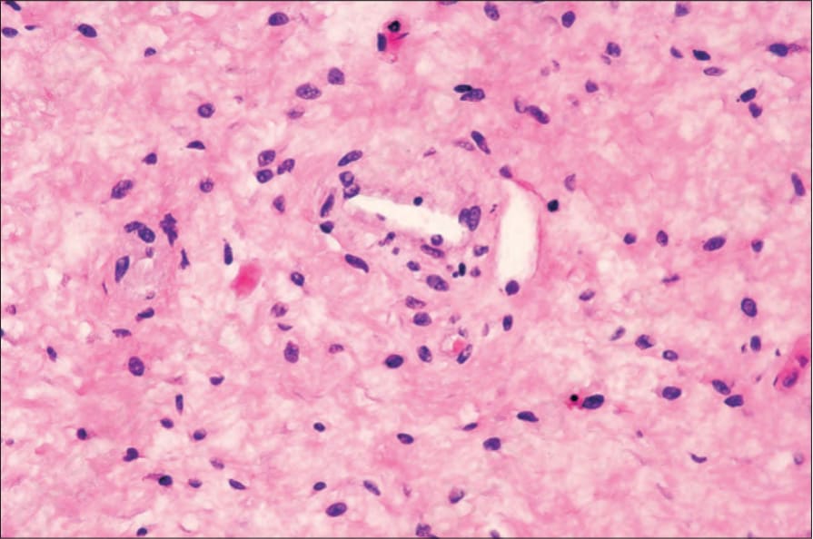
圖 12-290:侵襲性血管黏液瘤 (aggressive angiomyxoma):高倍視野顯示均一的細胞族群 (uniform cellular population)。無多形性 (pleomorphism)。By courtesy of M. Nucci, MD, Brigham and Women’s Hospital and Harvard Medical School, Boston, USA.
Fig. 12.290 Aggressive angiomyxoma: high-power view showing a uniform cellular population. There is no pleomorphism. By courtesy of M. Nucci, MD, Brigham and Women’s Hospital and Harvard Medical School, Boston, USA.
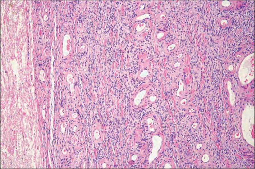
圖 12-291:細胞性血管纖維瘤 (cellular angiofibroma):此腫瘤的特徵為厚壁、玻璃樣變 (hyalinized) 的血管,伴隨密集的細胞性間質 (densely cellular stroma)。By courtesy of M. Nucci, MD, Brigham and Women’s Hospital and Harvard Medical School, Boston, USA.
Fig. 12.291 Cellular angiofibroma: the tumor is characterized by thick-walled, hyalinized blood vessels associated with a densely cellular stroma. By courtesy of M. Nucci, MD, Brigham and Women’s Hospital and Harvard Medical School, Boston, USA.