Aggressive angiomyxoma
Aggressive angiomyxoma
Histologic features Macroscopic examination reveals a soft, ill-defined, lobulated tumor with myxoid change. Microscopically, the lesion is infiltrative, with numerous small- and medium-sized blood vessels and a small number of tumor cells in a myxoid stroma (Figs 12.288–12.290). The blood vessels have thick walls, which are often hyalinized. Tumor cells are small, spindle-shaped, or stellate with ill-defined pale pink cytoplasm and vesicular nuclei. Cytological atypia is absent and mitotic figures are rare. Bundles of smooth muscle are frequently seen adjacent to blood vessels, a finding that can be highlighted by a desmin stain.10 Residual normal structures including glands and smooth muscle are often entrapped by the tumor. Scattered mast cells are often present. Multinucleated giant cells similar to those found in stromal polyps are occasionally found. Some cases overlap histologically with angiomyofibroblastoma (see above).
Clinical features This tumor presents as a slowly growing asymptomatic mass involving the pelvis and perineum.1–6 Exceptionally, the lesion can present in the retroperitoneum.7 It mainly affects females in the third or fourth decade of life, and it is exceptionally rare in children.8,9 Less than 5% of cases occur in males, with predilection for the scrotum, perineum, or groin.10–12 In males, lesions may mimic a hydrocele or an inguinal hernia.13,14 Tumors are often 10 cm or more in diameter and can sometimes attain a very large size. Genitourinary and anorectal symptoms usually ensue due to external compression by the tumor. In females, lesions present mainly in the vulva or perineum followed by the vagina and the pelvis. Because of its extensive infiltrative growth, complete surgical excision is often difficult; local recurrences are therefore frequent and occur in up to 30% of cases. Metastasis are exceptional.15,16
Rare case reports have been published of tumors displaying prominent reduction in size after treatment with gonadotrophin releasing hormone agonists.17,18
Immunohistochemically, tumor cells are positive for smooth-muscle actin and desmin. Positivity for estrogen and progesterone receptors is also seen, and in men androgen receptors are positive.5,7,19 Cytogenetic analysis of a number of aggressive angiomyxomas has often shown rearrangements of
555 Soft tissue tumors
chromosome 12q13–15. The latter results in an aberrant expression of HMGA2 (a member of the high-mobility-group protein family previously known as HMGIC).20–24 Interestingly, the area involved (12q14–15) is the same as that reported in a number of other tumors including leiomyoma and lipomatous neoplasms. Staining for HMGA2 may be useful to distinguish aggressive angiomyxoma from potential mimics, mainly angiomyofibroblastoma (see under the latter), but this marker, although sensitive, is not very specific.25,26
Electron microscopy shows cells with features of fibroblasts and myofibroblasts.5
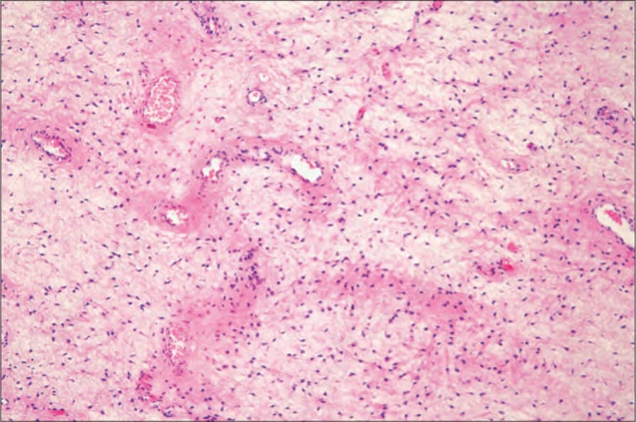
Fig. 12.288 Aggressive angiomyxoma: there are conspicuous blood vessels dispersed in a myxoid stroma. By courtesy of M. Nucci, MD, Brigham and Women’s Hospital and Harvard Medical School, Boston, USA.
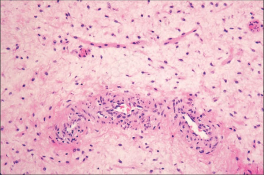
Fig. 12.289 Aggressive angiomyxoma: in this view, a smooth muscle bundle is evident in the upper field. By courtesy of M. Nucci, MD, Brigham and Women’s Hospital and Harvard Medical School, Boston, USA.
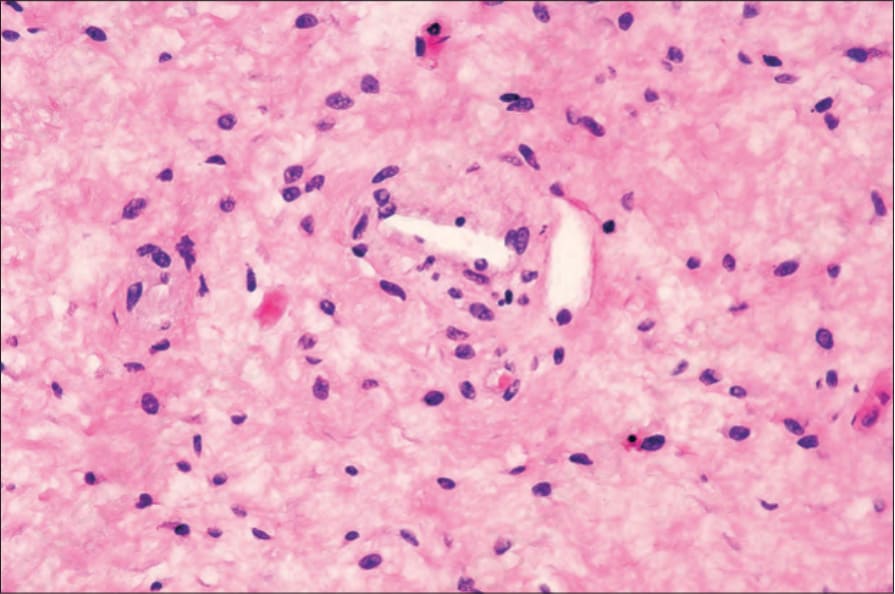
Fig. 12.290 Aggressive angiomyxoma: high-power view showing a uniform cellular population. There is no pleomorphism. By courtesy of M. Nucci, MD, Brigham and Women’s Hospital and Harvard Medical School, Boston, USA.
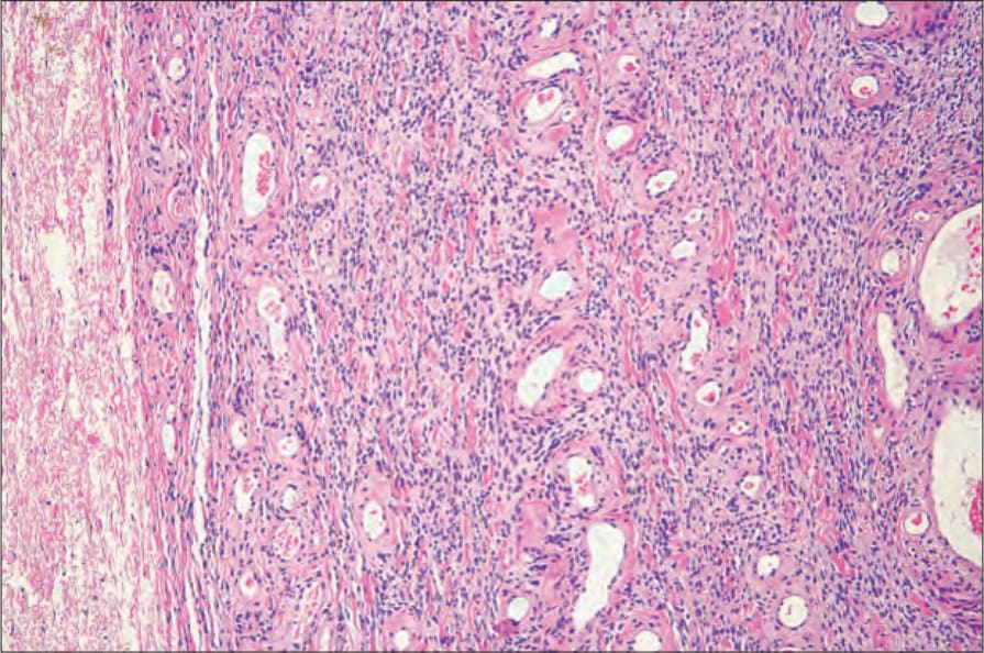
Fig. 12.291 Cellular angiofibroma: the tumor is characterized by thick-walled, hyalinized blood vessels associated with a densely cellular stroma. By courtesy of M. Nucci, MD, Brigham and Women’s Hospital and Harvard Medical School, Boston, USA.