臨床特徵 (Clinical Features)
- 唾石 (sialoliths) 或稱唾液腺結石 (salivary gland calculi) 好發於第六至第七個十年的病人。
- 位於小唾液腺者通常無症狀,除非合併感染;而位於大唾液腺者——尤其是 Wharton duct(唾石最常見的部位)——會在進食前出現疼痛、壓迫感與腫脹等症狀。
- 小唾液腺唾石最常見的部位為上唇黏膜 (upper lip mucosa) 與頰黏膜 (buccal mucosa),兩者合計佔所有小唾液腺結石 (minor salivary gland lithiasis) 的 75–90%。其表現為硬結節 (hard nodules),若感染可能滲流 (drain)。
致病機轉與組織學特徵 (Pathogenesis and Histologic Features)
- 唾石圍繞著一個由細菌、細胞碎屑及/或黏液栓 (mucous plug) 構成的中央核心 (central nidus) 形成。由於導管長而迂曲,唾石最常見於口腔底部 (floor of mouth) 的 Wharton duct。
- 唾石由位於囊狀擴張之排泄導管 (cystically dilated excretory duct) 內的同心層狀 (concentric lamellar) 與球狀鈣化 (globular calcifications) 構成,該導管的內襯呈現鱗狀、黏液細胞或纖毛細胞化生 (squamous, mucous cell or ciliated cell metaplasia),並伴有導管周圍發炎 (periductal inflammation)(Fig. 11.188)。唾石內或周圍可能存在細菌、發炎細胞及細胞碎屑。破裂 (rupture) 可導致急性與慢性發炎及/或異物反應 (foreign body reaction)。約四分之一的結石為未礦化 (unmineralized)。
鑑別診斷 (Differential Diagnosis)
- 靜脈石 (phleboliths) 與鈣化血栓 (calcified thrombi) 一般不具層狀構造 (not lamellated),且發生於內襯內皮細胞 (endothelial cells) 的血管腔 (vascular lumina) 內。
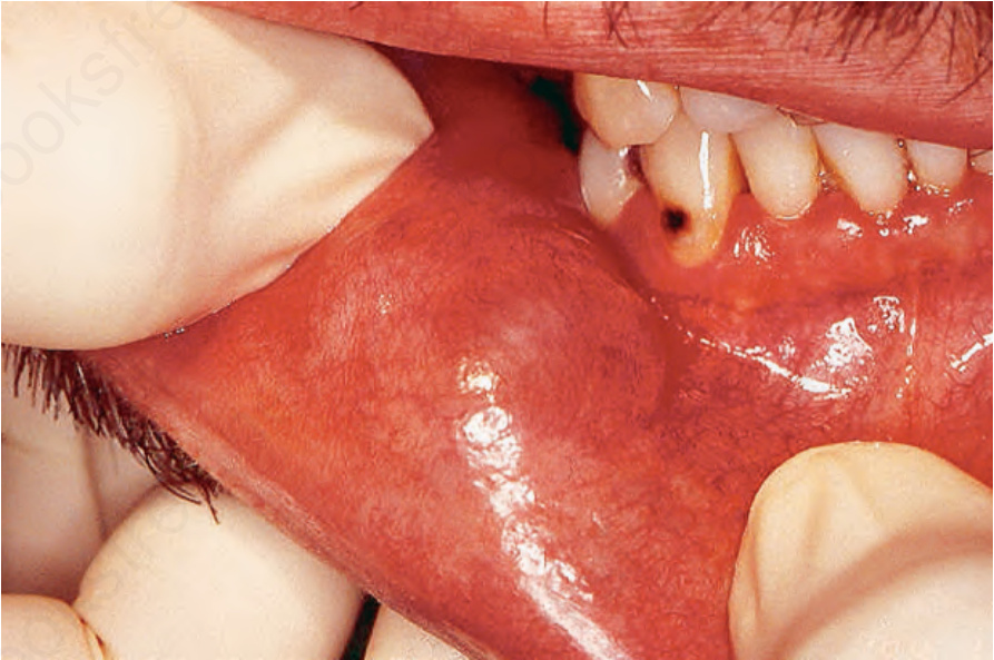
圖 11-179:黏液囊腫 (mucocele):注意下唇黏膜上呈藍色的無柄結節 (bluish sessile nodule),此為典型部位。
Fig. 11.179 Mucocele: note the bluish sessile nodule on the lower labial mucosa, a typical site.
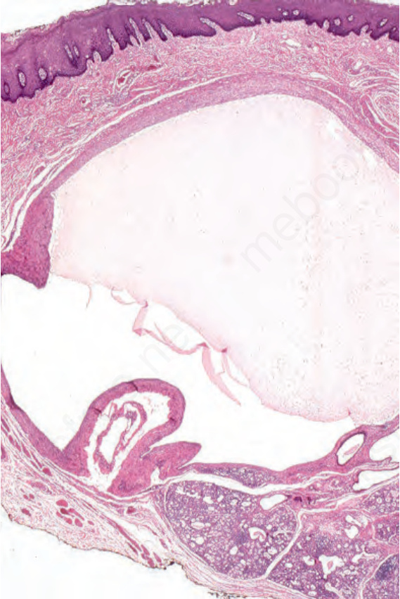
圖 11-180:黏液囊腫 (mucocele):可見一充滿黏液 (mucin) 的囊樣腔 (cyst-like cavity),周圍為肉芽組織 (granulation tissue),無內襯上皮 (lining epithelium)。
Fig. 11.180 Mucocele: there is a cyst-like cavity filled with mucin surrounded by granulation tissue without lining epithelium.
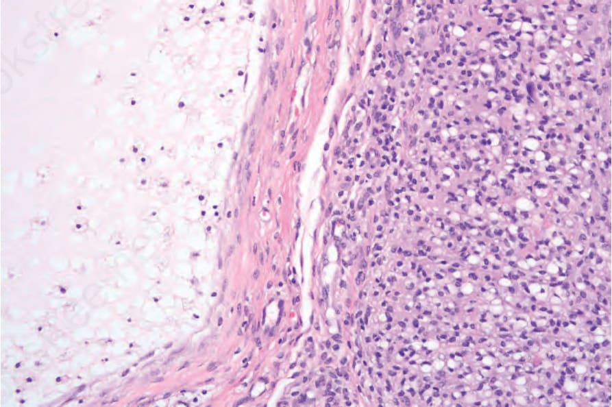
圖 11-181:黏液囊腫 (mucocele):黏液囊腫含有黏液 (mucin) 與噬黏液細胞 (muciphages),周圍包繞著含有噬黏液細胞與發炎細胞的肉芽組織 (granulation tissue);無內襯上皮存在。
Fig. 11.181 Mucocele: the mucocele contains mucin and muciphages and is surrounded by granulation tissue containing muciphages and inflammatory cells; there is no lining epithelium present.
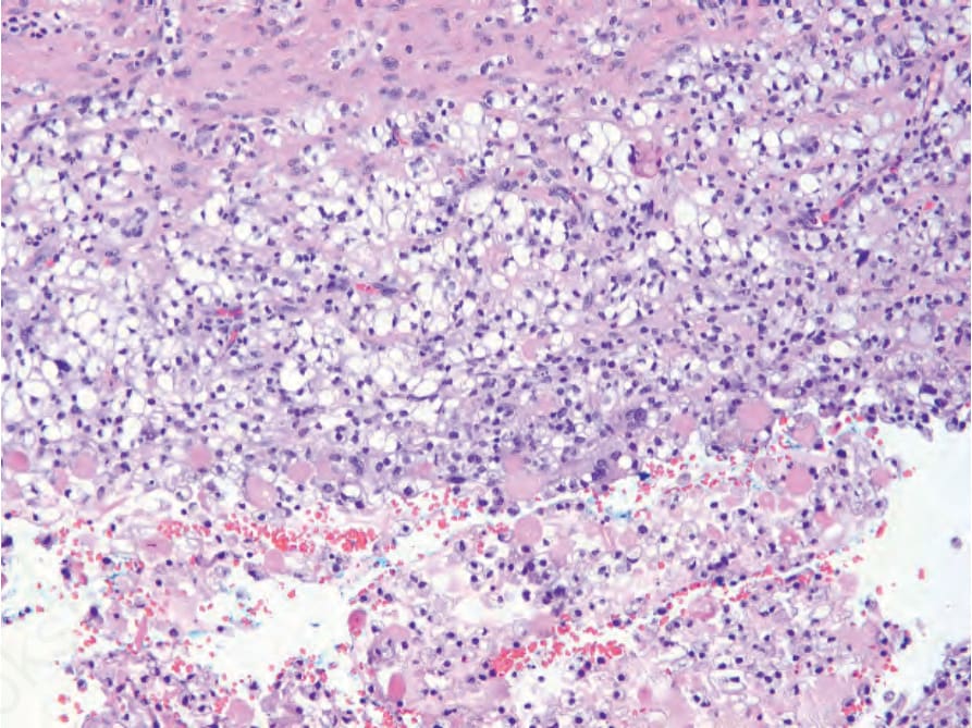
圖 11-182:黏液囊腫 (mucocele):黏液囊腫的囊壁可能含有透明細胞 (clear cells)。
Fig. 11.182 Mucocele: the wall of a mucocele may contain clear cells
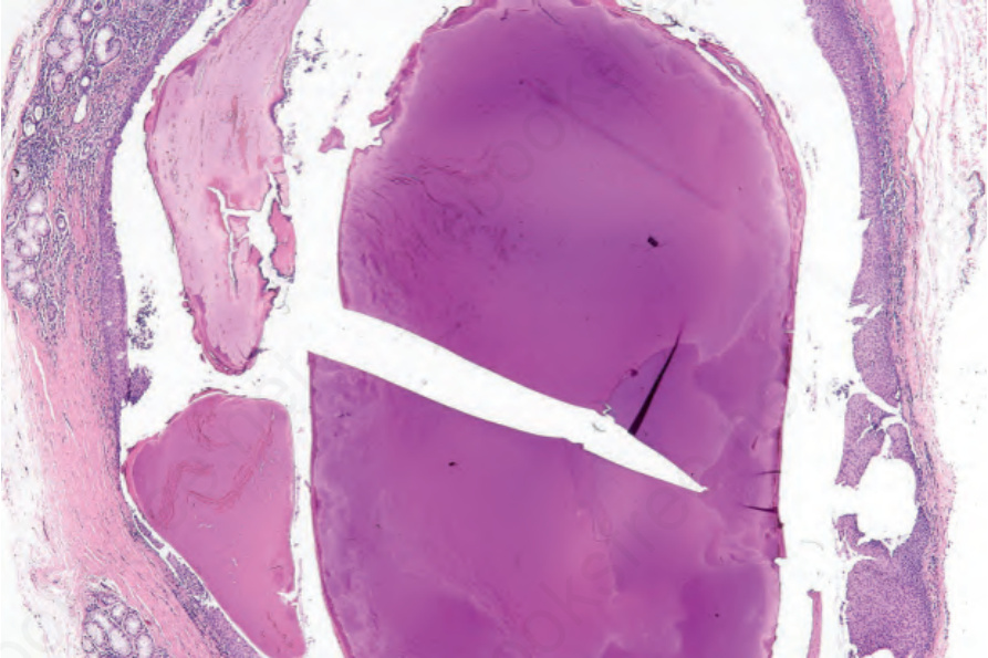
圖 11-188:唾石 (sialolith):唾石位於擴張的唾液導管 (ectatic salivary duct) 內,其內襯呈現鱗狀化生 (squamous metaplasia)。
Fig. 11.188 Sialolith: the sialolith is present within an ectatic salivary duct, the lining of which exhibits squamous metaplasia.