引言
自 1968 年首次將過氧化酶標記抗體法 (peroxidase-labeled antibody method) 應用於石蠟包埋組織以來,免疫組織化學 (immunohistochemistry, IHC) 已成為組織形態學評估的一項強大輔助檢查。IHC 在皮膚病理的診斷、預後、治療與致病機轉等方面有廣泛的應用,不僅應用於一系列的腫瘤性 (neoplastic)(表 2.2)、免疫性水疱性 (immunobullous) 與感染性疾病,也應用於反應性 (reactive) 與腫瘤性病變之間的區別。免疫組織學技術可以手動方式或在自動化平台上進行(圖 2.8)。自動化雖能提升染色的品質與再現性,但對於許多仍以手動執行 IHC 的實驗室而言,詳細而精確的 IHC 流程是達成最佳、可再現結果的關鍵。
表 2.1 內容:常用組織化學染色
下列內容對應表 2.1(染色/成分/結果)所示之常用組織化學染色:
A. 常規 (Routine)
- Hematoxylin-eosin(蘇木精-伊紅)— 細胞、結締組織 — 細胞核:藍色;細胞質:粉紅/紅色;細胞外基質 (extracellular matrix):紅/粉紅色。
B. 碳水化合物與醣綴合物 (Carbohydrates & glycoconjugates)
- Periodic acid-Schiff (PAS) — 中性黏蛋白 (neutral mucins)、肝醣 (glycogen) — 洋紅色 (magenta)。
- PAS-diastase — 肝醣、蛋白聚醣 (proteoglycans)、HA、抗澱粉酶之唾液黏蛋白 (resistant sialomucin) — 對澱粉酶消化具抗性 (resistant to diastase digestion)。
- Alcian blue, pH 2.5 — 不穩定之唾液黏蛋白 (labile sialomucin) — 藍色。
- Alcian blue, pH 1.0 — 硫酸黏蛋白 (sulfomucin)、抗性唾液黏蛋白 (resistant sialomucin) — 藍色。
- Mucicarmine — 唾液黏蛋白 (sialomucin)、硫酸黏蛋白 (sulfomucin) — 粉紅色。
- Colloidal iron — 唾液黏蛋白、硫酸黏蛋白、HA、蛋白聚醣 — 藍色。
- High iron diamine — 蛋白聚醣、硫酸黏蛋白 — 藍色。
- Toluidine blue — 硫酸黏蛋白 — 藍色。
- Hyaluronidase — HA — 對 HA 敏感 (sensitive to HA)。
C. 結締組織纖維 (Connective tissue fibers)
- Masson trichrome — 膠原蛋白 (collagen):藍色或綠色;肌肉、神經 (muscle, nerve):紅色。
- Verhöeff-van Gieson — 彈性纖維 (elastic fibers) — 黑色。
- Pinkus acid orcein — 彈性 (elastic) — 深棕色。
- Silver nitrate — 網狀纖維 (reticulum fibers) — 黑色。
D. 感染性染色 (Infective stains)
- Ziehl Neelsen — 抗酸桿菌 (acid fast bacilli) — 紅色。
- Fite-Faraco — (弱)抗酸桿菌 ((weakly) acid fast bacilli) — 紅色。
- PAS — 真菌、寄生蟲 (fungi, parasites) — 洋紅色 (magenta)。
- Mucicarmine — Cryptococcus sp — 紅色。
- Giemsa — Leishmania sp、Donovan bodies — 紅色/異染性紫色 (metachromatically purple)。
- Methenamine silver — 真菌、細菌 (fungi, bacteria) — 黑色。
- Grocott methenamine silver — 真菌 (fungi) — 黑色。
- Warthin Starry silver — 螺旋體、細菌 (spirochetes, bacteria) — 黑色。
- Dieterle and Steiner silver — 螺旋體、細菌 — 黑色。
E. 其他 (Other)
- Perl potassium ferrocyanide — 含鐵血黃素 (hemosiderin) — 藍色。
- Oil red O — 脂質 (lipids) — 紅色。
- Scarlet red — 脂質 — 紅色。
- Von Kossa — 磷酸鹽 (phosphate)(常以磷酸鈣 (calcium phosphate) 形式存在) — 黑色。
- Alizarin red S — 鈣 (calcium) — 橙紅色 (orange-red)。
- Congo red — 澱粉樣蛋白 (amyloid) — 蘋果綠色雙折射 (apple green birefringence)。
- Chloro-acetate esterase — 骨髓系細胞 (myeloid series) — 紅色顆粒 (red granules)。
HA, Hyaluronic acid(玻尿酸)。
表 2.2 內容:皮膚腫瘤的免疫組織化學診斷應用
下列內容對應表 2.2(染色/應用)所示之皮膚腫瘤診斷性免疫組織化學應用:
表皮與附屬器腫瘤 (Epidermal and appendageal neoplasms)
- AE1/AE3 — Pan-keratin(全角蛋白)。確認上皮譜系 (epithelial lineage)。
- CAM 5.2 — CKs 8, 18。確認上皮譜系。有助於確認腺體腫瘤 (glandular neoplasms)。
- MNF 116 — CKs 5, 6, 8, 17, 19。有助於診斷具單細胞浸潤 (single cell infiltration) 的 SCC。
- BerEP4 — 在 BCC 中為陽性,在 SCC 中為陰性。
- CK 7 — 用於確認乳房型與乳房外型 Paget 病 (mammary and extramammary Paget disease)。
- p63 — 區分原發性皮膚梭形細胞 SCC (primary cutaneous spindle SCC) 與間葉性梭形細胞腫瘤 (mesenchymal spindle cell tumors);以及區分原發性皮膚附屬器腫瘤與轉移性腺癌 (metastatic adenocarcinomas)。
- CD10 — 毛上皮瘤 (Trichoepithelioma):基質 (stroma) 與乳頭 (papillae) 為陽性,上皮為陰性。BCC:上皮為陽性,基質為陰性。
- bcl2 — 在 BCC 中為陽性,在 SCC 中為陰性。
血管增生性病變 (Vascular proliferations)
- ERG — 內皮細胞 (endothelial cells) 之細胞核染色。對內皮腫瘤具高特異性與特異性(優於 CD31)。
- CD31 — 對內皮腫瘤具高特異性與良好靈敏度。
- CD34 — 對內皮腫瘤具高靈敏度但低特異性。
- Fli-1 — 內皮細胞之細胞核染色。
- GLUT 1 — 在所有幼年型血管瘤 (juvenile hemangiomas) 的內皮細胞中為陽性。在先天性血管瘤 (congenital hemangiomas)(快速消退型先天性血管瘤 (rapidly involuting congenital hemangioma) 與不消退型先天性血管瘤 (noninvoluting congenital hemangioma)) 中通常為陰性。
黑色素細胞腫瘤 (Melanocytic tumors)
- S100 protein — 最廣泛使用的黑色素細胞標記。靈敏度高,但特異性不如其他黑色素細胞標記。
- Sox-10 — 新近引進的黑色素細胞標記,在神經腫瘤 (neural tumors) 中亦為陽性。對梭形細胞黑色素瘤 (spindle cell melanomas)(包括促結締組織增生性黑色素瘤 (desmoplastic melanoma))非常有用,並用於評估表皮內黑色素細胞增生(如惡性小痣 (lentigo maligna))(細胞核染色)。
- MITF-1 — 特異性低,但對評估表皮內黑色素細胞增生有用(細胞核染色)。
- HMB 45 — 特異性良好但靈敏度相對較低。在梭形細胞黑色素瘤中傾向為陰性。在 PEComa 中亦為陽性。
- Melan A / Mart 1 — 與 HMB45 具相似特異性。在梭形細胞黑色素瘤中傾向為陰性。
- Ki-67 — 黑色素瘤的增殖指數 (proliferation index) 較高 (13–35%),痣 (nevi) 則較低 (< 5%)。對評估某些黑色素細胞腫瘤有用,主要是痣樣黑色素瘤 (nevoid melanoma)。
神經外胚層與神經腫瘤 (Neuroectodermal and neural tumors)
- S100 protein — 在神經外胚層、神經元 (neuronal)、神經鞘 (nerve sheath)、軟骨樣 (chondroid) 腫瘤、某些汗腺腫瘤 (sweat gland tumors) 與肌上皮瘤 (myoepithelioma) 中為陽性。
- NSE — Merkel cell carcinoma(梅克爾細胞癌)。
- CK 20 — Merkel cell carcinoma。
- Neurofilament — Merkel cell carcinoma。
- Chromogranin — Merkel cell carcinoma。
- Synaptophysin — Merkel cell carcinoma。
- TTF1 — 在大多數 Merkel cell carcinoma 中為陰性。
肌源性/肌纖維母細胞分化 (Myogenic/myofibroblastic differentiation)
- MSA — 肌肉來源腫瘤 (tumors of muscle origin)。
- Desmin — 肌肉來源腫瘤(平滑肌與骨骼肌,罕見且局部出現於肌纖維母細胞腫瘤 (myofibroblastic tumors))。
- Myogenin — 在橫紋肌肉瘤 (rhabdomyosarcoma) 中為陽性。
- SMA — 在平滑肌腫瘤、血管球瘤 (glomus tumor)、肌周細胞瘤 (myopericytoma)、皮膚肌纖維瘤 (dermatomyofibroma) 中為陽性。
BCC, Basal cell carcinoma(基底細胞癌);CK, cytokeratin(細胞角蛋白);MSA, muscle specific actin(肌肉特異性肌動蛋白);PEComa, perivascular epithelioid cell tumor(血管周圍上皮樣細胞腫瘤);SCC, squamous cell carcinoma(鱗狀細胞癌);SMA, antismooth muscle actin(抗平滑肌肌動蛋白)。
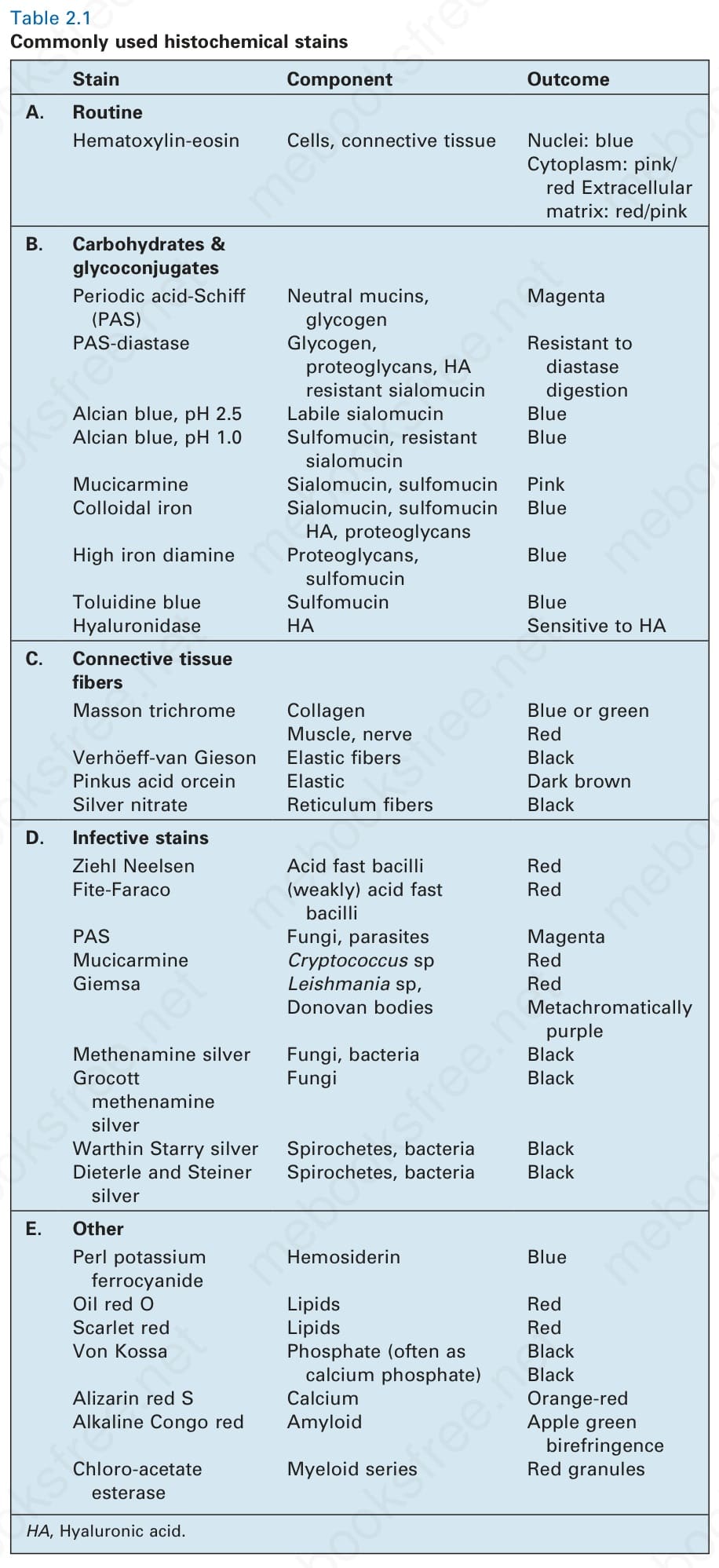
表 2-1:常用的組織化學染色 (commonly used histochemical stains)。
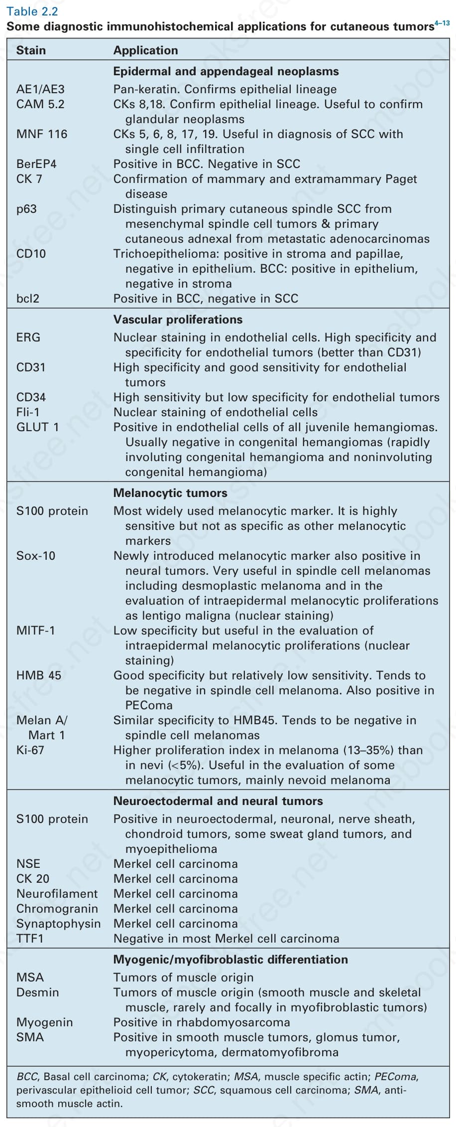
表 2-2:皮膚腫瘤的部分診斷性免疫組織化學應用 (some diagnostic immunohistochemical applications for cutaneous tumors)。
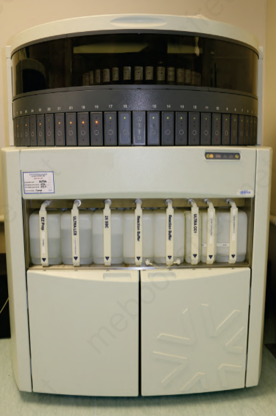
圖 2-8(圖說烘焙於圖內)。