Immunohistochemical techniques
Immunohistochemical techniques
Since the first practical application of antibodies using the peroxidase-labeled antibody method on paraffin-embedded tissues in 1968, immunohistochemistry (IHC) has emerged as a powerful supplementary investigation to histomorphological assessment.1–3 IHC has widespread dermatopathological diagnostic, prognostic, therapeutic, and pathogenetic applications, not only in a range of neoplastic (Table 2.2), immunobullous, and infective disease, but also in the distinction between reactive and neoplastic disorders.4–14 Immunohistological techniques can be performed manually or in automated platforms (Fig. 2.8). While automation allows enhanced quality and reproducibility of staining; detailed, exact IHC protocols are critical in the many laboratories that still perform manual IHC, to achieve optimal, reproducible results.
38 Specialized techniques in dermatopathology
Stain Component Outcome
A. Routine Hematoxylin-eosin Cells, connective tissue Nuclei: blue Cytoplasm: pink/
red Extracellular matrix: red/pink
B. Carbohydrates & glycoconjugates Periodic acid-Schiff
Stain Application
Epidermal and appendageal neoplasms AE1/AE3 Pan-keratin. Confirms epithelial lineage CAM 5.2 CKs 8,18. Confirm epithelial lineage. Useful to confirm
glandular neoplasms MNF 116 CKs 5, 6, 8, 17, 19. Useful in diagnosis of SCC with
single cell infiltration BerEP4 Positive in BCC. Negative in SCC CK 7 Confirmation of mammary and extramammary Paget
Neutral mucins,
Magenta
(PAS)
glycogen
PAS-diastase Glycogen,
Resistant to
proteoglycans, HA resistant sialomucin
diastase digestion Alcian blue, pH 2.5 Labile sialomucin Blue Alcian blue, pH 1.0 Sulfomucin, resistant
Blue
sialomucin
Mucicarmine Sialomucin, sulfomucin Pink Colloidal iron Sialomucin, sulfomucin
Blue
HA, proteoglycans
High iron diamine Proteoglycans,
Blue
sulfomucin
Toluidine blue Sulfomucin Blue Hyaluronidase HA Sensitive to HA
disease p63 Distinguish primary cutaneous spindle SCC from
mesenchymal spindle cell tumors & primary cutaneous adnexal from metastatic adenocarcinomas CD10 Trichoepithelioma: positive in stroma and papillae,
negative in epithelium. BCC: positive in epithelium, negative in stroma bcl2 Positive in BCC, negative in SCC
Vascular proliferations ERG Nuclear staining in endothelial cells. High specificity and
specificity for endothelial tumors (better than CD31) CD31 High specificity and good sensitivity for endothelial
tumors CD34 High sensitivity but low specificity for endothelial tumors Fli-1 Nuclear staining of endothelial cells GLUT 1 Positive in endothelial cells of all juvenile hemangiomas.
C. Connective tissue fibers Masson trichrome Collagen Blue or green Muscle, nerve Red Verhöeff-van Gieson Elastic fibers Black Pinkus acid orcein Elastic Dark brown Silver nitrate Reticulum fibers Black
D. Infective stains Ziehl Neelsen Acid fast bacilli Red Fite-Faraco (weakly) acid fast
Red
bacilli
PAS Fungi, parasites Magenta Mucicarmine Cryptococcus sp Red Giemsa Leishmania sp, Donovan bodies
Usually negative in congenital hemangiomas (rapidly involuting congenital hemangioma and noninvoluting congenital hemangioma)
Melanocytic tumors S100 protein Most widely used melanocytic marker. It is highly
sensitive but not as specific as other melanocytic markers Sox-10 Newly introduced melanocytic marker also positive in
neural tumors. Very useful in spindle cell melanomas including desmoplastic melanoma and in the evaluation of intraepidermal melanocytic proliferations as lentigo maligna (nuclear staining) MITF-1 Low specificity but useful in the evaluation of
Red Metachromatically
purple Methenamine silver Fungi, bacteria Black Grocott
Fungi Black
methenamine silver
Warthin Starry silver Spirochetes, bacteria Black Dieterle and Steiner
Spirochetes, bacteria Black
silver
E. Other Perl potassium
Hemosiderin Blue
ferrocyanide
Oil red O Lipids Red Scarlet red Lipids Red Von Kossa Phosphate (often as
intraepidermal melanocytic proliferations (nuclear staining) HMB 45 Good specificity but relatively low sensitivity. Tends to
be negative in spindle cell melanoma. Also positive in PEComa Melan A/
Similar specificity to HMB45. Tends to be negative in
Mart 1
spindle cell melanomas Ki-67 Higher proliferation index in melanoma (13–35%) than
in nevi (< 5%). Useful in the evaluation of some melanocytic tumors, mainly nevoid melanoma
Neuroectodermal and neural tumors S100 protein Positive in neuroectodermal, neuronal, nerve sheath,
chondroid tumors, some sweat gland tumors, and myoepithelioma NSE Merkel cell carcinoma CK 20 Merkel cell carcinoma Neurofilament Merkel cell carcinoma Chromogranin Merkel cell carcinoma Synaptophysin Merkel cell carcinoma TTF1 Negative in most Merkel cell carcinoma
Black Black Alizarin red S Calcium Orange-red Alkaline Congo red Amyloid Apple green
calcium phosphate)
birefringence Chloro-acetate
Myeloid series Red granules
esterase
HA, Hyaluronic acid.
Myogenic/myofibroblastic differentiation MSA Tumors of muscle origin Desmin Tumors of muscle origin (smooth muscle and skeletal
muscle, rarely and focally in myofibroblastic tumors) Myogenin Positive in rhabdomyosarcoma SMA Positive in smooth muscle tumors, glomus tumor,
myopericytoma, dermatomyofibroma
BCC, Basal cell carcinoma; CK, cytokeratin; MSA, muscle specific actin; PEComa, perivascular epithelioid cell tumor; SCC, squamous cell carcinoma; SMA, antismooth muscle actin.
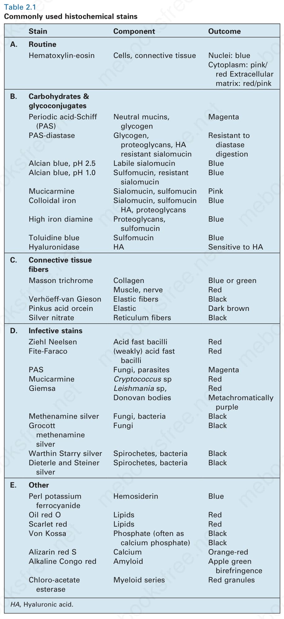
Table 2.1 Commonly used histochemical stains
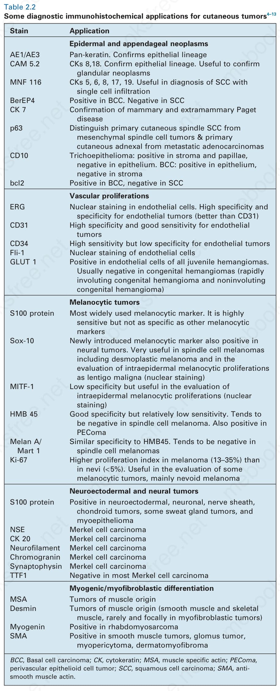
Table 2.2 Some diagnostic immunohistochemical applications for cutaneous tumors4–13
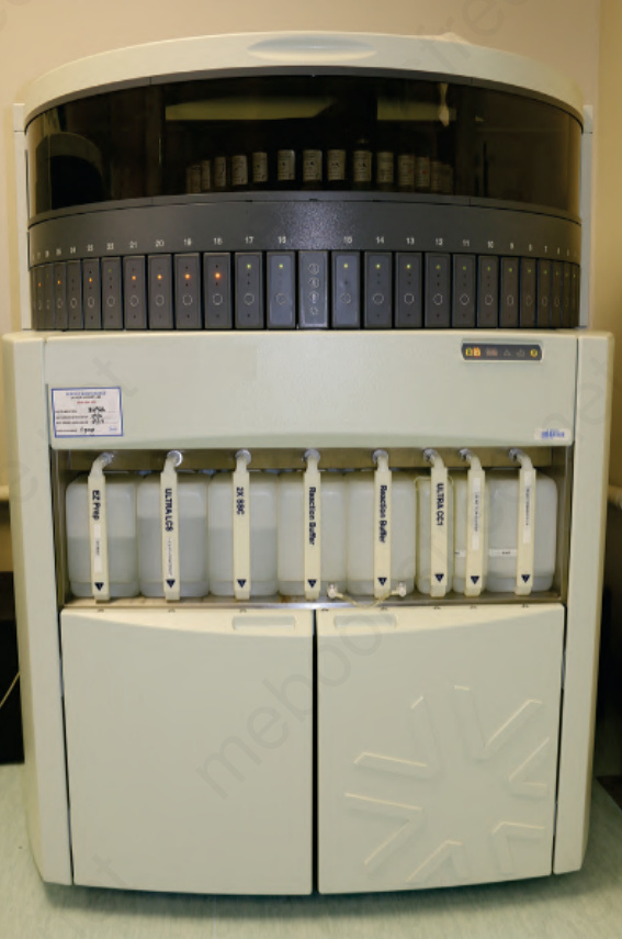
Fig. 2-8 (caption embedded in image / 圖說烘焙於圖內)